Description
Introduction
Congenital rubella syndrome (CRS) is a spectrum of clinical findings in a patient that results from primary maternal infection with rubella virus. Diagnosis is usually made clinically but imaging findings can prompt the diagnosis in appropriate clinical setting. The features of congenital rubella include sensorineural deafness, cataract, cardiac anomalies, mental retardation and microcephaly.
Case report
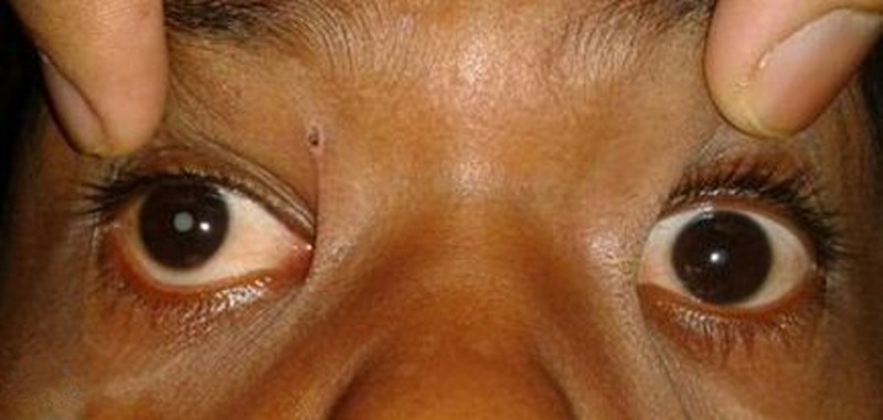
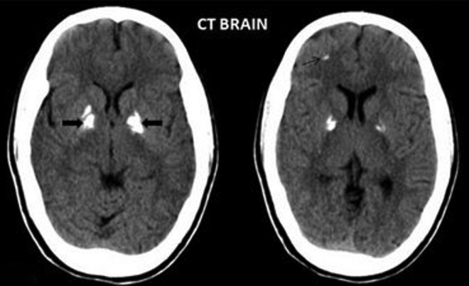
We report a case of 10-year-old girl who presented with a history of global developmental delay, intellectual disability and having been treated for patent ductus arteriosus. There was no maternal history of exposure to infection or vaccination during her pregnancy. The girl was the second child of a non-consanguineous marriage with no similar symptoms in her elder sibling. Clinical examination revealed mild facial asymmetry and hearing loss with bilateral central cataract (R>L; figure 1). CT of the brain (figure 2) showed bilateral dense, coarse calcifications of basal ganglia and a linear calcification was also noted in the right frontal lobe in the subcortical white matter.
Figure 1.

Clinical photograph of a 10-year-old girl shows bilateral cataract (right>left).
Figure 2.
CT of the brain reveals bilateral dense basal ganglia calcification (block arrow) and subcortical white matter calcification in the right frontal lobe (thin arrow).
Differential diagnosis
Basal ganglia and cortical calcifications are common features of all infections that constitute the toxoplasmosis, rubella, cytomegalovirus, herpes simplex virus (TORCH) syndrome. Cytomegalovirus and toxoplasmosis cause bilateral periventricular and subependymal calcifications.1 Interestingly, calcifications of toxoplasmosis may be resolved with therapy.2 Calcification pattern of TORCH infection is not specific for any infection.2 3
Endocrinal disorders such as hypoparathyroidism, pseudohypoparathyroidism, hypothyroidism and metabolic causes such as Leigh's disease, Fahr's disease, Cockayne's syndrome are well-known causes of basal ganglia calcifications.
Similarly carbon monoxide, lead poisoning and postchemotherapy/radiation therapy result in calcifications as well. However a normal variant of basal ganglia calcification is not uncommon.
Among the above causes cataract may be seen in toxoplasmosis, hypoparathyroidism, hypothyroidism, Cockayne's syndrome and postradiation therapy. However, the cataracts in CRS are often bilateral, central and may be lamellar, nuclear or membranous in nature.
Learning points.
Classical triad of congenital rubella syndrome is sensori neural deafness, cataract and cardiac anomalies.
Clinical manifestations of rubella are more severe if infection occurs in early trimester.
Bilateral periventricular, basal ganglia and brainstem calcifications are found.
Footnotes
Contributors: AKR acquired data, drafted the article, analysed and interpreted the data, revised the article critically for important intellectual content and finally approved the version for publishing. SN acquired data, drafted the article and finally approved the version for publishing. AEJ analysed and interpreted data, revised the article critically for important intellectual content, finally approved the version for publishing. MLP analysed and interpreted data, revised the article critically for important intellectual content and finally approved the version for publishing.
Competing interests: None.
Patient consent: Obtained.
Provenance and peer review: Not commissioned; externally peer reviewed.
References
- 1.KIroUglu Y, CcallI C, Karabulut N, et al. Intracranial calcifications on CT. Diagn Interv Radiol 2010;16:263–9 [DOI] [PubMed] [Google Scholar]
- 2.Appliedradiology.com. Articles: intracranial calcifications on applied radiology. http://www.appliedradiology.com/Issues/2009/11/Articles/Intracranial-calcifications/Intracranial-calcifications.aspx (accessed 6 Feb 2014)
- 3.Osborn A. Osborn's brain. Salt Lake City, UT: Amirsys Pub, 2013 [Google Scholar]


