

Abstract
Background:
The era of Open Access (OA) publication, a platform which serves to better disseminate scientific knowledge, is upon us, as more OA journals are in existence than ever before. The idea that peer-reviewed OA publication leads to higher rates of citation has been put forth and shown to be true in several publications. This is a significant benefit to authors and is in addition to another relatively less obvious but highly critical component of the OA charter, i.e. retention of the copyright by the authors in the public domain. In this study, we analyzed the citation rates of OA and traditional non-OA publications specifically for authors in the field of cytopathology.
Design:
We compared the citation patterns for authors who had published in both OA and traditional non-OA peer-reviewed, scientific, cytopathology journals. Citations in an OA publication (CytoJournal) were analyzed comparatively with traditional non-OA cytopathology journals (Acta Cytologica, Cancer Cytopathology, Cytopathology, and Diagnostic Cytopathology) using the data from web of science citation analysis site (based on which the impact factors (IF) are calculated). After comparing citations per publication, as well as a time adjusted citation quotient (which takes into account the time since publication), we also analyzed the statistics after excluding the data for meeting abstracts.
Results:
Total 28 authors published 314 publications as articles and meeting abstracts (25 authors after excluding the abstracts). The rate of citation and time adjusted citation quotient were higher for OA in the group where abstracts were included (P < 0.05 for both). The rates were also slightly higher for OA than non-OA when the meeting abstracts were excluded, but the difference was statistically insignificant (P = 0.57 and P = 0.45).
Conclusion
We observed that for the same author, the publications in the OA journal attained a higher rate of citation than the publications in the traditional non-OA journals in the field of cytopathology over a 5 year period (2007-2011). However, this increase was statistically insignificant if the meeting abstracts were excluded from the analysis. Overall, the rates of citation for OA and non-OA were slightly higher to comparable.
Keywords: Citations, impact, open access, publication
INTRODUCTION
It has been more than a decade since the publication of The Budapest Declaration, a landmark article, which was the result of a meeting of key players including many Nobel-laureates from the Open Access (OA) movement[1]. This declaration stated in part, “An old tradition and a new technology have converged to make possible an unprecedented public good.” They were speaking of using the internet and OA principles to disseminate scientific knowledge obtained through research to more people than ever before.[1,2,3,4]
In the time since this declaration, the scientific world has seen a steady increase in the acceptance of the OA publication charter as a robust and viable method of publication, thereby increasing the impact of OA on the scientific literature. This has increased the number of OA publications on the internet, which are available freely to anyone with internet access. Major societies, government agencies, top publishers, and consortiums in the scientific community have followed by publishing many additional declarations supporting the use of OA.[5]
One reason for the growth of OA in the medical community is the known advantage this platform has for both the readers and the authors. In 2001, Steve Lawrence reported in Nature a sentinel publication after analyzing 119,924 articles and concluded that free online availability of scientific publications increased citation rates.[6] Kurtz et al.,[7] Harnad et al.,[8] and others published similar results.[5,9,10]
Other than increasing the citation rates[11], an additional relatively less appreciated beneficial aspect of the OA charter is the retention of copyright by the intellectual property (IP) owner of the individual publication, that is its author/researcher[2,12]. The efforts, time, skills, talent, and many more assets, including variety of public resources, contributed by the ethical owner, the author (s) of the individual publications, are very important and deserve further consideration. Not to lose this IP to any group with restricted benefits to general academia and the public is a major benefit of the OA charter, which is achieved by applying the Creative Commons Attribution License[13], allowing retention of published material in the public domain. Authors are increasingly experiencing the benefits of this feature, which leads to more freedom in sharing and utilizing previously published unique materials such as images, figures, tables, etc., The benefit is applied to numerous academic activities including but not limited to writing reviews, chapters, books, and other teaching material, simply by citing the source of the original information.[14]
Authors have a better chance of becoming a renowned expert on their given subject, by seamless global distribution of their effort to anyone in the world with internet access. An additional advantage gained by authors, readers and the medical community as a whole, and perhaps the most important benefit of OA, is the advances in discovery and treatment as purported by the translational research model, which are made possible by barrier free dissemination of scientific knowledge.
No studies to date have looked at the impact of Open Access publishing on the citation rate in a small subspecialty field like cytopathology, where the majority of journals have been traditional-type publications. Our hypothesis, based on the findings reported previously concerning open access publishing,[5,6,7,8,9,10] is that for the same author publishing in both types of cytopathology journals, the publications in OA Cytopathology Journal such as CytoJournal under the Open Access charter will have a similar or higher citation rate (CR) as compared to the publications in the traditional non-OA cytopathology journals.
MATERIAL AND METHODS
The data in this study was collected solely from the Web of Science based on which impact factors (IF) are calculated.[15] The traditional non-OA journals analyzed are Acta Cytologica, Cancer Cytopathology, Cytopathology, and Diagnostic Cytopathology. This was compared with similar data for the publications in CytoJournal as OA cytopathology journal. The five years, 2007-2011, chosen arbitrarily are closer to the current year of 2013 with reasonable time needed for generation of citations for most of the journals and publications.
The authors selected for this study were those who fit the following criteria:
Those who published in CytoJournal at least two times within the time period of 2007- 2011
Those who published in non-open access traditional cytopathology journals (Acta Cytologica, Cancer Cytopathology, Cytopathology, and/or Diagnostic Cytopathology) at least twice within the time period of 2007-2011
Those who were not past or current editors/co-editors of the journals under study.
Each publication by these authors from the journals selected and the number of citations garnered by each publication as of August 22, 2013 was recorded using the Web of Science citation analysis site [Figure 1].[15] Web of Science was chosen as the database for our study for several reasons. It is a very large database with over 37 million records, and it includes all of the relevant and credible journals in the field of cytopathology. The database is also publisher neutral” giving equal treatment to commercial, OA, societal and university publications.[15] Citations of each publication for each author in both Open Access and traditional non-OA journals were noted and categorized by the publication year. The citations per publication (CPP) for the two journal types were compared.
Figure 1.
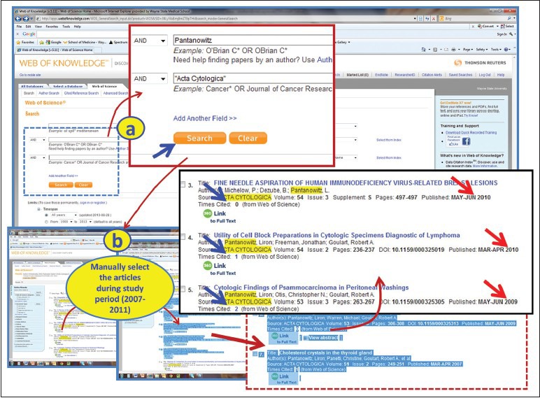
Use of the ‘Web of Sciences Database’ to harvest the raw data on number of citations for various publications for different authors publishing in cytopathology journals under study [Table 2]. (a) Add the name of the author and the journal. Click ‘Search’; (b) Manually select the publications during 2007 through 2011. Note the citation numbers and reference details [Table 2]
We designed another value to take into account the influence of the time factor after the publication (i.e. giving more power to publications which have had less time since publication). This metric, the ‘time adjusted citation quotient’ (Q value) was defined and calculated as follows:

N (Yr): Total number of citations for all publications under consideration in a specific year (Yr).
C (Yr): Average number of CPP in a specific year (Yr) by all cytopathology journals under study.
X: Total number of publications under consideration for that author from 2007 to 2011 for that journal category (OA or non-OA).
The Web of Science's citation analysis [Figure 2][15] was the source of the average number of CPP for that year in that journal i.e. C (2007), C (2008), C (2009), C (2010), and C (2011). Upon completion of the search for each of the cytopathology journal, C (Yr), the mean value for each year was calculated [Table 1]. The Q value for each publication in CytoJournal as OA cytopathology journal as well as the traditional non-OA cytopathology journals was then calculated by above formula. Minitab software[16] was used for statistical analysis.
Figure 2.

Use of ‘Web of Sciences citation analysis tool’ for finding the average number of citations per publication [C (Yr)] in a specific year for all the cytopathology journals under study. (a) Add the name of the journal; (b) Click ‘publication years’, choose the specific year of interest (2008); (c) Click ‘Create Citation Report’ link on the next screen; (d) Note the ‘Average Citations per Item’ C (Yr) for that journal for that year (2008) [Table 1]
Table 1.
Average number of citations per publication in a specific year [C (Yr)] for various cytopathology journals

To compare CRs for CytoJournal as an OA cytopathology journal versus non-OA cytopathology journals, by using freely available data on the web, a few of these authors were also analyzed arbitrarily by ‘Publish or Perish’ software which uses ‘Google scholar’ data.[17,18]
RESULTS
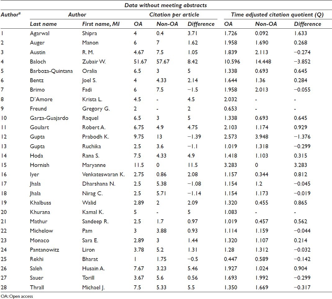
A total of 28 authors were identified as per the criteria who published papers or meeting abstracts in both OA and non-OA journals. When meeting abstracts were excluded 25 authors were considered. Overall, a total of 314 publications in cytopathology journals during 2007-2011 were evaluated based on the data from web of science citation analysis site (Impact factor is calculated based on this data) [Table 2].[19–325] Some publications were attributed to more than one author included in the study. The data shown in red in Table 2 indicate publications as meeting abstracts. Because OA cytopathology journal and some non-OA cytopathology journals did not publish meeting abstracts on regular basis and this feature may potentially impact the final comparison, we analyzed the data in two ways: First on all the data including the meeting abstracts, and then repeating the analysis after excluding the meeting abstracts.
Table 2.
Authors and citations (raw data)

In the group in which meeting abstracts were included, the combined number of publications per author ranged from 4 to 41 with an average of 15.6 and a median of 15. The number of publications per author in CytoJournal ranged from 2 to 9 with an average of 3.1 and a median of 2. The number of publications per author in traditional non-OA journals ranged from 2 to 32 with an average of 12.5 and a median of 15.
In the group in which meeting abstracts were excluded, the combined number of publications per author ranged from 3 to 27 with an average of 11.6 and a median of 10. The number of publications per author in CytoJournal ranged from 2 to 9 with an average of 3.1 and a median of 2. The number of publications per author in traditional non-OA journals ranged from 1 to 25 with an average of 8.4 and a median of 7.
The citations per publication (CPP) and the time adjusted quotions (Q values) were calculated for CytoJournal as an OA journal versus the traditional non-OA cytopathology Journals with the meetings abstracts included [Table 3]. Overall, the averages of both CPP and Q values were higher for OA Cytopathology Journal (cytojournal) than the traditional non-OA journals. To confirm our hypothesis, paired t-tests were run on both data sets (CPP and Q values) using Minitab software.[16]
Table 3.
Comparison of ‘Citations per publication’ and ‘time adjusted citation quotients’ (Q values) for CytoJournal as OA cytopathology journal versus non-OA cytopathology journals with meeting abstracts included [Figure 3]

For CPP, the following methodology was used. To make sure valid paired t-tests could be used, an Anderson-Darling normality test was run on the differences between CPP for CytoJournal as OA cytopathology journal and for the traditional non-OA journals. The normality test was passed and the paired t-test was run. A 95% confidence interval for the mean difference between [CytoJournal citations per publication] – [traditional non-OA citations per publication] was generated with the interval being (1.406, 3.824) with a P value of 0.001.
The same methodology was used to analyze the Q values. The 95% confidence interval for the mean difference between [CytoJournal Q value] - [traditional non-OA Q value] was (0.473, 1.084) with a P value of approximately 0.0001. The findings confirmed the hypothesis that the publications in OA cytopathology journal generated improved citation rate (CR) with higher CPP and Q values with statistically significant difference as compared to the publications in the traditional non-OA cytopathology journals [Figure 3a and b].
Figure 3.

With the second set of data, without the inclusion of meeting abstracts [Table 4], the same paired t-tests were run, with the null and alternative hypothesis similar to that for the first set of data. CPP with Open Access versus Non-Open Access showed a 95% confidence interval of (-1.038, 1.848) with a P value of 0.568. For the Q value, a 95% CI interval of (-0.309, 0.677) with a P value of 0.448. This analysis showed that CPP and Q values were also higher when meeting abstracts were taken out of the data set, but the difference was statistically insignificant [Figure 3c and d].
Table 4.
Comparison of ‘Citations per publication’ and ‘time adjusted citation quotients’ (Q values) for CytoJournal as OA cytopathology journal versus non-OA cytopathology journals without meeting abstracts [Figure 3c and d]
The results with ‘Publish or Perish’ software using ‘Google scholar’ data[17,18] also showed comparable pattern with higher citation rates for the publications in OA cytopathology journal than the traditional non-OA cytopathology journals. This data was unfiltered and included citations by all sorts of publication types.
DISCUSSION
A citation is defined as “a quoting of an authoritative source for substantiation”.[11] As almost all authors would like to be seen as “authoritative source” and their work as “substantial,” citations are a crucial metric in determining the success of both authors and journals. They are used in calculating such relied upon publication metrics in journology as impact factor and H-factor, which are used critically by many in evaluating the worthiness of a journal or an author[12]. Citations are indexed in several large databases on the World Wide Web, the largest of which is Thomson Scientific's Web of Science® which currently contains more than 40 million bibliographic records and 550 million citations from the past 100 years. We conducted the current study using the same data which is also used to calculate impact factor (IF)[15].
Since the Budapest declaration, several studies have examined the impact of the Open Access model of publication on the rates of citation for publications/authors. In 2001 in Nature, Steve Lawrence was the first to publish that free online availability of a publication greatly increased its impact on the scientific community. He analyzed CRs for 119,924 conference articles in computer science and related disciplines and excluded self-citations. He demonstrated the relationship of online availability as a function of the number of citations per article and the year of publication. The results were quite dramatic, showing a direct relationship between the factors, specifically a 157% increase in citations for articles that were free online compared to those which were not available free online.[6]
Our study showed that, in the field of cytopathology, authors who published in both OA cytopathology journal and traditional non-OA journals, accrued a relatively higher rate of citation per publication and time adjusted citation quotient for their publications in the OA journal with statistical difference (P < 0.01) [Table 3 and Figure 3a, b]. However, if meeting abstracts were excluded from the analysis, increase in CPP and Q values was statistically insignificant [Table 4 and Figure 3c, d] This data supports the prior published conclusions that the OA model is a legitimate platform for publication with comparable or even higher citation rates to traditional journals.
Kurtz et al. studied the increased CRs in OA publications but noted a possibility of selection bias. The suggested bias was that the most prominent authors are more likely to make their publications available in an OA model, artificially increasing the rate of citations.[7] As previously mentioned, cytopathology is a uniquely concise field, in which there are very limited numbers of Open Access journals, CytoJournal being one, but several traditional non-OA cytopathology journals. Because of this there are some authors who have publications in both OA and traditional journals, making comparison of the CRs possible while eliminating the above mentioned selection bias suggested by Kurtz et al. as a confounding factor.[7]
Another type of variable is the ‘early view bias,’ wherein a publication that is posted on an OA platform before final publication will have more time to accrue citations and thus skew the data towards citations in OA.[10] The OA journal used in our project (CytoJournal) does not post in pre-publication form, and thus our study was controlled against this type of bias.
Kurtz et al. also discussed another type of bias for which we were not able to control and that might influence the CRs of OA publications. This is a different type of selection bias, wherein the individual author selects their most important (and thus citable) publications for OA.[7] This type of bias is extremely subjective and difficult to prove or refute in a controlled study. However, it is important to highlight that the quality of the published material is generally the primary factor responsible for its overall impact and CR. Traditionally, the quality of published material is predominantly facilitated by the peer-review component of the editorial activity of the peer-reviewed journals. Thus, it is critical to understand and consider the quality of the peer-review process of any scientific journal irrespective of its OA status.
Some concerns have been raised in recent years regarding sprawling, low-quality journals (including some OA journals) which may have high turnover of many publications that have little relevance or contribution to today's scientific discoveries. This is an issue not applicable only to OA, but also to any journal irrespective of its status as an OA journal or traditional non-OA journal. The very core of any reputable scientific journal, with a quality-minded editorial board, is the high standard of the publications received and accepted after a vigorous peer review process with proactive participation by peer-reviewers.
We also evaluated, using a small cohort, the citation pattern of OA versus non-OA cytopathology publications with ‘Publish or Perish,’ software which uses ‘Google Scholar’ data on open, freely available platform.[17,18] As compared to the Web of Science, the inclusion of citations by Goggle Scholar is wider and includes many journals and other platforms which may cite the original work with a relatively liberal approach. The initial analysis with ‘Publish or Perish’ based on ‘Google Scholar’ data showed comparable results with relatively higher rates of citation for CytoJournal practicing OA publication model as compared to traditional non-OA cytopathology journals (without statistical significance).[332]
In summary, this study demonstrated that in the small subspecialty field of cytopathology, authors who published in both an Open Access journal (CytoJournal) and at least one traditional journal (Acta Cytologica, Cancer Cytopathology, Cytopathology, and/or Diagnostic Cytopathology) accrued a comparable or slightly higher CR for OA publications as compared to the traditional non-OA cytopathology journals over a five year period from 2007-2011 [Figure 3].
COMPETING INTEREST STATEMENT BY ALL AUTHORS
No competing interest to declare by any of the authors.
AUTHORSHIP STATEMENT BY ALL AUTHORS
All authors declare that we qualify for authorship as defined by ICMJE http://www.icmje.org/#author.
ETHICS STATEMENT BY ALL AUTHORS
This study did not require approval from Institutional Review Board (IRB) (or its equivalent) as it is based on analysis of published data on web.
EDITORIAL/PEER-REVIEW STATEMENT
CytoJournal editorial team thanks the Academic editor: LaVentra E. Danquah, MIS, MLIS, (laventra@wayne. edu) Coordinator for Instruction, Liaison, and Outreach Services, Shiffman Medical Library, Mazurek Medical Education Commons, Wayne State University, 320 E. Canfield, Detroit, MI 48201, (313.577.9083) for completing the peer-review process for the manuscript of this article.
ACKNOWLEDGMENTS
We thank Anushree Shidham for the copy-editing support. We thank Sandra Martin, Director of the Shiffman Medical Library and Learning Resources Centers, Wayne State University School of Medicine; Distinguished Member of the Academy of Health Information Professionals for her guidance to harvest the raw data on citations for all the authors in this study using ‘Web of Sciences Database’.
Abbreviations (in alphabetical order) used:
C, average number of CPP for a particular journal; C(Yr), C for a specific year; CPP, Citations Per Publication; CR, Citation Rate; IF, Impact Factor; IP, Intellectual Property; N/A, not applicable; OA, Open Access; Q value, time adjusted citation quotient; Yr, year.
Contributor Information
Nora K. Frisch, Email: nfrisch@med.wayne.edu.
Romil Nathan, Email: romilnathan@gmail.com.
Yasin K. Ahmed, Email: yahme@med.wayne.edu.
Vinod B. Shidham, Email: vshidham@med.wayne.edu.
REFERENCES
- 1.Timeline of the open access movement. [Last accessed on 2014 Feb 24]. Available from: http://legacy.earlham.edu/~peters/fos/timeline.htm .
- 2.Open access explained!-PHD animation. [Last accessed on 2014 Feb 24]. Available from: http://www.youtube.com/watch?v=L5rVH1KGBCY .
- 3.Science in the Open, The online home of Cameron Neylon, 14 February 2012. On the 10th Anniversary of the Budapest Declaration. [Last accessed on 2013 Aug 25]. Available from: http://www.cameronneylon.net/blog/on-the-10th-anniversary-of-the-budapest-declaration/. (Archived by WebCite® at http://www.webcitation.org/6ISSTKfrP)
- 4.Berlin declaration on open access to knowledge in the sciences and humanities. [Last accessed on 2014 Feb 24]. Available from: http://www.freeknowledge.eu/documents/reference/berlin .
- 5.Testa J, McVeigh ME. The impact of open access journals: A citation study from Thomson ISI. [Last accessed on 2013 Aug 25]. Available from: http://www.lib.uiowa.edu/scholarly/documents/ISI_impact-oa-journals.pdf .
- 6.Lawrence S. Free online availability substantially increases a paper's impact. Nature. 2001;411:521. doi: 10.1038/35079151. [DOI] [PubMed] [Google Scholar]
- 7.Kurtz MJ, Eichhorn G, Accomazzi A, Grant C, Demleitner M, Henneken E, et al. The effect of use and access on citations. Inf Process Manage. 2005. p. 41. Available from: https://www.cfa.harvard.edu/~kurtz/IPM-abstract.html .
- 8.Harnad S, Brody T. Comparing the impact of open access (OA) vs. Non-OA articles in the same journals. D-Lib Mag. 2004. p. 10. Available from: http://www.dlib.org/dlib/june04/harnad/06harnad.html .
- 9.Gargouri Y, Hajjem C, Larivière V, Gingras Y, Carr L, Brody T, et al. Self-selected or mandated, open access increases citation impact for higher quality research. PLoS One. 2010;5:e13636. doi: 10.1371/journal.pone.0013636. [DOI] [PMC free article] [PubMed] [Google Scholar]
- 10.Craig ID, Plume AM, McVeigh ME, Pringle J, Amin M. Do open access articles have greater citation impact? A critical review of the literature. J Informetr. 2007;1:239–48. [Google Scholar]
- 11.The free dictionary. [Last accessed on 2013 Aug 28]. Available from: http://www.thefreedictionary.com .
- 12.Shidham VB, Sandweiss L, Atkinson BF. First CytoJournal peer-reviewer's retreat in 2006-Open access, peer-review, and impact factor. Cytojournal. 2006;3:5. doi: 10.1186/1742-6413-3-5. [DOI] [PMC free article] [PubMed] [Google Scholar]
- 13.The creative commons attribution license. [Last accessed on 2014 Feb 24]. Available from: http://www.creativecommons.org/licenses/by/2.0 .
- 14.Shidham VB, Pitman MB, Demay RM. How to write an article: Preparing a publishable manuscript! Cytojournal. 2012;9 doi: 10.4103/1742-6413.92545. [DOI] [PMC free article] [PubMed] [Google Scholar]
- 15.Web of Science: The world's most trusted citation index covering the leading scholarly literature. [Last accessed on 2013 Aug 28]. Available from: http://www.thomsonreuters.com/web-of-science .
- 16.Minitab (Version 16) [Software] 2013. [Last accessed on 2013 Aug 28]. Available from: http://www.minitab.com/en-US/products/minitab/default.aspx .
- 17.Harzing AW. Publish or perish. 2007. [Last accessed on 2013 Aug 29]. Available from: http://www.harzing.com/pop.htm .
- 18.Google Scholar. [Last accessed on 2013 Aug 29]. Available from: http://www.scholar.google.com .
- 19.Arora R, Agarwal S, Mathur SR, Verma K, Iyer VK, Aron M. Utility of a limited panel of calretinin and Ber-EP4 immunocytochemistry on cytospin preparation of serous effusions: A cost-effective measure in resource-limited settings. Cytojournal. 2011;8:14. doi: 10.4103/1742-6413.83233. [DOI] [PMC free article] [PubMed] [Google Scholar]
- 20.Chitragar S, Agarwal S, Iyer VK, Mathur SR, Karak AK, Chharchhodawala T, et al. Cyto-morphological features of extramedullary acute megakaryoblastic leukemia on fine needle aspiration and cerebrospinal fluid cytology: A case report. Cytojournal. 2011;8:17. doi: 10.4103/1742-6413.85496. [DOI] [PMC free article] [PubMed] [Google Scholar]
- 21.Sharma M, Agarwal S, Wadhwa N, Mishra K, Gadre DJ. Spectrum of cytomorphology of tuberculous lymphadenitis and changes during anti-tubercular treatment. Cytopathology. 2007;18:180–3. doi: 10.1111/j.1365-2303.2007.00441.x. [DOI] [PubMed] [Google Scholar]
- 22.Nayak A, Iyer VK, Agarwal S, Agarwala S. Fine needle aspiration cytology of fetal rhabdomyomatous and teratoid Wilms tumor. Acta Cytol. 2010;54:563–8. doi: 10.1159/000325178. [DOI] [PubMed] [Google Scholar]
- 23.Agarwal S, Mathur SR, Ray R, Sharma SC, Sinha P. Cytopathological diagnosis of hyalinizing trabecular tumour, a rare thyroid neoplasm. Cytopathology. 2010;21:133–4. doi: 10.1111/j.1365-2303.2009.00698.x. [DOI] [PubMed] [Google Scholar]
- 24.Iyer V, Nayak A, Agarwal S, Agarwala S. Cytomorphologic spectrum of pediatric renal tumors on aspiration cytology: A study of 120 cases. Acta Cytol. 2010;54:416. [Google Scholar]
- 25.Mathur SR, Arora R, Agarwal S, Verrna K, Iyer VK, Aron M. diagnostic utility of a limited panel of markers in distinguishing reactive mesothelial proliferations from adenocarcinomas. Acta Cytol. 2010;54:471. [Google Scholar]
- 26.Agarwal S, Gupta R, Iyer VK, Mathur SR, Ray R. Cytopathological diagnosis of alveolar soft part sarcoma, a rare soft tissue neoplasm. Cytopathology. 2011;22:318–22. doi: 10.1111/j.1365-2303.2010.00796.x. [DOI] [PubMed] [Google Scholar]
- 27.Agarwal S, Mathur SR, Ray R, Sharma SC, Sinha P. Cytopathological diagnosis of hyalinising trabecular tumour, a rare thyroid neoplasm. Cytopathology. 2011;22:65–6. doi: 10.1111/j.1365-2303.2010.00744.x. [DOI] [PubMed] [Google Scholar]
- 28.Brimo F, Popradi G, Michel RP, Auger M. Primary effusion lymphoma involving three body cavities. Cytojournal. 2009;6:21. doi: 10.4103/1742-6413.56361. [DOI] [PMC free article] [PubMed] [Google Scholar]
- 29.Renshaw AA, Brimo F, Auger M. Surrogate indicators of sensitivity in gynecologic cytology: Can they be used to improve the measurement of sensitivity in the laboratory? Cytojournal. 2009;6:19. doi: 10.4103/1742-6413.56359. [DOI] [PMC free article] [PubMed] [Google Scholar]
- 30.Brimo F, Michel RP, Khetani K, Auger M. Primary effusion lymphoma: A series of 4 cases and review of the literature with emphasis on cytomorphologic and immunocytochemical differential diagnosis. Cancer. 2007;111:224–33. doi: 10.1002/cncr.22691. [DOI] [PubMed] [Google Scholar]
- 31.Auger M. A duo of diagnostic challenges in fine needle aspirations of the thyroid: The suspicious for follicular neoplasm” and “suspicious for papillary carcinoma categories. Acta Cytol. 2007;51:274. [Google Scholar]
- 32.Alkuwari E, Khetan K, Dendukuri N, Wang L, Auger M. Quantitative assessment of nuclear grooves in neoplastic and nonneoplastic lesions in fine needle aspiration samples of the thyroid: A retrospective cytohistologic study of 94 cases. Acta Cytol. 2007;51:284. [PubMed] [Google Scholar]
- 33.Auger M, Aikuwari E. Accuracy of fine needle aspiration cytology of axillary lymph nodes-A study of 132 cases with cyto-histological correlation. Cancer Cytopathol. 2007;111:414. [Google Scholar]
- 34.Alkuwari E, Auger M. Accuracy of fine-needle aspiration cytology of axillary lymph nodes in breast cancer patients: A study of 115 cases with cytologic-histologic correlation. Cancer. 2008;114:89–93. doi: 10.1002/cncr.23344. [DOI] [PubMed] [Google Scholar]
- 35.Deschenes M, Renshaw AA, Auger M. Measuring the significance of workload on performance of cytotechnologists in gynecologic cytology: A study using rapid prescreening. Cancer. 2008;114:149–54. doi: 10.1002/cncr.23497. [DOI] [PubMed] [Google Scholar]
- 36.Deschênes M, Michel RP, Tabah R, Auger M. Fine-needle aspiration cytology of Castleman disease: Case report with review of the literature. Diagn Cytopathol. 2008;36:904–8. doi: 10.1002/dc.20934. [DOI] [PubMed] [Google Scholar]
- 37.Brimo F, Ouad L, Brodeur J, Charbonneau M, Auger M. Unusual microbial organisms seen in two cervical smears. Diagn Cytopathol. 2009;37:836–8. doi: 10.1002/dc.21114. [DOI] [PubMed] [Google Scholar]
- 38.Brimo F, Renshaw AA, Deschenes M, Charbonneau M, Auger M. Improvement in the routine screening performance of cytotechnologists over time: A study using rapid prescreening. Cancer. 2009;117:311–7. doi: 10.1002/cncy.20042. [DOI] [PubMed] [Google Scholar]
- 39.Moriarty AT, Laucirica R, Auger M, Souers RJ, Chmara BA, Wilbur DC. Granulomatous inflammation: An underestimated cause of false positive diagnoses in lung fine needle aspirates observations from the college of American pathologists non gynecologic cytopathology (NGC) interlaboratory comparison program. Cancer Cytopathol. 2009;117:350. doi: 10.5858/2009-0491-CPR2.1. [DOI] [PubMed] [Google Scholar]
- 40.Kushner YB, Brimo F, Schwartzman K, Auger M. A rare case of pulmonary cryptococcal inflammatory myofibroblastic tumor diagnosed by fine needle aspiration cytology. Diagn Cytopathol. 2010;38:447–51. doi: 10.1002/dc.21259. [DOI] [PubMed] [Google Scholar]
- 41.Orellana ME, Brimo F, Auger M, Galic J, Deschenes J, Burnier MN. Cytopathological diagnosis of adult retinoblastoma in a vitrectomy specimen. Diagn Cytopathol. 2010;38:59–64. doi: 10.1002/dc.21135. [DOI] [PubMed] [Google Scholar]
- 42.Renshaw AA, Auger M, Birdsong G, Cibas ES, Henry M, Hughes JH, et al. ASC/SIL ratio for cytotechnologists: A survey of its utility in clinical practice. Diagn Cytopathol. 2010;38:180–3. doi: 10.1002/dc.21167. [DOI] [PubMed] [Google Scholar]
- 43.Auger M, Vielh P. FNAC of the breast: The (cyto) pathologist input. Acta Cytol. 2010;54:371–2. [Google Scholar]
- 44.Schwartz M, Laucirica R, Booth C, Auger M, Thomas R, Thomas N, et al. Cytology of spontaneous nipple discharge: Is it worth it. Performance of nipple discharge preparations in the college of American pathologists inter-laboratory comparison program in non-gynecologic cytology? Cancer Cytopathol. 2010;118:317–8. doi: 10.5858/arpa.2012-0231-CP. [DOI] [PubMed] [Google Scholar]
- 45.Auger M. Rapid prescreening in gynecologic cytology: A more efficient quality assurance method. Cancer Cytopathol. 2011;119:357–60. doi: 10.1002/cncy.20189. [DOI] [PubMed] [Google Scholar]
- 46.Austin RM, Benstein B, Bentz J, Bigner S, Freund GG, Rocco GL, et al. Market survey predictions on the future of US Pap testing. Cytojournal. 2009;6:17. doi: 10.4103/1742-6413.55885. [DOI] [PMC free article] [PubMed] [Google Scholar]
- 47.Austin RM, Zhao C. Test group biases and ethical concerns mar New England Journal of Medicine articles promoting HPV screening for cervical cancer in rural India. Cytojournal. 2009;6:12. doi: 10.4103/1742-6413.53466. [DOI] [PMC free article] [PubMed] [Google Scholar]
- 48.Al-Abbadi MA, Bloom LI, Fatheree LA, Haack LA, Minkowitz G, Wilbur DC, et al. Adequate reimbursement is crucial to support cost-effective rapid on-site cytopathology evaluations. Cytojournal. 2010;7:22. doi: 10.4103/1742-6413.71740. [DOI] [PMC free article] [PubMed] [Google Scholar]
- 49.Bandyopadhyay S, Austin M, Zhao C. Hybrid capture 2 hrHPV DNA detection in ThinPrep Pap test vials is a very useful ancillary test in women with atypical squamous cells, cannot exclude HSIL (ASC-H) Cancer Cytopathol. 2007;111:379–80. [Google Scholar]
- 50.Flanangan MB, Dabbs DJ, Mauser N, White S, Austin RM, Chivukula M. Replacement of conventional Pap smears (CPS) with liquid based cytology (LBC) and imaged-LBC increases detection of abnormals. Cancer Cytopathol. 2007;111:387–8. [Google Scholar]
- 51.Kapali M, Agaram NP, Dabbs D, Kanbour A, White S, Austin RM. Routine endometrial sampling of asymptomatic premenopausal women shedding normal endometrial cells in Papanicolaou tests is not cost effective. Cancer. 2007;111:26–33. doi: 10.1002/cncr.22424. [DOI] [PubMed] [Google Scholar]
- 52.O’Connor SM, Austin RM, Carter G, White S, Dabbs DJ, Chivukula M. HPV triage of ASC-H with follow-up cervical Intraepithelial neoplasia 3 (CIN 3) lesions is more efficient than routine colposcopic referral. Cancer Cytopathology. 2007;111:375. [Google Scholar]
- 53.Zhao C, Elishaev E, Yuan KH, Yu J, Austin RM. Very low human Papillomavirus DNA prevalence in mature women with negative computer-imaged liquid-based Pap tests. Cancer. 2007;111:292–7. doi: 10.1002/cncr.22949. [DOI] [PubMed] [Google Scholar]
- 54.Austin RM, McCoy DR. The confused pathology expert: The ongoing challenge of monitoring physician expert witness testimony. Diagn Cytopathol. 2007;35:749–55. doi: 10.1002/dc.20729. [DOI] [PubMed] [Google Scholar]
- 55.Austin RM, Felix JC, Alonzo TA. Improved diagnostic reproducibility with the MonoPrep (R) Pap test. Cancer Cytopathol. 2008;114:387. [Google Scholar]
- 56.Austin RM, Felix JC, Alonzo TA. Improvement in specimen adequacy using the MonoPrep Pap test system. Cancer Cytopathol. 2008;114:386–7. [Google Scholar]
- 57.Austin RM, Onisko A, Druzdzel MJ. The Pittsburgh Cervical Cancer Screening Model (PCCSM) Cancer Cytopathol. 2008;114:345. [Google Scholar]
- 58.Kanbour-Shakir A, Onisko A, Austin M. High Risk Hpv test results preceding over 500 cases of biopsy proven high grade cervical squamous dysplasia (Cin 2/3) Cancer Cytopathol. 2008;114:345–6. [Google Scholar]
- 59.Zhao C, Austin RM. Adjunctive high-risk human papillomavirus DNA testing is a useful option for disease risk assessment in patients with negative Papanicolaou tests without an endocervical/transformation zone sample. Cancer. 2008;114:242–8. doi: 10.1002/cncr.23598. [DOI] [PubMed] [Google Scholar]
- 60.Zhao CQ, Austin RM. Detection rates for high risk HPV DNA in women 50 and older with negative and abnormal pap test results. Cancer Cytopathol. 2008;114:366–7. [Google Scholar]
- 61.Zhao CQ, Austin RM. High risk HPV DNA detection rates in over 26,000 cytology negative imaged liquid-based pap test samples. Cancer Cytopathol. 2008;114:369–70. [Google Scholar]
- 62.Zhao C, Austin RM, Pan J, Barr N, Martin SE, Raza A, et al. Clinical significance of atypical glandular cells in conventional pap smears in a large, high-risk U.S. west coast minority population. Acta Cytol. 2009;53:153–9. doi: 10.1159/000325117. [DOI] [PubMed] [Google Scholar]
- 63.Zhao CQ, Kalposi-Novak P, Austin RM. histological follow-up findings in adolescents with HSIL cytology results. Cancer Cytopathol. 2009;117:383–4. [Google Scholar]
- 64.Austin RM. Computer-assisted Papanicolaou imaging: Another valuable tool in the challenge of Papanicolaou test screening for glandular neoplasia. Cancer Cytopathol. 2010;118:65–7. doi: 10.1002/cncy.20068. [DOI] [PubMed] [Google Scholar]
- 65.Chivukula M, Austin M, Matsko J, Duwe A, Freidman T, Mauser N, et al. Use of dual-stain for P16 and Ki-67 in the interpretation of abnormal pap cytology results: A prospective study. Cancer Cytopathol. 2010;118:333–4. [Google Scholar]
- 66.Austin RM. Exhortations to abandon the Pap test as a routine initial cervical screening test are still premature and carry significant risks. Diagn Cytopathol. 2010;38:783–7. doi: 10.1002/dc.21350. [DOI] [PubMed] [Google Scholar]
- 67.Heider A, Austin RM, Zhao C. HPV test results stratify risk for histopathologic follow-up findings of high-grade cervical intra-epithelial neoplasia in women with low-grade squamous intra-epithelial lesion Pap results. Acta Cytol. 2011;55:48–53. doi: 10.1159/000320877. [DOI] [PubMed] [Google Scholar]
- 68.Gao FF, Austin RM, Zhao C. Histopathologic follow-up and human papillomavirus DNA test results in 290 patients with high-grade squamous intraepithelial lesion Papanicolaou test results. Cancer Cytopathol. 2011;119:377–86. doi: 10.1002/cncy.20176. [DOI] [PubMed] [Google Scholar]
- 69.Gao FF, Khalbuss WE, Austin RM, Monaco SE. Cytomorphology of crystal storing histiocytosis in the breast associated with lymphoma: A case report. Acta Cytol. 2011;55:302–6. doi: 10.1159/000324558. [DOI] [PubMed] [Google Scholar]
- 70.Baloch ZW, Barroeta JE, Walsh J, Gupta PK, Livolsi VA, Langer JE, et al. Utility of thyroglobulin measurement in fine-needle aspiration biopsy specimens of lymph nodes in the diagnosis of recurrent thyroid carcinoma. Cytojournal. 2008;5:1. doi: 10.1186/1742-6413-5-1. [DOI] [PMC free article] [PubMed] [Google Scholar]
- 71.Baloch ZW, Cibas ES, Clark DP, Layfield LJ, Ljung BM, Pitman MB, et al. The National Cancer Institute Thyroid fine needle aspiration state of the science conference: A summation. Cytojournal. 2008:5. doi: 10.1186/1742-6413-5-6. [DOI] [PMC free article] [PubMed] [Google Scholar]
- 72.Griffin AC, Schwartz LE, Baloch ZW. Utility of on-site evaluation of endobronchial ultrasound-guided transbronchial needle aspiration specimens. Cytojournal. 2011;8:20. doi: 10.4103/1742-6413.90081. [DOI] [PMC free article] [PubMed] [Google Scholar]
- 73.Deveci MS, Deveci G, LiVolsi VA, Gupta PK, Baloch ZW. Concordance between thyroid nodule sizes measured by ultrasound and gross pathology examination: Effect on patient management. Diagn Cytopathol. 2007;35:579–83. doi: 10.1002/dc.20714. [DOI] [PubMed] [Google Scholar]
- 74.Baloch ZW, LiVolsi VA, Asa SL, Rosai J, Merino MJ, Randolph G, et al. Diagnostic terminology and morphologic criteria for cytologic diagnosis of thyroid lesions: A synopsis of the National Cancer Institute Thyroid Fine-Needle Aspiration State of the Science Conference. Diagn Cytopathol. 2008;36:425–37. doi: 10.1002/dc.20830. [DOI] [PubMed] [Google Scholar]
- 75.Khayyata S, Barroeta JE, Livolsi VA, Baloch ZW. Papillary hyperplastic nodule: Pitfall in the cytopathologic diagnosis of papillary thyroid carcinoma. Cancer Cytopathol. 2008;114:441. doi: 10.4158/EP.14.7.863. [DOI] [PubMed] [Google Scholar]
- 76.Bongiovanni M, Bloom L, Krane JF, Baloch ZW, Powers CN, Hintermann S, et al. Cytomorphologic features of poorly differentiated thyroid carcinoma: A multi-institutional analysis of 40 cases. Cancer. 2009;117:185–94. doi: 10.1002/cncy.20023. [DOI] [PubMed] [Google Scholar]
- 77.Khayyata S, Yun S, Pasha T, Jian B, McGrath C, Yu G, et al. Value of P63 and CK5/6 in distinguishing squamous cell carcinoma from adenocarcinoma in lung fine-needle aspiration specimens. Diagn Cytopathol. 2009;37:178–83. doi: 10.1002/dc.20975. [DOI] [PubMed] [Google Scholar]
- 78.Baloch ZW, Pasha T, Wong L, Shiina N, Zhang P, Tomaszewski J, et al. ProEx C expression in urothelial carcinoma: A tissue microarray and cytology study. Cancer Cytopathol. 2009;117:370. [Google Scholar]
- 79.Faquin WC, Baloch ZW. Fine-needle aspiration of follicular patterned lesions of the thyroid: Diagnosis, management, and follow-up according to National Cancer Institute (NCI) recommendations. Diagn Cytopathol. 2010;38:731–9. doi: 10.1002/dc.21292. [DOI] [PubMed] [Google Scholar]
- 80.Layfield LJ, Cibas ES, Baloch Z. Thyroid fine needle aspiration cytology: A review of the National Cancer Institute state of the science symposium. Cytopathology. 2010;21:75–85. doi: 10.1111/j.1365-2303.2010.00750.x. [DOI] [PubMed] [Google Scholar]
- 81.Ponce-Camacho MA, Diaz de Leon-Medina R, Miranda-Maldonado I, Garza-Guajardo R, Hernandez-Salazar J, Barboza-Quintana O. A 5-year-old girl with a congenital ganglioneuroma diagnosed by fine needle aspiration biopsy: A case report. Cytojournal. 2008;5:5. doi: 10.1186/1742-6413-5-5. [DOI] [PMC free article] [PubMed] [Google Scholar]
- 82.Gomez-Macías GS, Garza-Guajardo R, Segura-Luna J, Barboza-Quintana O. Inadequate fine needle aspiration biopsy samples: Pathologists versus other specialists. Cytojournal. 2009;6:9. doi: 10.4103/1742-6413.52831. [DOI] [PMC free article] [PubMed] [Google Scholar]
- 83.Barboza-Quintana O, Garza-Guajardo R, Assad-Morel C, Méndez-Olvera N. Pseudomycetoma for microsporum canis: Report of a case diagnosed by fine needle aspiration biopsy. Acta Cytol. 2007;51:424–8. doi: 10.1159/000325759. [DOI] [PubMed] [Google Scholar]
- 84.Barboza-Quintana O, Alvarado-Bernal YL, Flores-Gutiérrez JP, Ancer-Rodríguez J, Garza-Guajardo R. Diagnosis of carcinosarcoma metastatic to the umbilicus by fine needle aspiration biopsy: A case report. Acta Cytol. 2010;54:819–22. [PubMed] [Google Scholar]
- 85.Smith GD, Riding M, Oswald K, Bentz JS. Integrating a FISH imaging system into the cytology laboratory. Cytojournal. 2010;7:3. doi: 10.4103/1742-6413.62258. [DOI] [PMC free article] [PubMed] [Google Scholar]
- 86.Smith GD, Willmore-Payne C, Chadwick BE, Bentz JS. Real time PCR and high resolution melting analysis platforms to detect EGFR mutations and predict response to tyrosine kinase inhibitor (TKI) therapy in NSCLC. Cancer Cytopathol. 2007;111:409. [Google Scholar]
- 87.Layfield LJ, Skripenova S, Bentz J, Smock C. Cytologic fine needle aspiration findings by vegetant intravascular hemangioendothelioma. Acta Cytol. 2008;52:273–5. doi: 10.1159/000325502. [DOI] [PubMed] [Google Scholar]
- 88.Collins BT, Layfield LJ, Gopez EV, Bentz JS. utilization of the indeterminate/atypia of undetermined significance category in thyroid aspiration cytology. How often is it used. Cancer Cytopathol. 2008;114:440. [Google Scholar]
- 89.Smith GD, Bentz JS. Fishing to detect urinary and other cancers: Do imaging systems help? Cancer Cytopathol. 2008;114:361. doi: 10.1002/cncy.20066. [DOI] [PubMed] [Google Scholar]
- 90.Layfield LJ, Bentz J. Giant-cell containing neoplasms of the pancreas: An aspiration cytology study. Diagn Cytopathol. 2008;36:238–44. doi: 10.1002/dc.20785. [DOI] [PubMed] [Google Scholar]
- 91.Ducatman BS, Bentz JS, Moriarty AT, Souers RJ, Fatheree LA, Booth CN, et al. Performance in gynecologic cytology proficiency testing: What have we learned? Cancer Cytopathol. 2009;117:352–3. [Google Scholar]
- 92.Laucirica R, Bentz JS, Clayton AC, Souers RS, Chmara BA, Moriarty AT. Performance characteristics of mucinous (Colloid) carcinoma of the breast in fine needles aspirates: Observations from the college of American pathologists interlaboratory comparison program in nongynecologic cytopathology (CAP NGC) Cancer Cytopathol. 2009;117:359. doi: 10.5858/arpa.2010-0652-CP. [DOI] [PubMed] [Google Scholar]
- 93.Smith GD, Collins BT, Bentz JS. Bioview duet-Assisted fluorescence in situ hybridization for gastrointestinal malignancy. Cancer Cytopathol. 2009;117:367–8. [Google Scholar]
- 94.Teman CJ, Wilson AR, Collins BT, Bentz JS. Cytologic detection of endometrial adenocarcinoma: Correlation with adenocarcinoma grade and papanicolaou preparation type. Cancer Cytopathol. 2009;117:379–80. [Google Scholar]
- 95.Agarwal AM, Bentz JS, Hungerford R, Abraham D. Parathyroid fine-needle aspiration cytology in the evaluation of parathyroid adenoma: Cytologic findings from 53 patients. Diagn Cytopathol. 2009;37:407–10. doi: 10.1002/dc.21020. [DOI] [PubMed] [Google Scholar]
- 96.Fischer A, Clayton A, Bentz J, Wasserman P, Henry M, Souers R, et al. Performance of direct smears compared to liquid-based preparations of thyroid FNA samples: Analysis of 47,076 responses in the college of American pathologists interlaboratory comparison program in non-gynecologic cytopathology. Cancer Cytopathol. 2010;118:307–8. [Google Scholar]
- 97.Smith GD, Bentz JS. “FISHing” to detect urinary and other cancers: Validation of an imaging system to aid in interpretation. Cancer Cytopathol. 2010;118:56–64. doi: 10.1002/cncy.20066. [DOI] [PubMed] [Google Scholar]
- 98.Smith GD, Chadwick BE, Adler DG, Bentz JS. Comparison of ThinPrep UroCyte and cytospin slide preparations for gastrointestinal specimens: Evaluation and retrospective performance review. Diagn Cytopathol. 2010;38:902–12. doi: 10.1002/dc.21357. [DOI] [PubMed] [Google Scholar]
- 99.Smith GD, Zhou L, Rowe LR, Jarboe EA, Collins BT, Bentz JS, et al. Allele-specific PCR with competitive probe blocking for sensitive and specific detection of BRAF V600E in thyroid fine-needle aspiration specimens. Acta Cytol. 2011;55:576–83. doi: 10.1159/000333453. [DOI] [PubMed] [Google Scholar]
- 100.Brimo F, Nahal A. Malignant epithelioid hemangioendothelioma with spindle phenotype: Report of an unusual case diagnosed by fine needle aspiration cytology. Acta Cytol. 2008;52:721–4. doi: 10.1159/000325629. [DOI] [PubMed] [Google Scholar]
- 101.Shidham VB, Varsegi G, D’Amore K. Two-color immunocytochemistry for evaluation of effusion fluids for metastatic adenocarcinoma. Cytojournal. 2010;7:1. doi: 10.4103/1742-6413.59887. [DOI] [PMC free article] [PubMed] [Google Scholar]
- 102.Shidham VB, Mehrotra R, Varsegi G, D’Amore KL, Hunt B, Narayan R. p16 immunocytochemistry on cell blocks as an adjunct to cervical cytology: Potential reflex testing on specially prepared cell blocks from residual liquid-based cytology specimens. Cytojournal. 2011;8:1. doi: 10.4103/1742-6413.76379. [DOI] [PMC free article] [PubMed] [Google Scholar]
- 103.D’Amore KL, Basir Z. Anal screening cytology and histomorphologic correlation: A Single center experience. Cancer Cytopathol. 2009;117:385–6. [Google Scholar]
- 104.Shidham VB, D’Amore K, Varsegi G. Objective and definitive subcategorization of LSIL with p16(INK4a) immunocytochemistry on cell 104. Block sections of cervical cytology specimens. Cancer Cytopathol. 2009;117:390–1. [Google Scholar]
- 105.Davis-Devine S, Day SJ, Anderson A, French A, Madison-Henness D, Mohar N, et al. Collection of the BD SurePath Pap Test with a broom device plus endocervical brush improves disease detection when compared to the broom device alone or the spatula plus endocervical brush combination. Cytojournal. 2008;6:4. doi: 10.4103/1742-6413.45495. [DOI] [PMC free article] [PubMed] [Google Scholar]
- 106.Freund GG, Saccomanno G, Boreson N, Davis-Devine S, Day SJ. High-risk HPV is found within the no further review tier using the focal point slide profilier. Cancer Cytopathol. 2007;111:370. [Google Scholar]
- 107.Blunier TE, Patel A, Shears R, Day SJ, Davis-Devine S, Risley N, et al. The relationship between digene high-risk HPV HC2 DNA test relative light unit (RLU)/Cutoff (CO) value and cervical biopsy proven dysplasia using the BD surepath (TM) liquid-based pap test for specimen collection. Cancer Cytopathol. 2009;117:380–1. [Google Scholar]
- 108.Van Dyke J, Davis-Devine S, Mentock E, Blunier T, Gaudier F, Freund G. Clinical utility of FNA-derived cell block when compared to simultaneously collected core biopsy. Cancer Cytopathol. 2010;118:391. [Google Scholar]
- 109.Pantanowitz L, Hornish M, Goulart RA. Informatics applied to cytology. Cytojournal. 2008;5:16. doi: 10.4103/1742-6413.44773. [DOI] [PMC free article] [PubMed] [Google Scholar]
- 110.Pantanowitz L, Hornish M, Goulart RA. The impact of digital imaging in the field of cytopathology. Cytojournal. 2009;6:6. doi: 10.4103/1742-6413.48606. [DOI] [PMC free article] [PubMed] [Google Scholar]
- 111.Pantanowitz L, Kuperman M, Goulart RA. Clinical history of HIV infection may be misleading in cytopathology. Cytojournal. 2010:7. doi: 10.4103/1742-6413.64375. [DOI] [PMC free article] [PubMed] [Google Scholar]
- 112.Setia N, Goulart RA, Leiman G, Otis CN, Modem R, Pantanowtiz L. Cytomorphology of cervicovaginal melanoma: ThinPrep versus conventional Papanicolaou tests. Cytojournal. 2010;7:25. doi: 10.4103/1742-6413.75666. [DOI] [PMC free article] [PubMed] [Google Scholar]
- 113.Hollowell ML, Goulart RA, Gang DL, Otis CN, Prior J, Sachs BF, et al. Cytologic features of müllerian papilloma of the cervix: Mimic of malignancy. Diagn Cytopathol. 2007;35:607–11. doi: 10.1002/dc.20712. [DOI] [PubMed] [Google Scholar]
- 114.Kandil D, Leiman G, Allegretta M, Trotman W, Pantanowitz L, Goulart R, et al. Glypican-3 immunocytochemistry in liver fine-needle aspirates: A novel stain to assist in the differentiation of benign and malignant liver lesions. Cancer. 2007;111:316–22. doi: 10.1002/cncr.22954. [DOI] [PubMed] [Google Scholar]
- 115.Kandil D, Leiman G, Trotman W, Allegretta M, Pantanowitz L, Goulart R, et al. Glypican-3: Comparison of a novel stain in immunocytochemistry and immunohistochemistry in hepatocellular carcinoma. Acta Cytol. 2007;51:331. [Google Scholar]
- 116.Pantanowitz L, Panetti C, Goulart RA, Cooper R. Cholesterol crystals in the thyroid gland. Acta Cytol. 2007;51:249–51. [PubMed] [Google Scholar]
- 117.Assaad M, Hornish A, Florence R, Goulart R. Companson of conventional smears versus ThinPrep (R) in thyroid fine needle aspiration biopsy: Diagnostic categories and surgical correlation. Cancer Cytopathol. 2007;111:397–8. [Google Scholar]
- 118.Goulart RA, Bugbee AC, Lemon L, Metzler RL. Community performance characteristics of validated slides in proficiency testing: The American society for clinical pathology GYN PT (TM) program. Cancer Cytopathol. 2007;111:364–5. [Google Scholar]
- 119.Pantanowitz L, Hornish M, Florence R, Goulart R. Vaginal Pap tests status-post hysterectomy. Diagn Cytopathol. 2007;35:539–40. doi: 10.1002/dc.20691. [DOI] [PubMed] [Google Scholar]
- 120.Modem R, Goulart R, Pantanowitz L. Utility of fine needle aspiration biopsy in the diagnosis of thyroid lymphoma. Cancer Cytopathol. 2007;111:399. [Google Scholar]
- 121.Pantanowitz L, Hornish M, Goulart R. Outcome of clinically based large-scale screening for Chlamydia trachomatis infection using the ThinPrep (R) Pap test collection vial. Cancer Cytopathol. 2007;111:383–4. [Google Scholar]
- 122.Pantanowitz L, Otis CN, Goulart RA. Cytologic findings of psammocarcinoma in peritoneal washings. Cancer Cytopathol. 2007;111:405. doi: 10.1159/000325305. [DOI] [PubMed] [Google Scholar]
- 123.Pantanowitz L, Otis CN, Goulart RA. Immunocytochemical evaluation of p16(INK4A) in ThinPrep (R) Pap tests diagnosed as atypical squamous cells, cannot exclude high-grade squamous intraepithelial lesion. Cancer Cytopathol. 2007;111:362. [Google Scholar]
- 124.Pinco J, Goulart R, Otis C, Garb J, Pantanowitz L. Impact of digital image manipulation in cytopathology. Cancer Cytopathol. 2007;111:430. [Google Scholar]
- 125.Goulart RA, Hornish M, Panetti C, Pantanowitz L. HPV DNA test results for cytotechnologist ASC-US downgraded to NILM following cytopathologist review: A potential laboratory quality indicator. Cancer Cytopathol. 2008;114:428. [Google Scholar]
- 126.Henninger B, Hornish M, Pantanowitz L, Goulart RA. A novel computer program for assessment of graduated diagnostic competency of cytopathology fellows. Cancer Cytopathol. 2008;114:402–3. [Google Scholar]
- 127.Panetti C, Hornish M, Goulart RA, Pantanowitz L. The effect of uterine cervical polyps on cervical cytology. Cancer Cytopathol. 2008;114:397. [Google Scholar]
- 128.Pantanowitz L, Otis CN, Goulart RA. Cytologic findings of psammocarcinoma in peritoneal washings. Acta Cytol. 2009;53:263–7. doi: 10.1159/000325305. [DOI] [PubMed] [Google Scholar]
- 129.Pantanowitz L, Warren M, Goulart RA. Anthracotic pigment in pleural fluid: A case report. Acta Cytol. 2009;53:306–8. doi: 10.1159/000325313. [DOI] [PubMed] [Google Scholar]
- 130.Henninger B, Hornish M, Cao QJ, Pantanowitz L, Goulart R. Morphologic pap test findings in HPV negative women age 30 years and older: What information will be lost with HPV only primary screening? Cancer Cytopathol. 2009;117:357–8. [Google Scholar]
- 131.Henninger B, Goulart RA, Otis CN, Pantanowitz L. Melamed-wolinska intracytoplasmic urothelial cell bodies: Evaluation with red blood cell markers glycophorin-C and GLUT-1. Cancer Cytopathol. 2009;117:373–4. [Google Scholar]
- 132.Pantanowitz L, Goulart RA. Added value of cytomorphology in pulmonary specimens submitted for the detection of pneumocystis. Cancer Cytopathol. 2009;117:402–3. [Google Scholar]
- 133.Setia N, Modem R, Goulart RA, Leiman G, Otis CN, Pantanowitz L. Cytomorphology of cervicovaginal melanoma: ThinPrep versus conventional papanicolaou tests. Cancer Cytopathol. 2009;117:391–2. doi: 10.4103/1742-6413.75666. [DOI] [PMC free article] [PubMed] [Google Scholar]
- 134.Pantanowitz L, Freeman J, Goulart RA. Utility of cell block preparations in cytologic specimens diagnostic of lymphoma. Acta Cytol. 2010;54:236–7. doi: 10.1159/000325019. [DOI] [PubMed] [Google Scholar]
- 135.Filomena C, Covell J, Metzler R, Goulart R. Morphologic features which affect validation and proficiency test performance of biopsy-proven HSIL pap tests. Cancer Cytopathol. 2010;118:306. [Google Scholar]
- 136.Pantanowitz L, Hornish M, Cao QJ, Goulart RA. HPV data can be used as a cytopathology laboratory quality indicator. Diagn Cytopathol. 2010;38:159–60. doi: 10.1002/dc.21186. [DOI] [PubMed] [Google Scholar]
- 137.Wang Y, Goulart RA, Pantanowitz L. Oil red O staining in cytopathology. Diagn Cytopathol. 2011;39:272–3. doi: 10.1002/dc.21390. [DOI] [PubMed] [Google Scholar]
- 138.Jian B, Kolansky AS, Baloach ZW, Gupta PK. Entamoeba gingivalis pulmonary abscess-diagnosed by fine needle aspiration. Cytojournal. 2008;5:12. doi: 10.4103/1742-6413.43179. [DOI] [PMC free article] [PubMed] [Google Scholar]
- 139.Gupta PK. Progression from on-site to point-of-care fine needle aspiration service: Opportunities and challenges. Cytojournal. 2010;7:6. doi: 10.4103/1742-6413.63195. [DOI] [PMC free article] [PubMed] [Google Scholar]
- 140.Shi Y, Griffin AC, Zhang PJ, Palmer JN, Gupta P. Sinus histiocytosis with massive lymphadenopathy (Rosai-Dorfman Disease): A case report and review of 49 cases with fine needle aspiration cytology. Cytojournal. 2011;8:3. doi: 10.4103/1742-6413.76731. [DOI] [PMC free article] [PubMed] [Google Scholar]
- 141.Park JY, Malik A, Dumoff KL, Gupta PK. Case report and review of lupus erythematosus cells in cytology fluids. Diagn Cytopathol. 2007;35:806–9. doi: 10.1002/dc.20758. [DOI] [PubMed] [Google Scholar]
- 142.Young NA, Greening SE, Gupta P, Bibbo M, Ehya H. The declining Pap test: An omen of extinction or an opportunity for reform? Acta Cytol. 2008;52:277–8. doi: 10.1159/000325506. [DOI] [PubMed] [Google Scholar]
- 143.Neumann T, Meyer M, Patten FW, Johnson FL, Erozan YS, Frable WJ, et al. Premalignant and malignant cells in sputum from lung cancer patients. Cancer. 2009;117:473–81. doi: 10.1002/cncy.20052. [DOI] [PubMed] [Google Scholar]
- 144.Gupta PK. University of Pennsylvania aspiration cart (Penn-A-Cart): An innovative journey in fine needle aspiration service. Acta Cytol. 2010;54:165–8. doi: 10.1159/000325002. [DOI] [PubMed] [Google Scholar]
- 145.Gupta R, Mathur SR, Gupta SD, Durgapal P, Iyer VK, Das CJ, et al. Hepatic epithelioid hemangioendothelioma: A diagnostic pitfall in aspiration cytology. Cytojournal. 2010;6:25. doi: 10.4103/1742-6413.58951. [DOI] [PMC free article] [PubMed] [Google Scholar]
- 146.Gupta R, Mathur SR, Iyer VK, Kumar AS, Seth A. Cytomorphologic consideration in malignant ascites with renal cell carcinoma: A report of two cases. Cytojournal. 2010;7:4. doi: 10.4103/1742-6413.62256. [DOI] [PMC free article] [PubMed] [Google Scholar]
- 147.Gupta R, Dhingra K, Singh S, Nigam S, Jain S. Multicystic nephroma: A case report. Acta Cytol. 2007;51:651–3. doi: 10.1159/000325819. [DOI] [PubMed] [Google Scholar]
- 148.Singh S, Gupta R, Mandal AK. Pilomatrixoma: A potential diagnostic pitfall in aspiration cytology. Cytopathology. 2007;18:260–2. doi: 10.1111/j.1365-2303.2006.00354.x. [DOI] [PubMed] [Google Scholar]
- 149.Gupta R, Singh S. Cytologic diagnosis of fibrous hamartoma of infancy: A case report of a rare soft tissue lesion. Acta Cytol. 2008;52:201–3. doi: 10.1159/000325480. [DOI] [PubMed] [Google Scholar]
- 150.Mathur SR, Aron M, Gupta R, Sharma MC, Arora VK. Malignant mesothelioma of tunica vaginalis: A report of 2 cases with preoperative cytologic diagnosis. Acta Cytol. 2008;52:740–3. doi: 10.1159/000325635. [DOI] [PubMed] [Google Scholar]
- 151.Gupta R, Mathur SR, Arora VK, Sharma SG. Cytologic features of extragonadal germ cell tumors: A study of 88 cases with aspiration cytology. Cancer. 2008;114:504–11. doi: 10.1002/cncr.23983. [DOI] [PubMed] [Google Scholar]
- 152.Gupta R, Mathur SR, Agarwala S, Kaushal S, Srivastav A. Primary soft tissue hydatidosis: Aspiration cytological diagnosis in two cases. Diagn Cytopathol. 2008;36:884–6. doi: 10.1002/dc.20936. [DOI] [PubMed] [Google Scholar]
- 153.Srinivasan R, Gautam U, Gupta R, Rajwanshi A, Vasistha RK. Synovial sarcoma: Diagnosis on fine-needle aspiration by morphology and molecular analysis. Cancer. 2009;117:128–36. doi: 10.1002/cncy.20006. [DOI] [PubMed] [Google Scholar]
- 154.Gupta R, Jain R, Singh S, Gupta K, Kudesia M. Sclerosing polycystic adenosis of parotid gland: A cytological diagnostic dilemma. Cytopathology. 2009;20:130–2. doi: 10.1111/j.1365-2303.2007.00537.x. [DOI] [PubMed] [Google Scholar]
- 155.Gupta N, Gupta R, Bakshi J, Rajwanshi A. Fine needle aspiration cytology in a case of fibrous dysplasia of jaw. Diagn Cytopathol. 2009;37:920–2. doi: 10.1002/dc.21139. [DOI] [PubMed] [Google Scholar]
- 156.Gupta N, Gupta R, Rajwanshi A, Bakshi J. Multinucleated giant cells in HIV-associated benign lymphoepithelial cyst-like lesions of the parotid gland on FNAC. Diagn Cytopathol. 2009;37:203–4. doi: 10.1002/dc.20991. [DOI] [PubMed] [Google Scholar]
- 157.Gupta R, Dey P, Jain V, Gupta N. Cervical tuberculosis detection in Papanicolaou-stained smear: Case report with review of literature. Diagn Cytopathol. 2009;37:592–5. doi: 10.1002/dc.21082. [DOI] [PubMed] [Google Scholar]
- 158.Gupta R, Mathur SR, Singh P, Agarwala S, Gupta SD. Cellular mesoblastic nephroma in an infant: Report of the cytologic diagnosis of a rare paediatric renal tumor. Diagn Cytopathol. 2009;37:377–80. doi: 10.1002/dc.21028. [DOI] [PubMed] [Google Scholar]
- 159.Gupta R, Singh S, Gupta K, Kudesia M. Clear-cell hidradenoma in a child: A diagnostic dilemma for the cytopathologist. Diagn Cytopathol. 2009;37:531–3. doi: 10.1002/dc.21073. [DOI] [PubMed] [Google Scholar]
- 160.Gopal K, Singh S, Gupta R. Fine needle aspiration cytology in bilateral adrenal masses in a 58-year-old man. Acta Cytol. 2010;54:234–6. doi: 10.1159/000325018. [DOI] [PubMed] [Google Scholar]
- 161.Kalra S, Gupta R, Singh S, Gupta K, Kudesia M. Primary cutaneous Ewing's sarcoma/primitive neuroectodermal tumor: Report of the first case diagnosed on aspiration cytology. Acta Cytol. 2010;54:193–6. doi: 10.1159/000325008. [DOI] [PubMed] [Google Scholar]
- 162.Mathur SR, Gupta R, Seith A, Agarwala S, Subramanian S, Gupta SD. Aspiration cytology of mesenchymal hamartoma of the chest wall in an infant: A case report. Acta Cytol. 2010;54:63–5. doi: 10.1159/000324969. [DOI] [PubMed] [Google Scholar]
- 163.Sharma S, Kotru M, Gupta R. Isolated amyloidosis of cervical lymph nodes. Acta Cytol. 2010;54:1078–80. [PubMed] [Google Scholar]
- 164.Gupta S, Gupta R, Bansal B, Singh S, Gupta K, Kudesia M. Significance of incidental detection of filariasis on aspiration smears: A case series. Diagn Cytopathol. 2010;38:517–20. doi: 10.1002/dc.21264. [DOI] [PubMed] [Google Scholar]
- 165.Gupta S, Gupta R, Singh S, Gupta K, Kudesia M. Nuclear morphometry and texture analysis of B-cell non-Hodgkin lymphoma: Utility in subclassification on cytosmears. Diagn Cytopathol. 2010;38:94–103. doi: 10.1002/dc.21154. [DOI] [PubMed] [Google Scholar]
- 166.Marchevsky AM, Walts AE, Bose S, Gupta R, Fan X, Frishberg D, et al. Evidence-based evaluation of the risks of malignancy predicted by thyroid fine-needle aspiration biopsies. Diagn Cytopathol. 2010;38:252–9. doi: 10.1002/dc.21185. [DOI] [PubMed] [Google Scholar]
- 167.Ahuja A, Iyer VK, Gupta R, Suri V, Mathur SR, Arora R. Fine needle aspiration cytology of anaplastic meningioma. Cytopathology. 2011;22:276–7. doi: 10.1111/j.1365-2303.2010.00810.x. [DOI] [PubMed] [Google Scholar]
- 168.Gupta R, Dey P, Vasishtha RK. Fine needle aspiration cytology in malignant mesothelioma of the tunica vaginalis testis. Cytopathology. 2011;22:66–8. doi: 10.1111/j.1365-2303.2010.00764.x. [DOI] [PubMed] [Google Scholar]
- 169.Singh G, Gupta R, Kakkar A, Iyer VK, Kashyap S, Bakhshi S, et al. Fine needle aspiration cytology of metastatic ocular medulloepithelioma. Cytopathology. 2011;22:343–5. doi: 10.1111/j.1365-2303.2010.00821.x. [DOI] [PubMed] [Google Scholar]
- 170.Gupta S, Gupta R, Singh S, Pant L. Solitary intrascrotal neurofibroma: A case diagnosed on aspiration cytology. Diagn Cytopathol. 2011;39:843–6. doi: 10.1002/dc.21558. [DOI] [PubMed] [Google Scholar]
- 171.Thrall MJ, Russell DK, Bonfiglio TA, Hoda RS. Use of the ThinPrep Imaging System does not alter the frequency of interpreting Papanicolaou tests as atypical squamous cells of undetermined significance. Cytojournal. 2008;5:10. doi: 10.1186/1742-6413-5-10. [DOI] [PMC free article] [PubMed] [Google Scholar]
- 172.Yakoushina TV, Lavi E, Hoda RS. Pituitary carcinoma diagnosed on fine needle aspiration: Report of a case and review of pathogenesis. Cytojournal. 2010;7:14. doi: 10.4103/1742-6413.67108. [DOI] [PMC free article] [PubMed] [Google Scholar]
- 173.Avant CC, Hitchcock T, Colello CJ, Hoda RS. Strongyloides stercoralis in a bronchoalveolar lavage processed as ThinPrep. Diagn Cytopathol. 2007;35:503–4. doi: 10.1002/dc.20679. [DOI] [PubMed] [Google Scholar]
- 174.Hitchcock T, Avant C, Colello C, Hoda RS. Chaetomium fungal elements in a conventional vaginal smear. Diagn Cytopathol. 2007;35:376–8. doi: 10.1002/dc.20644. [DOI] [PubMed] [Google Scholar]
- 175.Hoda RS. Non-gynecologic cytology on liquid-based preparations: A morphologic review of facts and artifacts. Diagn Cytopathol. 2007;35:621–34. doi: 10.1002/dc.20698. [DOI] [PubMed] [Google Scholar]
- 176.Kaplan JS, Hoda RS. Clear cell variant of fibrolamellar hepatocellular carcinoma: Diagnosis of recurrence by fine-needle aspiration. Diagn Cytopathol. 2007;35:459–62. doi: 10.1002/dc.20635. [DOI] [PubMed] [Google Scholar]
- 177.Russell DK, Warner JN, Giampoli EJ, Bonfiglio TA, Hoda RS. Prevalence of anal intraepithelial neoplasia with respect to high risk human papillomavirus and human immunodeficiency virus (HIV) infection. Cancer Cytopathol. 2008;114:398. [Google Scholar]
- 178.Warner JN, Russell DK, Plavnicky JS, Thrall M, Giampoli EJ, Bonfiglio TA, et al. The value of anal cytology as a screening tool in a high risk patient population. Cancer Cytopathol. 2008;114:398–9. [Google Scholar]
- 179.Wagner DG, Russell DK, Benson JM, Hoda RS, Bonfiglio TA. Cellient (TM) automated cell block versus traditional cell block preparation: A comparison of morphologic features and immunohistochemical staining. Cancer Cytopathol. 2009;117:396. doi: 10.1002/dc.21457. [DOI] [PubMed] [Google Scholar]
- 180.Yakoushina TV, Wagner D, Benson J, Russell D, Gu E, Bonfiglio TA, et al. Incidence of significant cervical lesions in low-risk women after supracervical hysterectomy for benign uterine disease is low. Cancer Cytopathol. 2009;117:392–3. [Google Scholar]
- 181.Yakoushina TV, Medina IM, Hoda RS. “String of pearls” appearance of blue blobs in postmenopausal atrophy on ThinPrep Pap test. Diagn Cytopathol. 2009;37:738–9. doi: 10.1002/dc.21008. [DOI] [PubMed] [Google Scholar]
- 182.Hoda RS, Hoda SA. Fine needle aspiration cytology of breast: Correlation with needle core biopsy. Acta Cytol. 2010;54:369. [Google Scholar]
- 183.Hoda R, Vakil B, Ma GY, Romain-Delva E, Tolentino C, Erroll M. Pap tests that are negative on computer-imaged ThinPrep (R) cytology but positive for HPV on hybrid-capture test: Study of 344 cases. Cancer Cytopathol. 2010;118:337–8. [Google Scholar]
- 184.Loukeris K, Salvatore SP, Hoda RS. Pulmonary zygomycoses in an immunosuppressed patient. Diagn Cytopathol. 2011;39:37. doi: 10.1002/dc.21317. [DOI] [PubMed] [Google Scholar]
- 185.Johnykutty S, Miller CH, Hoda RS, Giampoli EJ. Fine-needle aspiration of dedifferentiated acinic cell carcinoma: Report of a case with cyto-histological correlation. Diagn Cytopathol. 2009;37:763–8. doi: 10.1002/dc.21116. [DOI] [PubMed] [Google Scholar]
- 186.Wagner DG, Weisensel J, Mentrikoski MJ, Leo SD, Bonfiglio TA, Hoda RS. ThinPrep Pap test of endocervical adenocarcinoma with lymph node metastasis: Report of a case in a 17-year-old woman. Diagn Cytopathol. 2010;38:633–8. doi: 10.1002/dc.21257. [DOI] [PubMed] [Google Scholar]
- 187.Wagner DG, Russell DK, Benson JM, Schneider AE, Hoda RS, Bonfiglio TA. Cellient™ automated cell block versus traditional cell block preparation: A comparison of morphologic features and immunohistochemical staining. Diagn Cytopathol. 2011;39:730–6. doi: 10.1002/dc.21457. [DOI] [PubMed] [Google Scholar]
- 188.Arora R, Mathur SR, Aron M, Verma K, Iyer VK, Arora VK, et al. Oncocytic carcinoid tumor of the lung: A case report of diagnostic pitfall in filter membrane preparation of bronchial washings. Acta Cytol. 2007;51:907–10. doi: 10.1159/000325868. [DOI] [PubMed] [Google Scholar]
- 189.Sharma SG, Mathur SR, Aron M, Iyer VK, Arora VK, Verma K. Chromophobe renal cell carcinoma with calcification: Report of a case with rare finding on aspiration smears. Diagn Cytopathol. 2008;36:647–50. doi: 10.1002/dc.20861. [DOI] [PubMed] [Google Scholar]
- 190.Das P, Iyer VK, Mathur SR, Ray R. Anaplastic large cell lymphoma: A critical evaluation of cytomorphological features in seven cases. Cytopathology. 2010;21:251–8. doi: 10.1111/j.1365-2303.2009.00675.x. [DOI] [PubMed] [Google Scholar]
- 191.Gupta R, Iyer VK, Mirdha BR, Guleria R, Kumar L, Agarwal SK. Role of cytology and polymerase chain reaction based detection of Pneumocystis jirovecii infection in bronchoalveolar lavage fluid. Acta Cytol. 2010;54:296–302. doi: 10.1159/000325038. [DOI] [PubMed] [Google Scholar]
- 192.Sigamani E, Iyer VK, Agarwala S. Fine needle aspiration cytology of infantile haemangioendothelioma of the liver: A report of two cases. Cytopathology. 2010;21:398–402. doi: 10.1111/j.1365-2303.2010.00739.x. [DOI] [PubMed] [Google Scholar]
- 193.Iyer V, Chopra A, Agarwala S. Diagnosis of pediatric liver tumors on fine needle aspiration cytology and ancillary techniques: A study of 72 cases. Acta Cytol. 2010;54:415. [Google Scholar]
- 194.Iyer V, Das P, Mathur S, Ray R. Retrospective and prospective analysis of ALK-1 immunocytochemistry in cytodiagnosis of anaplastic large cell lymphoma: A study of 12 cases. Acta Cytol. 2010;54:410–1. [Google Scholar]
- 195.Arava S, Sharma A, Iyer VK, Mathur SR. Iodamoeba butschlii in a routine cervical smear. Cytopathology. 2010;21:342–3. doi: 10.1111/j.1365-2303.2009.00717.x. [DOI] [PubMed] [Google Scholar]
- 196.Gupta C, Iyer VK, Kaushal S, Agarwala S, Mathur SR. Fine needle aspiration cytology of undifferentiated embryonal sarcoma of the liver. Cytopathology. 2010;21:414–6. doi: 10.1111/j.1365-2303.2009.00732.x. [DOI] [PubMed] [Google Scholar]
- 197.Kaushal S, Iyer VK, Mathur SR, Ray R. Fine needle aspiration cytology of medullary carcinoma of the thyroid with a focus on rare variants: A review of 78 cases. Cytopathology. 2011;22:95–105. doi: 10.1111/j.1365-2303.2010.00747.x. [DOI] [PubMed] [Google Scholar]
- 198.Nayak A, Iyer VK, Agarwala S. The cytomorphologic spectrum of Wilms tumour on fine needle aspiration: A single institutional experience of 110 cases. Cytopathology. 2011;22:50–9. doi: 10.1111/j.1365-2303.2010.00741.x. [DOI] [PubMed] [Google Scholar]
- 199.Li S, Yan Z, Jhala N, Jhala D. Fine needle aspiration diagnosis of Rosai-Dorfman disease in an osteolytic lesion of bone. Cytojournal. 2010;7:12. doi: 10.4103/1742-6413.65058. [DOI] [PMC free article] [PubMed] [Google Scholar]
- 200.Crowe A, Knight CS, Jhala D, Bynon SJ, Jhala NC. Diagnosis of metastatic fibrolamellar hepatocellular carcinoma by endoscopic ultrasound-guided fine needle aspiration. Cytojournal. 2011;8:2. doi: 10.4103/1742-6413.76495. [DOI] [PMC free article] [PubMed] [Google Scholar]
- 201.Eltoum IA, Chhieng DC, Jhala D, Jhala NC, Crowe DR, Varadarajulu S, et al. Cumulative sum procedure in evaluation of EUS-guided FNA cytology: The learning curve and diagnostic performance beyond sensitivity and specificity. Cytopathology. 2007;18:143–50. doi: 10.1111/j.1365-2303.2007.00433.x. [DOI] [PubMed] [Google Scholar]
- 202.Jhala D, Grizzle WE, Jhala N. Clear cells with cytoplasmic vacuolization mimicking macrophages in pancreatic adenocarcinoma in fine needle aspirates obtained by EUS-FNA: A diagnostic caveat. Cancer Cytopathology. 2007;111(5):416. [Google Scholar]
- 203.Bakdounes K, Jhala N, Jhala D. Diagnostic usefulness and challenges in the diagnosis of mesothelioma by endoscopic ultrasound guided fine needle aspiration. Diagn Cytopathol. 2008;36:503–7. doi: 10.1002/dc.20811. [DOI] [PubMed] [Google Scholar]
- 204.Jhala N, Siegal GP, Jhala D. Large, clear cytoplasmic vacuolation: An under-recognized cytologic clue to distinguish solid pseudopapillary neoplasms of the pancreas from pancreatic endocrine neoplasms on fine-needle aspiration. Cancer. 2008;114:249–54. doi: 10.1002/cncr.23595. [DOI] [PubMed] [Google Scholar]
- 205.Meara RS, Jhala N, Eltoum I, Arnoletti JP, Jhala D. Fine needle aspiration of an axillary lymph node in a patient suspected of having metastatic cancer of unknown primary. Cytopathology. 2008;19:192–6. doi: 10.1111/j.1365-2303.2007.00434.x. [DOI] [PubMed] [Google Scholar]
- 206.Knight CS, Eloubeidi MA, Crowe R, Jhala NC, Jhala DN, Chhieng DC, et al. Utility of endoscopic ultrasound-guided fine needle aspiration in diagnosis of perirectal lesions and in staging of colorectal carcinoma. Cancer Cytopathol. 2008;114:359. [Google Scholar]
- 207.Snowden C, Eloubeidi M, Crowe R, Jhala N, Jhala D, Kulesza P, et al. Endoscopic ultrasound-guided fine needle aspirations of pancreatic and peri-pancreatic spindle cell lesions. Cancer Cytopathol. 2009;117:409–10. [Google Scholar]
- 208.Taniyama K, Jhala DN, Oshita S, Saito A, Kuraoka K, Kawakami Y, et al. Atypical squamous cells and HPV of the uterine cervix in Japan and Asia. Cancer Cytopathol. 2009;117:388–9. [Google Scholar]
- 209.Apewokin S, Steciuk M, Griffin S, Jhala D. Strongyloides hyperinfection diagnosed by bronchoalveolar lavage in an immunocompromized host. Cytopathology. 2010;21:345–7. doi: 10.1111/j.1365-2303.2010.00765.x. [DOI] [PubMed] [Google Scholar]
- 210.Bean SM, Baker A, Eloubeidi M, Eltoum I, Jhala N, Crowe R, et al. Endoscopic ultrasound-guided fine-needle aspiration of intrathoracic and intra-abdominal spindle cell and mesenchymal lesions. Cancer Cytopathol. 2011;119:37–48. doi: 10.1002/cncy.20120. [DOI] [PubMed] [Google Scholar]
- 211.Steciuk M, Jhala D, Haber M, Jhala N. Endoscopic ultrasound-guided fine needle aspiration: A powerful modality in the diagnosis of aggressive systemic mastocytosis. Cytopathology. 2011;22:130–2. doi: 10.1111/j.1365-2303.2010.00748.x. [DOI] [PubMed] [Google Scholar]
- 212.Taniyama K, Jhala DN, Katayama H, Kuraoka K, Naito Z, Rangdaeng S, et al. Multinational comparison of diagnostic clues for uterine cervical lesions among cytotechnologists in Asian countries. Diagn Cytopathol. 2011;39:489–94. doi: 10.1002/dc.21418. [DOI] [PubMed] [Google Scholar]
- 213.Al-Maghraby HQ, Khalbuss WE, Rao UN, Cieply K, Dacic S, Monaco SE. Fine needle aspiration biopsy diagnosis of dedifferentiated liposarcoma: Cytomorphology and MDM2 amplification by FISH. Cytojournal. 2010;7:5. doi: 10.4103/1742-6413.62257. [DOI] [PMC free article] [PubMed] [Google Scholar]
- 214.Kastenbaum HA, Khalbuss WE, Felgar RE, Stoller R, Monaco SE. The spectrum of coincident entities with small lymphocytic lymphoma/chronic lymphocytic leukemia (SLL/CLL) diagnosed by cytology. Cytojournal. 2010;7:20. doi: 10.4103/1742-6413.70966. [DOI] [PMC free article] [PubMed] [Google Scholar]
- 215.Monaco SE, Schuchert MJ, Khalbuss WE. Diagnostic difficulties and pitfalls in rapid on-site evaluation of endobronchial ultrasound guided fine needle aspiration. Cytojournal. 2010;7:9. doi: 10.4103/1742-6413.64385. [DOI] [PMC free article] [PubMed] [Google Scholar]
- 216.Arya P, Khalbuss WE, Monaco SE, Pantanowitz L. Salivary duct carcinoma with striking neutrophil-tumor cell cannibalism. Cytojournal. 2011;8:15. doi: 10.4103/1742-6413.84222. [DOI] [PMC free article] [PubMed] [Google Scholar]
- 217.Gilbert CM, Monaco SE, Cooper ST, Khalbuss WE. Endoscopic ultrasound-guided fine-needle aspiration of metastases to the pancreas: A study of 25 cases. Cytojournal. 2011;8:7. doi: 10.4103/1742-6413.79779. [DOI] [PMC free article] [PubMed] [Google Scholar]
- 218.Khalbuss WE, Yang H, Lian Q, Elhosseiny A, Pantanowitz L, Monaco SE. The cytomorphologic spectrum of small-cell carcinoma and large-cell neuroendocrine carcinoma in body cavity effusions: A study of 68 cases. Cytojournal. 2011;8:18. doi: 10.4103/1742-6413.86816. [DOI] [PMC free article] [PubMed] [Google Scholar]
- 219.Lang TU, Khalbuss WE, Monaco SE, Pantanowitz L. Solitary Tracheobronchial Papilloma: Cytomorphology and ancillary studies with histologic correlation. Cytojournal. 2011;8:6. doi: 10.4103/1742-6413.77286. [DOI] [PMC free article] [PubMed] [Google Scholar]
- 220.Purgina B, Jaffe R, Monaco SE, Khalbuss WE, Beasley HS, Dunn JA, et al. Cytomorphology of Erdheim-Chester disease presenting as a retroperitoneal soft tissue lesion. Cytojournal. 2011;8:22. doi: 10.4103/1742-6413.91242. [DOI] [PMC free article] [PubMed] [Google Scholar]
- 221.Yildiz-Aktas IZ, Monaco SE, Khalbuss WE, Parwani AV, Jaffe TM, Pantanowitz L. Testicular touch preparation cytology in the evaluation of male infertility. Cytojournal. 2011;8:24. doi: 10.4103/1742-6413.91244. [DOI] [PMC free article] [PubMed] [Google Scholar]
- 222.Khalbuss WE, Bajestani S, D’Agostino HJ. Cytomorphology of a solitary left chest wall mass: An unusual presentation from unknown primary hepatocellular carcinoma. Diagn Cytopathol. 2007;35:586–9. doi: 10.1002/dc.20459. [DOI] [PubMed] [Google Scholar]
- 223.Khalbuss WE, Fischer G, Bazooband A. Imprint cytology of epithelioid hepatic angiomyolipoma: Mimicry of hepatocellular carcinoma. Acta Cytol. 2007;51:670–2. [PubMed] [Google Scholar]
- 224.Khalbuss WE, Fischer G, Tutuncuoglu SO. Cytomorphology of basaloid (basal cell) carcinoma of the lung. Acta Cytol. 2007;51:834–6. [PubMed] [Google Scholar]
- 225.Henry CA, Parwani AV, Khalbuss WE. Bone and soft tissue lymphomas and leukemias diagnosed by fine needle aspiration biopsies. Our experience with 123 cases. Cancer Cytopathol. 2008;114:430–1. [Google Scholar]
- 226.Khalbuss WE, Parwani AV. Diagnostic utility and accuracy of fine needle aspiration cytology in soft tissue and bone lesions: A retrospective review of 862 cases. Cancer Cytopathol. 2008;114:431–2. [Google Scholar]
- 227.Domfeh AB, Teot LA, Khalbuss W, Cai GP, Monaco SE. Fine needle aspiration biopsy of the omentum is a sensitive method for detecting malignancy: A review of 42 cases and correlation with peritoneal fluid cytology. Cancer Cytopathol. 2009;117:364–5. [Google Scholar]
- 228.Gilbert CM, Parwani AV, Khalbuss WE. Digital imaging and its applications for cytopathology. Cancer Cytopathol. 2009;117:398. [Google Scholar]
- 229.Kastenbaum HA, Khalbuss WE, Monaco SE. Coincident entities with chronic lymphocytic leukemia/small lymphocytic lymphoma (CLL/SLL) diagnosed by fine needle aspiration biopsy. Cancer Cytopathol. 2009;117:403–4. [Google Scholar]
- 230.Khalbuss WE, Teot LA, Monaco SE. Diagnostic accuracy and limitations of fine-needle aspiration cytology of bone and soft tissue lesions: A review of 1114 cases with cytological-histological correlation. Cancer Cytopathol. 2010;118:24–32. doi: 10.1002/cncy.20058. [DOI] [PubMed] [Google Scholar]
- 231.Hanson J, Dvorakova M, Bihlmeyer S, Quintana L, Khalbuss W, Naud S, et al. Secondary liver tumors diagnosed by fine needle aspiration: Updating old data in the proper patient cohort. Cancer Cytopathol. 2010;118:377–80. [Google Scholar]
- 232.Kamat S, Felgar R, Khalbuss W, Monaco S. Fine needle aspiration diagnosis of Hodgkin lymphoma: The utility of ancillary studies and core biopsy. Cancer Cytopathol. 2010;118:372. [Google Scholar]
- 233.Khalbuss W, Monaco S. Cytomorphological spectrum and ancillary studies in liver fine needle aspiration biopsies of non-epithelial neoplasms: A study of 57 cases. Cancer Cytopathol. 2010;118:380. [Google Scholar]
- 234.Yang HT, Monaco S, Lian QA, Khalbuss W. The cytomorphological spectrum of small cell carcinomas and large cell neuroendocrine carcinomas in serous effusion. Our experience with 68 cases. Cancer Cytopathol. 2010;118:321. [Google Scholar]
- 235.Gao FF, Khalbuss WE, Austin RM, Monaco SE. Cytomorphology of crystal storing histiocytosis in the breast associated with lymphoma: A case report. Acta Cytol. 2011;55:302–6. doi: 10.1159/000324558. [DOI] [PubMed] [Google Scholar]
- 236.Pantanowitz L, Parwani AV, Khalbuss WE. Digital imaging for cytopathology: Are we there yet? Cytopathology. 2011;22:73–4. doi: 10.1111/j.1365-2303.2011.00852.x. [DOI] [PubMed] [Google Scholar]
- 237.Naqvi AH, Abraham JL, Kellman RM, Khurana KK. Calcium pyrophosphate dihydrate deposition disease (CPPD)/Pseudogout of the temporomandibular joint-FNA findings and microanalysis. Cytojournal. 2008;5:8. doi: 10.1186/1742-6413-5-8. [DOI] [PMC free article] [PubMed] [Google Scholar]
- 238.Shah SS, Faquin WC, Izquierdo R, Khurana KK. FNA of misclassified primary malignant neoplasms of the thyroid: Impact on clinical management. Cytojournal. 2009;6:1. doi: 10.4103/1742-6413.45191. [DOI] [PMC free article] [PubMed] [Google Scholar]
- 239.Steele LA, Johnson J, Khurana KK. Can we eliminate cytologic screening and use positive HPV digene test alone as a basis for cervical biopsy. Cancer Cytopathol. 2007;111:358–9. [Google Scholar]
- 240.Walsh B, Zhou Y, Khurana K. Comparative analysis of abnormal cells found in 22 random fields versus 22 targeted fields in slides with prior HGSIL diagnosis using the ThinPrep imaging system. Cancer Cytopathol. 2008;114:381. [Google Scholar]
- 241.Deng FM, Hsu J, Khurana KK. Transmucosal fine needle aspiration of oral and pharyngeal lesions. Cancer Cytopathol. 2009;117:435–6. [Google Scholar]
- 242.Leung AH, Roy A, Khurana KK. Utility of repeat endoscopic ultrasound-guided fine needle aspiration in evaluation of suspicious pancreatic lesions. Cancer Cytopathol. 2009;117:408. [Google Scholar]
- 243.Mead K, Izquierdo R, Steele C, Khurana K. Assessing the utility of fine-needle aspiration in male patients with thyroid nodules. Cancer Cytopathol. 2010;118:351–2. [Google Scholar]
- 244.Rong R, Pavlovitz B, Roy A, Khurana K. Utility of telecytopathology for rapid preliminary diagnosis of endoscopic ultrasound-guided pancreatic fine needle aspiration biopsies. Cancer Cytopathol. 2010;118:381. [Google Scholar]
- 245.Arora VK, Aggarwal S, Mathur S, Rath GK, Julka PK. Pleural mesothelioma: An unusual case diagnosed on pleural fluid cytology and immunocytochemistry. Diagn Cytopathol. 2009;37:509–12. doi: 10.1002/dc.21063. [DOI] [PubMed] [Google Scholar]
- 246.Das P, Kulkarni K, Mathur SR, Kaushal S, Ahuja A, Sarkar C, et al. Cytomorphology of posttraumatic testicular mesothelial hyperplasia in a child. Acta Cytol. 2010;54:1089–93. [PubMed] [Google Scholar]
- 247.Okonda S, Wright C, Michelow P. The status of cervical cytology in Swaziland, Southern Africa: A descriptive study. Cytojournal. 2009;6:14. doi: 10.4103/1742-6413.54916. [DOI] [PMC free article] [PubMed] [Google Scholar]
- 248.Michelow P, Hartman I, Schulze D, Lamla-Hillie S, Williams S, Levin S, et al. Atypical squamous cells, cannot exclude high grade squamous intraepithelial (ASC-H) in HIV-positive women. Cytojournal. 2010;7:8. doi: 10.4103/1742-6413.64376. [DOI] [PMC free article] [PubMed] [Google Scholar]
- 249.Michelow P, Meyers T, Dubb M, Wright C. The utility of fine needle aspiration in human immunodeficiency virus-positive children. Acta Cytol. 2007;51:330. [Google Scholar]
- 250.Michelow P, Meyers T, Dubb M, Wright C. The utility of fine needle aspiration in HIV positive children. Cytopathology. 2008;19:86–93. doi: 10.1111/j.1365-2303.2007.00474.x. [DOI] [PubMed] [Google Scholar]
- 251.Dubb M, Michelow P. Cytologic features of hidradenoma in fine needle aspiration biopsies. Acta Cytol. 2009;53:179–82. doi: 10.1159/000325121. [DOI] [PubMed] [Google Scholar]
- 252.Dubb M, Michelow P. Fine needle aspiration cytology of pilomatrixoma and differential diagnoses. Acta Cytol. 2009;53:683–8. [PubMed] [Google Scholar]
- 253.Dubb M, Michelow P, Grayson W. Cytologic features of trichoblastoma in fine needle aspiration biopsies. Acta Cytol. 2009;53:679–82. doi: 10.1159/000325410. [DOI] [PubMed] [Google Scholar]
- 254.Firnhaber C, Zungu K, Levin S, Michelow P, Montaner LJ, McPhail P, et al. Diverse and high prevalence of human papillomavirus associated with a significant high rate of cervical dysplasia in human immunodeficiency virus-infected women in Johannesburg, South Africa. Acta Cytol. 2009;53:10–7. doi: 10.1159/000325079. [DOI] [PMC free article] [PubMed] [Google Scholar]
- 255.Schroeter L, Louw M, Michelow P, Wright C. Application of cytomorphologic features and immunocytochemistry to cervicovaginal smears. A scoring system to distinguish between endometrial and endocervical adenocarcinoma. Acta Cytol. 2009;53:630–6. doi: 10.1159/000325403. [DOI] [PubMed] [Google Scholar]
- 256.Michelow P, Dezube BJ, Pantanowitz L. Results from a series of fine needle aspirations in South African HIV plus patients. Cancer Cytopathol. 2009;117:395. doi: 10.1002/cncy.20083. [DOI] [PubMed] [Google Scholar]
- 257.Dubb M, Michelow P. Cytologic features of chondroid syringoma in fine needle aspiration biopsies: A report of 3 cases. Acta Cytol. 2010;54:183–6. doi: 10.1159/000325005. [DOI] [PubMed] [Google Scholar]
- 258.Michelow P, Dezube BJ, Pantanowitz L. Fine needle aspiration of breast masses in HIV-infected patients: Results from a large series. Cancer Cytopathol. 2010;118:218–24. doi: 10.1002/cncy.20083. [DOI] [PubMed] [Google Scholar]
- 259.Leiman G, Chhieng D, de Boer B, Klijanienko J, Michelow P, Schalper J. Concluding congress slide seminar: The international cytologic all-stars finale. Acta Cytol. 2010;54:386. [Google Scholar]
- 260.Michelow P, Dezube B, Pantanowitz L. Fine needle aspiration of human immunodeficiency virus-related breast lesions. Acta Cytol. 2010;54:497. [Google Scholar]
- 261.Neethling G, Louw M, Schubert P, Michelow P, Wright CA. Autofluorescence on papanicolaou stained smears: A rapid ancillary diagnostic technique. Acta Cytol. 2010;54:398. [Google Scholar]
- 262.Razack R, Michelow P, Wessels G, Poole J, Stefan C, Wright CA. An inter-institutional study of the value of fine needle aspiration biopsy in pediatric oncology in resource limited countries. Acta Cytol. 2010;54:407. [Google Scholar]
- 263.Pantanowitz L, Michelow P. Review of human immunodeficiency virus (HIV) and squamous lesions of the uterine cervix. Diagn Cytopathol. 2011;39:65–72. doi: 10.1002/dc.21364. [DOI] [PubMed] [Google Scholar]
- 264.Monaco SE, Dabbs DJ, Kanbour-Shakir A. Pleomorphic lobular carcinoma in pleural fluid: Diagnostic pitfall for atypical mesothelial cells. Diagn Cytopathol. 2008;36:657–61. doi: 10.1002/dc.20866. [DOI] [PubMed] [Google Scholar]
- 265.Monaco SE, Teot LA, Felgar RE, Surti U, Cai G. Fluorescence in situ hybridization studies on direct smears: An approach to enhance the fine-needle aspiration biopsy diagnosis of B-cell non-Hodgkin lymphomas. Cancer. 2009;117:338–48. doi: 10.1002/cncy.20040. [DOI] [PubMed] [Google Scholar]
- 266.Monaco SE, Navolotskaia O, Cai G. Fine-needle aspiration biopsy diagnosis of prostate carcinoma metastatic to the breast. Diagn Cytopathol. 2009;37:388–90. doi: 10.1002/dc.21051. [DOI] [PubMed] [Google Scholar]
- 267.Kanbour-Shakir A, Colaizzi A, Anisa K, Ibrahim A, Monaco S. The role of axillary lymph node FNA in breast cancer staging prior to chemotherapy with histologic correlation. Cancer Cytopathol. 2010;118:315. doi: 10.1159/000334659. [DOI] [PubMed] [Google Scholar]
- 268.Setia N, Ghobrial P, Liron P. Eosinophilic ascites due to severe eosinophilic ileitis. Cytojournal. 2010;7:19. doi: 10.4103/1742-6413.70408. [DOI] [PMC free article] [PubMed] [Google Scholar]
- 269.Assaad MW, Pantanowitz L, Otis CN. Diagnostic accuracy of image-guided percutaneous fine needle aspiration biopsy of the mediastinum. Diagn Cytopathol. 2007;35:705–9. doi: 10.1002/dc.20738. [DOI] [PubMed] [Google Scholar]
- 270.Modem RR, Otis CN, Florence RR, Pantanowitz L. Intestinal type adenocarcinoma in situ of the cervix. Diagn Cytopathol. 2007;35:584–5. doi: 10.1002/dc.20678. [DOI] [PubMed] [Google Scholar]
- 271.Pantanowitz L. Black thyroid. Diagn Cytopathol. 2007;35:135–6. doi: 10.1002/dc.20590. [DOI] [PubMed] [Google Scholar]
- 272.Pantanowitz L, Otis CN. Myxoid liposarcoma. Diagn Cytopathol. 2007;35:283–4. doi: 10.1002/dc.20615. [DOI] [PubMed] [Google Scholar]
- 273.Pantanowitz L, Otis CN. Cystitis glandularis. Diagn Cytopathol. 2008;36:181–2. doi: 10.1002/dc.20774. [DOI] [PubMed] [Google Scholar]
- 274.Kuperman MB, Florence RR, Visintainer PF, Pantanowitz L, Otis CN. Diagnostic utility of adding GLUT-1 to an immunocytochemical panel for malignant mesothelioma. Cancer Cytopathol. 2009;117:361–2. [Google Scholar]
- 275.Rekhi B, Ajit D, Joseph SK, Gawas S, Deodhar KK. Evaluation of atypical squamous cells on conventional cytology smears: An experience from a screening program practiced in limited resource settings. Cytojournal. 2010;7:15. doi: 10.4103/1742-6413.67110. [DOI] [PMC free article] [PubMed] [Google Scholar]
- 276.Rekhi B, Kane SV. Microfilariae coexisting with a follicular lesion in thyroid aspirate smears in an uncommon case of a retrosternal thyroid mass, clinically presenting as malignancy. Cytojournal. 2011;8:4. doi: 10.4103/1742-6413.76732. [DOI] [PMC free article] [PubMed] [Google Scholar]
- 277.Rekhi B, Gorad BD, Chinoy R. Scope of FNAC in soft tissue tumors: Five years of experience from a tertiary cancer referral center in India. Acta Cytol. 2007;51:343. doi: 10.1186/1742-6413-4-20. [DOI] [PMC free article] [PubMed] [Google Scholar]
- 278.Khurana A, Rekhi B, Kane SV, Shukla P, Ramadwar M. Pancreatic tuberculosis masquerading as pancreatic carcinoma in two cases: Role of fine needle aspiration cytology in diagnosis. Cytopathology. 2007;18:380–3. doi: 10.1111/j.1365-2303.2007.00535.x. [DOI] [PubMed] [Google Scholar]
- 279.Ajit D, Gawas S, Rekhi B. ASC and SIL on pap smears-A tmh experience in an opportunistic screening programme. Cancer Cytopathol. 2008;114:446. [Google Scholar]
- 280.Kumar R, Rekhi B, Shirazi N, Pais A, Amare P, Gawde D, et al. Spectrum of cytomorphological features, including literature review, of an extraskeletal myxoid chondrosarcoma with t (9;22)(q22;q12) (TEC/EWS) results in one case. Diagn Cytopathol. 2008;36:868–75. doi: 10.1002/dc.20931. [DOI] [PubMed] [Google Scholar]
- 281.Rekhi B, Kane SV, D’Cruz A. Cytomorphology of anaplastic giant cell type of medullary thyroid carcinoma-A diagnostic dilemma in an elderly female: A case report. Diagn Cytopathol. 2008;36:136–8. doi: 10.1002/dc.20754. [DOI] [PubMed] [Google Scholar]
- 282.Roy P, Rekhi B, Chinoy RF. Panorama of cytomorphological findings of filariasis in the contralateral breast, clinically mimicking a carcinoma in a known case: A case report. Diagn Cytopathol. 2008;36:794–6. doi: 10.1002/dc.20867. [DOI] [PubMed] [Google Scholar]
- 283.Rekhi B, Kane SV, Chaturvedi P. Cytomorphology of a metastatic basaloid squamous carcinoma from an intranasal tumor: A diagnostic mimic for a neuroendocrine tumor – A rare case report. Diagn Cytopathol. 2009;37:546–8. doi: 10.1002/dc.21079. [DOI] [PubMed] [Google Scholar]
- 284.Rekhi B, Desai S, Jambhekar NA. Epithelioid variant of a pleomorphic liposarcoma displaying arborizing vascular arrangements on cytology smears: A case report of an interesting cytomorphologic pattern in an uncommon tumor. Acta Cytol. 2010;54:963–7. [PubMed] [Google Scholar]
- 285.Rekhi B, Sridhar E, Viswanathan S, Shet TM, Jambhekar NA. ALK+anaplastic large cell lymphoma with cohesive, perivascular arrangements on cytology, mimicking a soft tissue sarcoma: A report of 2 cases. Acta Cytol. 2010;54:75–8. doi: 10.1159/000324972. [DOI] [PubMed] [Google Scholar]
- 286.Uke M, Rekhi B, Ajit D, Jambhekar NA. The use of p63 as an effective immunomarker in the diagnosis of pulmonary squamous cell carcinomas on de-stained bronchial lavage cytological smears. Cytopathology. 2010;21:56–63. doi: 10.1111/j.1365-2303.2009.00678.x. [DOI] [PubMed] [Google Scholar]
- 287.Rekhi B, Pathuthara S, Ajit D, Kane SV. “Signet-ring” cells - A caveat in the diagnosis of a diffuse peritoneal mesothelioma occurring in a lady presenting with recurrent ascites: An unusual case report. Diagn Cytopathol. 2010;38:435–9. doi: 10.1002/dc.21248. [DOI] [PubMed] [Google Scholar]
- 288.Ruben I, Dighe S, Ajit D, Gujral S, Jambhekar NA, Rekhi B. Pulmonary non-Hodgkin's lymphoma (NHL) of diffuse large B-cell type with simultaneous humeral involvement in a young lady: An uncommon presentation with cytologic implications. Diagn Cytopathol. 2010;38:217–20. doi: 10.1002/dc.21180. [DOI] [PubMed] [Google Scholar]
- 289.Rekhi B, Ingle A, Patil B, Jambhekar NA. Cytomorphological spectrum of a nodular hidradenoma in a young girl presenting with an inguinal soft tissue mass. Cytopathology. 2011;22:135–7. doi: 10.1111/j.1365-2303.2010.00792.x. [DOI] [PubMed] [Google Scholar]
- 290.Rekhi B, Ramadwar M, Shukla P. Cytomorphological spectrum of amoebic liver abscess in an uncommon case, clinically masquerading as malignancy. Diagn Cytopathol. 2011;39:827–9. doi: 10.1002/dc.21524. [DOI] [PubMed] [Google Scholar]
- 291.Saleh HA, Clayman L, Masri H. Fine needle aspiration biopsy of intraoral and oropharyngeal mass lesions. Cytojournal. 2008;5:4. doi: 10.1186/1742-6413-5-4. [DOI] [PMC free article] [PubMed] [Google Scholar]
- 292.Saleh HA, Hammoud J, Zakaria R, Khan AZ. Comparison of Thin-Prep and cell block preparation for the evaluation of Thyroid epithelial lesions on fine needle aspiration biopsy. Cytojournal. 2008;5:3. doi: 10.1186/1742-6413-5-3. [DOI] [PMC free article] [PubMed] [Google Scholar]
- 293.Saleh HA, Feng J, Tabassum F, Al-Zohaili O, Husain M, Giorgadze T. Differential expression of galectin-3, CK19, HBME1, and Ret oncoprotein in the diagnosis of thyroid neoplasms by fine needle aspiration biopsy. Cytojournal. 2009;6:18. doi: 10.4103/1742-6413.55894. [DOI] [PMC free article] [PubMed] [Google Scholar]
- 294.Al-Abbadi MA, Abuhammour W, Harahsheh A, Abdel-Haq NM, Hasan RA, Saleh HA. Conjunctival changes in children with Kawasaki disease: Cytopathologic characterization. Acta Cytol. 2007;51:370–4. doi: 10.1159/000325749. [DOI] [PubMed] [Google Scholar]
- 295.Halloush RA, Khasawneh FA, Saleh HA, Soubani AO, Piskorowski TJ, Al-Abbadi MA. Fine needle aspiration cytology of lung lesions: A clinicopathological and cytopathological review of 150 cases with emphasis on the relation between the number of passes and the incidence of pneumothorax. Cytopathology. 2007;18:44–51. doi: 10.1111/j.1365-2303.2007.00410.x. [DOI] [PubMed] [Google Scholar]
- 296.Palmieri B, Sblendorio V, Saleh F, Al-Sebeih K. Securebox: A multibiopsy sample container for specimen identification and transport. Acta Cytol. 2008;52:691–6. doi: 10.1159/000325623. [DOI] [PubMed] [Google Scholar]
- 297.Saleh H, Kapadia R. Aspiration biopsy cytology of extraabdominal desmoid tumor concurrently occurring in a patient with tumoral calcinosis. Diagn Cytopathol. 2008;36:624–7. doi: 10.1002/dc.20862. [DOI] [PubMed] [Google Scholar]
- 298.Saleh HA, Hammoud J, Bassily N. The utility of thinprep monolayer preparation in the evaluation of thyroid lesions by fine needle aspiration biopsy: Comparison with conventional smear method. Cancer Cytopathol. 2008;114:437–8. doi: 10.1159/000325113. [DOI] [PubMed] [Google Scholar]
- 299.Saleh HA, Hammoud J, Zakaria R. Comparison of thin-prep and cell block preparation in thyroid fine needle aspiration biopsy. Cancer Cytopathol. 2008;114:439. doi: 10.1186/1742-6413-5-3. [DOI] [PMC free article] [PubMed] [Google Scholar]
- 300.Saleh HA, Aulicino M, Saleh SM, Khan AZ. The utility of immunocytochemical panel in discriminating hepatocellular carcinoma from metastatic adenocarcinoma on aspiration biopsy cytology of the liver. Cancer Cytopathol. 2009;117:406. doi: 10.1002/dc.20982. [DOI] [PubMed] [Google Scholar]
- 301.Saleh HA, Giorgadze T, Al-Zohaili O. Differential expression of galectin-3, ck19, hbme1 and ret oncoprotein in the diagnosis of thyroid neoplasms by fine needle aspiration biopsy. Cancer Cytopathol. 2009;117:424. doi: 10.4103/1742-6413.55894. [DOI] [PMC free article] [PubMed] [Google Scholar]
- 302.Saleh HA, Saleh SM, Makki H. Best practice immunocytochemical panel in differentiating reactive mesothelial cells from metastatic adenocarcinoma in serous effusions. Cancer Cytopathol. 2009;117:364. doi: 10.1002/dc.21006. [DOI] [PubMed] [Google Scholar]
- 303.Jin B, Saleh H. Pitfalls in the diagnosis of adult rhabdomyoma by fine needle aspiration: Report of a case and a brief literature review. Diagn Cytopathol. 2009;37:483–6. doi: 10.1002/dc.21033. [DOI] [PubMed] [Google Scholar]
- 304.Saleh HA, Baker H. Aspiration biopsy cytology of tumoral calcinosis: A case report. Acta Cytol. 2009;53:323–6. doi: 10.1159/000325318. [DOI] [PubMed] [Google Scholar]
- 305.Saleh HA, Hammoud J, Shah MB. Aspiration biopsy cytology of ectopic thyroid tissue in the lateral chest wall: A case report. Acta Cytol. 2009;53:313–5. doi: 10.1159/000325315. [DOI] [PubMed] [Google Scholar]
- 306.Saleh H, Bassily N, Hammoud MJ. Utility of a liquid-based, monolayer preparation in the evaluation of thyroid lesions by fine needle aspiration biopsy: Comparison with the conventional smear method. Acta Cytol. 2009;53:130–6. doi: 10.1159/000325113. [DOI] [PubMed] [Google Scholar]
- 307.Arabi H, Shah M, Saleh H. Aspiration biopsy cytomorphology of primary pulmonary germ cell tumor metastatic to the brain. Diagn Cytopathol. 2009;37:715–9. doi: 10.1002/dc.21089. [DOI] [PubMed] [Google Scholar]
- 308.Saleh HA, Aulicino M, Zaidi SY, Khan AZ, Masood S. Discriminating hepatocellular carcinoma from metastatic carcinoma on fine-needle aspiration biopsy of the liver: The utility of immunocytochemical panel. Diagn Cytopathol. 2009;37:184–90. doi: 10.1002/dc.20982. [DOI] [PubMed] [Google Scholar]
- 309.Saleh HA, El-Fakharany M, Makki H, Kadhim A, Masood S. Differentiating reactive mesothelial cells from metastatic adenocarcinoma in serous effusions: The utility of immunocytochemical panel in the differential diagnosis. Diagn Cytopathol. 2009;37:324–32. doi: 10.1002/dc.21006. [DOI] [PubMed] [Google Scholar]
- 310.David D, Clayman L, Saleh H. Value of fine-needle aspiration biopsy in initial evaluation of floor of the mouth masses: Report of a case of low-grade mucoepidermoid carcinoma. Diagn Cytopathol. 2010;38:81–4. doi: 10.1002/dc.21163. [DOI] [PubMed] [Google Scholar]
- 311.Jin B, Pickens A, Shah MB, Turrisi A, Saleh H. Primary large cell neuroendocrine carcinoma of the vagina: Cytomorphology of previously unreported case. Diagn Cytopathol. 2010;38:925–8. doi: 10.1002/dc.21343. [DOI] [PubMed] [Google Scholar]
- 312.Eilers S, Facik M, Mukunyadzi P, Wakely P, Spiczka AW, LaPolice P, et al. Cytopathologic interpretative variation of nodular goiter of the thyroid on fine needle aspiration cytology: Synopsis of 1050 responses from the ASCP NonGyn assessment program. Cancer Cytopathol. 2010;118:347–8. [Google Scholar]
- 313.Beraki E, Sauer T. Determination of HER-2 status on FNAC material from breast carcinomas using in situ hybridization with dual chromogen visualization with silver enhancement (dual SISH) Cytojournal. 2010;7:21. doi: 10.4103/1742-6413.70968. [DOI] [PMC free article] [PubMed] [Google Scholar]
- 314.Sauer T. Fine-needle aspiration cytology of extra mammary metastatic lesions in the breast: A retrospective study of 36 cases diagnosed during 18 years. Cytojournal. 2010;7:10. doi: 10.4103/1742-6413.65056. [DOI] [PMC free article] [PubMed] [Google Scholar]
- 315.Sauer T, Ebeltoft K, Pedersen MK, Kåresen R. Liquid based material from fine needle aspirates from breast carcinomas offers the possibility of long-time storage without significant loss of immunoreactivity of estrogen and progesterone receptors. Cytojournal. 2010;7:24. doi: 10.4103/1742-6413.75665. [DOI] [PMC free article] [PubMed] [Google Scholar]
- 316.Bofin AM, Nygård JF, Skare GB, Dybdahl BM, Westerhagen U, Sauer T. Papanicolaou smear history in women with low-grade cytology before cervical cancer diagnosis. Cancer. 2007;111:210–6. doi: 10.1002/cncr.22865. [DOI] [PubMed] [Google Scholar]
- 317.Sauer T. Breast cytopathology: Fine needle aspiration cytology of mammographic microcalcifications with emphasis on the cytologic features of ductal carcinoma in situ. Acta Cytol. 2007;51:259. [Google Scholar]
- 318.Sauer T, Keramzadeh M. Characteristic cytologic features of histologic grade 1 breast carcinomas in fine needle aspirates. Acta Cytol. 2007;51:309–10. [Google Scholar]
- 319.Karimzadeh M, Sauer T. Diagnostic accuracy of fine-needle aspiration cytology in histological grade 1 breast carcinomas: Are we good enough? Cytopathology. 2008;19:279–86. doi: 10.1111/j.1365-2303.2008.00543.x. [DOI] [PubMed] [Google Scholar]
- 320.Sauer T, Karimzadeh M. Characteristic cytological features of histological grade one (G1) breast carcinomas in fine needle aspirates. Cytopathology. 2008;19:287–93. doi: 10.1111/j.1365-2303.2007.00521.x. [DOI] [PubMed] [Google Scholar]
- 321.Anshu, Herbert A, Cochand-Priollet B, Cross P, Desai M, Dina R, et al. Survey of medical training in cytopathology carried out by the journal Cytopathology. Cytopathology. 2010;21:147–56. doi: 10.1111/j.1365-2303.2010.00761.x. [DOI] [PubMed] [Google Scholar]
- 322.Beraki E, Sauer T. Determination of her-2 status on fnac material from breast carcinomas using duoSISH. Acta Cytol. 2010;54:414. doi: 10.4103/1742-6413.70968. [DOI] [PMC free article] [PubMed] [Google Scholar]
- 323.Ebeltoft K, Pedersen MK, Sauer T, Karesen R. LIQUID-based material from fine needle aspirates of breast carcinomas is an optimal pre-procedural handling of the tumor cells and offers the possibility of long time storage without significant loss of antigenicity of estrogen and progesterone receptors. Acta Cytol. 2010;54:442. doi: 10.4103/1742-6413.75665. [DOI] [PMC free article] [PubMed] [Google Scholar]
- 324.Kocjan G, Cochand-Priollet B, de Agustin PP, Bourgain C, Chandra A, Daneshbod Y, et al. Diagnostic terminology for reporting thyroid fine needle aspiration cytology: European Federation of Cytology Societies thyroid working party symposium, Lisbon 2009. Cytopathology. 2010;21:86–92. doi: 10.1111/j.1365-2303.2010.00751.x. [DOI] [PubMed] [Google Scholar]
- 325.Thrall MJ, Giampoli EJ. Routine review of ascites fluid from patients with cirrhosis or hepatocellular carcinoma is a low-yield procedure: An observational study. Cytojournal. 2009;6:16. doi: 10.4103/1742-6413.54919. [DOI] [PMC free article] [PubMed] [Google Scholar]
- 326.Thrall M, Kjeldahl K, Gulbahce HE, Pambuccian SE. Liquid-based Papanicolaou test (SurePath) interpretations before histologic diagnosis of endometrial hyperplasias and carcinomas-study of 272 cases classified by the 2001 Bethesda System. Cancer Cytopathol. 2007;111:217–23. doi: 10.1002/cncr.22770. [DOI] [PubMed] [Google Scholar]
- 327.Alsharif M, Andrade RS, Stelow EB, Thrall MJ, Pambuccian S. Endobronchial ultrasound-guided (EBUS) transbronchial fine needle aspiration (FNA): The university of Minnesota initial experience. Cancer Cytopathol. 2007;111:408–9. [Google Scholar]
- 328.Thrall MJ, Miller L, McKeon D, Stelow EB, Gulbahce HE, Pambuccian SE. The effect of changing the threshold for atypical squamous cells under the Bethesda 2001 classification system. Cancer Cytopathol. 2007;111:369. [Google Scholar]
- 329.Thrall MJ, Pambuccian SE, Stelow EB, McKeon DM, Miller L, Savik K, et al. Impact of the more restrictive definition of atypical squamous cells introduced by the 2001 Bethesda system on the sensitivity and specificity of the papanicolaou test-A 5-year follow-up study of papanicolaou tests originally interpreted as ASCUS, reclassified according to Bethesda 2001 criteria. Cancer Cytopathol. 2008;114:171–9. doi: 10.1002/cncr.23538. [DOI] [PubMed] [Google Scholar]
- 330.Thrall MJ, Giampoli EJ. Routine review of ascites fluid from patients with cirrhosis or hepatocellular carcinoma is a low-yield procedure. Cancer Cytopathol. 2008;114:354–5. doi: 10.4103/1742-6413.54919. [DOI] [PMC free article] [PubMed] [Google Scholar]
- 331.Thrall MJ, Smith DA, Mody DR. Women>or=30 years of age with low grade squamous intraepithelial lesion (LSIL) have low positivity rates when cotested for high-risk human papillomavirus: Should we reconsider HPV triage for LSIL in older women? Diagn Cytopathol. 2010;38:407–12. doi: 10.1002/dc.21209. [DOI] [PubMed] [Google Scholar]
- 332.Smith D, Fairley T, Thrall M. A pilot study of rapid prescreening in liquid-based papanicolaou tests. Cancer Cytopathol. 2010;118:342–3. [Google Scholar]