
Fig. 3. The plaque morphology.
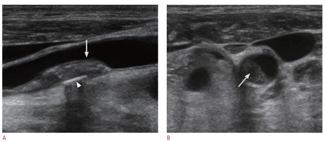
A. A gray-scale image of a longitudinal scan of the distal common carotid artery shows plaque with mixed echogenicity (arrow). A calcification is visible (arrowhead). The plaque surface is smooth. B. A transverse scan of the plaque at the distal common carotid artery shows central low echogenicity (arrow). The more lucent plaque is known to be associated with a higher risk of the stroke.