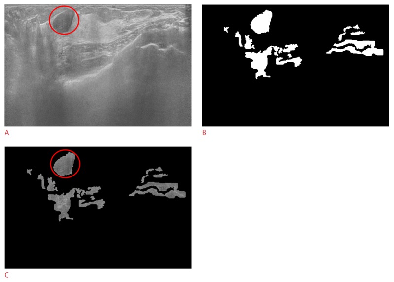
Figure 5. True positive case in a 46-year-old female with a pathologically proven fibroadenoma in her right breast.

A. The original automated breast ultrasonogram shows a 1.84-cm circumscribed hypoechoic nodule (fibroadenoma). B. The final mask after the adjusted Otsu's threshold and mor-phological operations shows mass candidates in white. C. The gray-scaled frame with the final mask reveals the true positive object (circle) and the false positives (some of which will be removed from the final mask after support vector machine classification).
