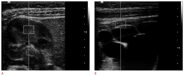
Figure 1. Shear wave velocity measurements in a two-month-old girl with hydronephrosis.

Figures show acoustic radiation force impulse measurements with a 4-9-MHz linear transducer using a 5 mm × 6 mm region of interest including both the renal cortex and the medulla with a normal right kidney in the axial view (A) and a hydronephrotic left kidney in the axial view (B). Note that the region of interest includes a calyx in the hydronephrotic kidney, which can cause a partial volume effect.
