Abstract
N-(p-Coumaryol) tryptamine (CT), a phenolic amide, has been reported to exhibit anti-oxidant and anti-inflammatory activities. However, the underlying mechanism by which CT exerts its pharmacological properties has not been clearly demonstrated. The objective of this study is to elucidate the anti-inflammatory mechanism of CT in lipopolysaccharide (LPS)-challenged RAW264.7 macrophage cells. CT significantly inhibited LPS-induced extracellular secretion of pro-inflammatory mediators such as nitric oxide (NO) and PGE2, and protein expressions of iNOS and COX-2. In addition, CT significantly suppressed LPS-induced secretion of pro-inflammatory cytokines such as TNF-α and IL-1β. To elucidate the underlying anti-inflammatory mechanism of CT, involvement of MAPK and Akt signaling pathways was examined. CT significantly attenuated LPS-induced activation of JNK/c-Jun, but not ERK and p38, in a concentration-dependent manner. Interestingly, CT appeared to suppress LPS-induced Akt phosphorylation. However, JNK inhibition, but not Akt inhibition, resulted in the suppression of LPS-induced responses, suggesting that JNK/c-Jun signaling pathway significantly contributes to LPS-induced inflammatory responses and that LPS-induced Akt phosphorylation might be a compensatory response to a stress condition. Taken together, the present study clearly demonstrates CT exerts anti-inflammatory activity through the suppression of JNK/c-Jun signaling pathway in LPS-challenged RAW264.7 macrophage cells.
Keywords: N-(p-Coumaroyl) tryptamine, RAW 264.7 cells, Lipopolysaccharide, iNOS, COX-2, JNK, c-Jun
INTRODUCTION
The c-Jun N-terminal kinase (JNK) of mitogen-activated protein (MAP) kinases has been reported to be implicated in the pathogenesis of various inflammatory disorders including sepsis (Ip and Davis, 1998; Supinski et al., 2009). JNK activation contributes to the development of inflammation-induced cell dysfunction and activation of caspases in several organs through the activation of transcription factor c-Jun (Cho and Choi, 2002; Wang et al., 2004). Recently, we demonstrated that inhibition of JNK activation with aromadendrin significantly attenuates lipopolysaccharide (LPS)-induced inflammatory responses in RAW264.7 cells (Lee et al., 2013).
Although macrophages play essential roles in the mobilization of the host defense against bacterial infection (Rehman et al., 2012), aberrant activation of macrophages has been also reported to play pathogenic roles in various inflammatory disorders including sepsis (Kim et al., 2012). In pathogenic conditions, abnormally activated macrophages produce excessive amount of a variety of pro-inflammatory mediators and cytokines that eventually aggravates the inflammatory conditions (Itharat and Hiransai, 2012). LPS, a component of the outer membrane of Gram-negative bacteria, is the most common cause of macrophage activation (Rietschel and Brade, 1992). LPS-induced activation of macrophages has been reported to cause a wide range of pro-inflammatory responses including secretion of pro-inflammatory mediators, expression of adhesion molecules and coagulation factors, phagocytosis, and cytoskeletal rearrangement (Sweet and Hume, 1996). Therefore, the suppression of aberrant macrophage activation might be a valuable therapeutic target for the treatment of inflammatory disorders.
N-(p-Coumaryol) tryptamine (CT) and its derivatives such as N-(p-coumaroyl) serotonin have been reported to exhibit various pharmacological activities including anti-oxidant, anti-inflammatory, and growth-promoting properties (Takii et al., 1999; Takii et al., 2003). It has been reported that the antioxidant effect of CT and its derivatives was due to radical scavenging activity (Zhang et al., 1997). However, the underlying mechanism by which CT exerts its pharmacological activity has not been clearly demonstrated. CT has been reported to be present in various medicinal herbs including Safflower oil cake (Carthamus tinctorius L.) (Takii et al., 1999; Takii et al., 2003) and Ravensara anisata (Andrianaivoravelona et al., 1999). CT, used in the present study, was isolated from the stem of Zea mays (Sim et al., 2014).
The objective of the present study was to examine whether CT possesses the anti-inflammatory activity in LPS-challenged RAW264.7 cells and to understand its underlying mechanism by which CT exerts the anti-inflammatory property in order to provide a valuable pharmacological agent that could suppress aberrantly activated macrophages in inflammation-related conditions.
MATERIALS AND METHODS
Reagents and cell culture
Bacterial lipopolysaccharide (LPS) from Escherichia coli serotype 055:B5 was purchased from Sigma-Aldrich (St. Louis, MO, USA). N-(p-Coumaryol) tryptamine (CT) (Fig. 1) was isolated and identified from Zea mays (Fig. 1) (Sim et al., 2014). CT was dissolved in dimethyl sulfoxide (DMSO) and added to the cell culture at the desired concentrations. The macrophage RAW264.7 cells were maintained in Dulbecco’s modified Eagle’s medium (DMEM; Gibco BRL, Grand Island, NY, USA) containing 5% heat-inactivated fetal bovine serum and penicillin-streptomycin (Gibco BRL) at 37°C, 5% CO2. In all experiments, cells were incubated in the presence of the indicated concentrations of CT before the addition of LPS (200 ng/ml).
Fig. 1.
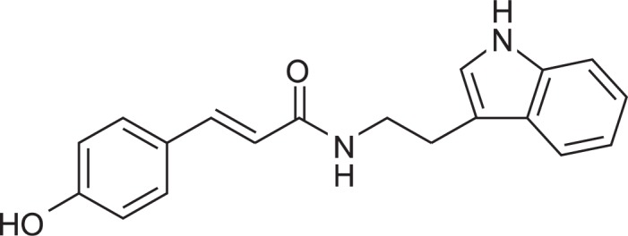
Chemical structure of N-(p-coumaryol) tryptamine.
Cell viability
Cell viability was determined by 3-(4,5-dimethylthiazol-2-yl)-2,5-diphenyltetrazolium bromide (MTT) assay. RAW 264.7 macrophage cells were seeded at 5×10⁵ cells per well and incubated with CT at various concentrations for 24 hr at 37°C. After incubation, MTT (0.5 mg/ml in PBS) was added to each well, and the cells were incubated for 3 hr at 37°C and 5% CO2. The resulting formazan crystals were dissolved in dimethyl sulfoxide (DMSO). Absorbance was determined at 540 nm. The results were expressed as a percentage of surviving cells over control cells.
Nitrite quantification assay
The production of NO was estimated by measuring the amount of nitrite, a stable metabolite of NO, using the Griess reagent as described (Lee et al., 2012). After CT-pretreated RAW264.7 macrophage cells were stimulated with LPS in 12-well plates for 24 hr, 100 μL of the cell supernatant was mixed with an equal volume of Griess reagent. Light absorbance was read at 540 nm. The results were expressed as a percentage of released NO from LPS-stimulated RAW 264.7 cells. To prepare a standard curve, sodium nitrite was used to prepare a standard curve.
ELISA assay for cytokines
The RAW264.7 macrophage cells were treated with CT in the absence or presence of LPS. After 24 hr incubation, TNF-α and IL-1β levels in culture media were quantified using monoclonal anti-TNF-α or IL-1β antibodies according to the manufacturer’s instruction (R&D Systems).
Western blot analysis
The RAW 264.7 macrophage cells were incubated with CT for 1 hr prior to LPS treatment. Cells were washed with PBS and lysed in PRO-PREP lysis buffer (iNtRON Biotechnology, Seongnam, Korea). Equal amounts of protein were separated on 10% SDS-polyacrylamide gel. Proteins were transferred to Hypond PVDF membrane (Amersham Biosciences, Piscataway, NJ, USA) and blocked in 5% skim milk in TBST for 1 hr at room temperature. Specific antibodies against inducible NO synthase (iNOS), COX-2, extracellular signal-regulated kinase (ERK), phosphorylated (p)-ERK, p38, p-p38, c-Jun N-terminal kinase (1:1,000; Cell signaling Technology), Akt, p-Akt (1:1,000; Cell signaling Technology), and β-actin (1:2,500; Sigma) were diluted in 5% skim milk. After thoroughly washing with TBST, horseradish peroxidase-conjugated secondary antibodies were applied. The blots were developed by the enhanced chemiluminescence detection (Amersham Biosciences).
Statistical analysis
All values shown in the figures are expressed as the mean ± SD obtained from at least three independent experiments. Statistical significance was analyzed by two-tailed Student’s t-test. Data with values of p<0.05 were considered as statistically significant. Single (* and #) and double (** and ##) marks represent statistical significance in p<0.05 and p<0.01, respectively.
RESULTS
N-(p-Coumaryol) tryptamine (CT) inhibits NO and PGE2 secretion in LPS-stimulated RAW 264.7 macrophage cells
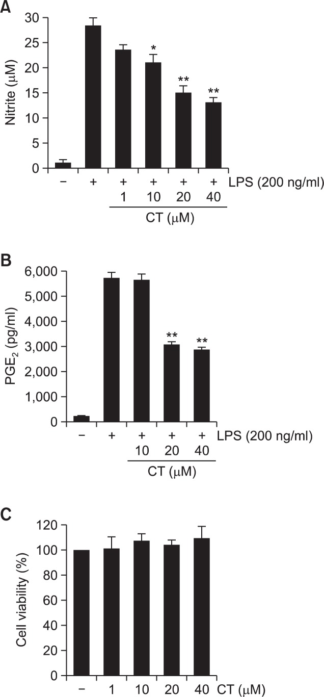
Given the previous reports that inflammatory mediators such as NO and PGE2 play key roles in the progression of inflammation (Ock et al., 2009; Lee et al., 2012), the effects of CT on the extracellular release of NO and PGE2 were examined in LPS-stimulated RAW264.7 macrophages. Cells were incubated with CT (1, 10, 20, or 40 μM) for 1 hr prior to LPS treatment (200 ng/ml). LPS showed markedly increased NO and PGE2 production in RAW264.7 cells. However, CT significantly inhibited extracellular release of NO and PGE2 in LPS-stimulated RAW264.7 cells in concentration dependent manners (Fig. 2A, B). In addition, CT showed negligible cytotoxicity in concentration ranges used in the study (Fig. 2C).
Fig. 2.
Effects of N-(p-coumaryol) tryptamine on LPS-induced extracellular release of NO (A) and PGE2 (B) in RAW264.7 macrophage cells. RAW264.7 cells were pretreated with indicated concentrations of N-(p-coumaryol) tryptamine for 1 hr before incubation with LPS (200 ng/ml) for 24 hr. The level of nitrite and PGE2 were measured using Griess reagent and ELISA assay, respectively. N-(p-Coumaryol) tryptamine significantly suppressed LPS-induced extracellular release of NO and PGE2. (C) Effect of N-(p-coumaryol) tryptamine on the viability of RAW264.7 cells. No significant cell death was observed with N-(p-coumaryol) tryptamine concentrations used in the present study. The data were obtained from three independent experiments and expressed as mean ± S.D. (n=3). *p<0.05 and **p<0.01 indicate statistically significant differences from treatment with LPS alone. CT stands for N-(p-coumaryol) tryptamine.
CT inhibits LPS-induced expressions of iNOS and COX-2
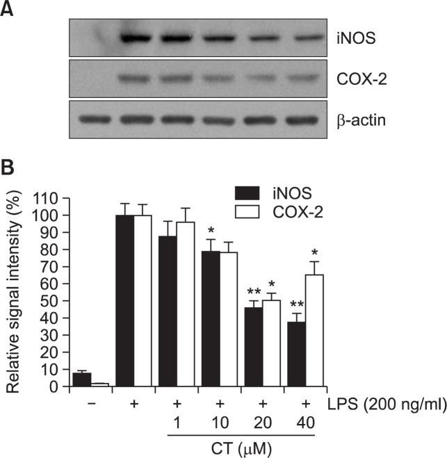
As CT inhibited LPS-induced extracellular release of NO and PGE2 in RAW264.7 cells (Fig. 2), whether the attenuated production of NO and PGE2 was attributable to downregulation of iNOS and COX-2 expression was examined in the absence or presence of CT. LPS treatment resulted in the significantly increased expression of iNOS and COX-2 proteins in RAW264.7 cells. Pretreatment of CT resulted in a significant suppression in LPS-induced iNOS and COX-2 overexpression levels in a concentration-dependent manner (Fig. 3), indicating that attenuated release of NO and PGE2 is due to decreased expression of their responsible genes by CT.
Fig. 3.
Effects of N-(p-coumaryol) tryptamine on LPS-induced expression of iNOS and COX-2 in RAW264.7cells. (A) The cell lysates were subjected to SDS-PAGE, and then protein levels of iNOS and COX-2 were determined by Western blot analysis. N-(p-Coumaryol) tryptamine significantly attenuated LPS-induced over expression of iNOS and COX-2. Images are representative of three independent experiments that shows reproducible results. (B) Quantitative analysis of immunoblots of iNOS and COX-2. N-(p-Coumaryol) tryptamine significantly suppressed LPS-induced iNOS and COX-2 expression. The data were obtained from three independent experiments and expressed as mean ± S.D. (n=3). *p<0.05 and **p<0.01 indicate statistically significant differences from treatment with LPS alone. CT stands for N-(p-coumaryol) tryptamine.
CT attenuates LPS-induced release of pro-inflammatory cytokines such as IL-1β and TNF-α
To examine the effects of CT on the extracellular release of pro-inflammatory cytokines such as IL-1β and TNF-α, secretion of these cytokines was measured using ELISA assay in LPS-stimulated RAW264.7 cells. LPS treatment resulted in excessive extracellular release of IL-1β and TNF-α in RWA 264.7 cells. However, CT significantly attenuated LPS-induced extracellular release of IL-1β and TNF-α in a concentration-dependent manner (Fig. 4).
Fig. 4.
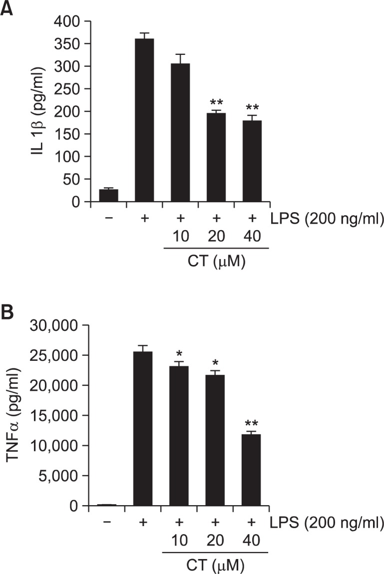
Effect of N-(p-coumaryol) tryptamine on LPS-induced extracellular secretion of IL-1β and in RAW264.7 macrophage cells. RAW264.7 cells were pretreated with indicated concentrations of N-(p-coumaryol) tryptamine for 1 hr, then incubated with LPS (200 ng/ml) for 24 hrs. The concentrations of IL-1β (A) and TNF-α (B) in collected cell culture media were measured by ELISA assay as described in the methods. N-(p-Coumaryol) tryptamine meaningfully reduced LPS-stimulated IL-1β and TNF-α cytokines in aconcentration-dependent manner. The values are expressed as mean ± SD for three independent experiments. *p<0.05 and **p<0.01 indicate statistically significant differences from treatments with LPSalone.
CT inhibits the phosphorylation of JNK/c-Jun in LPS-stimulated RAW264.7 cells
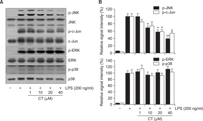
MAP kinase signaling pathways have been reported to be implicated in the LPS-induced inflammatory responses (Guha and Mackman, 2001; Rushworth et al., 2005). In the present study, to understand the underlying signaling mechanism by which CT exhibits its anti-inflammatory activity, the effect of CT on LPS-stimulated phosphorylation of JNK, ERK, and p38 kinase in RAW264.7 cells was examined. Cells were pretreated with CT at indicated concentrations (1, 10, 20, and 40 μM), and then were treated with LPS (200 ng/ml) for 30 min. CT significantly suppressed the LPS-induced phosphorylation of JNK and transcription factor c-Jun, a downstream target of JNK, in a concentration-dependent manner in RAW 264.7 cells (Fig. 5). However, no significant attenuation of LPS-induced ERK and p38 phosphorylation was observed (Fig. 5), suggesting that CT exerts its anti-inflammatory action through the suppression of JNK/c-Jun signaling pathway in RAW264.7 cells.
Fig. 5.
Effect of N-(p-coumaryol) tryptamine on LPS-induced activation of MAPK signaling pathway in RAW264.7 macrophage cells. (A) representative immunoblots, (B) quantitative analysis of immunoblots. Cellswere challenged with 200 ng/ml LPS in the absence or presence of N-(p-coumaryol) tryptamine. LPS-induced increased phosphorylation of JNK and c-jun was significantly attenuated with N-(p-coumaryol) tryptamine treatment. However, phosphorylation of ERK and p38 was not affected with N-(p-coumaryol) tryptamine treatment, suggesting that JNK/c-jun signaling might play a key role in the LPS-induced activation of RAW264.7 cells. Images are representative of three independent experiments that shows reproducible results. The values are expressed as mean ± SD for three independent experiments. *p<0.05 and **p<0.01 indicate statistically significant differences from treatments with LPS alone. CT stands for N-(p-coumaryol) tryptamine.
CT appears to inhibit LPS-induced Akt phosphorylation in LPS-stimulated RAW 264.7 cells
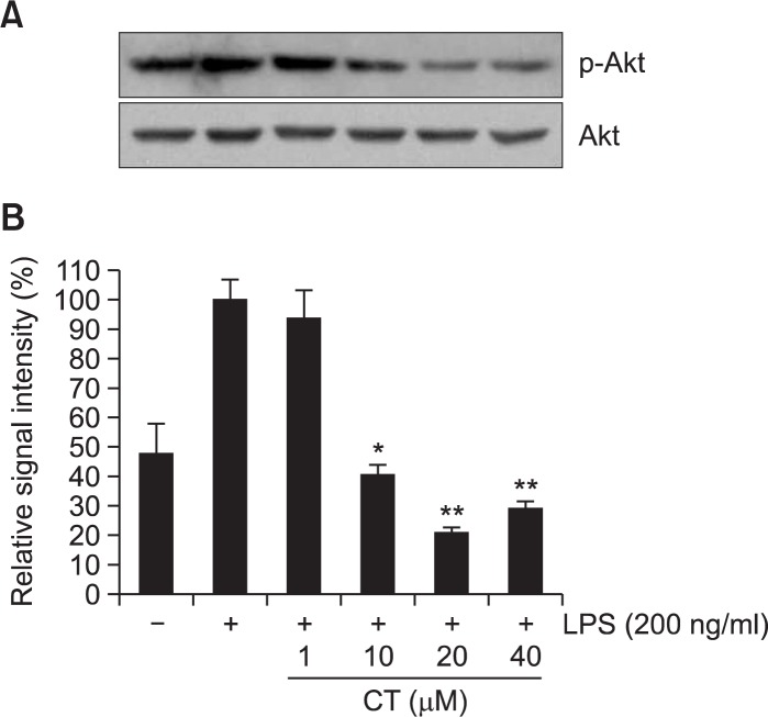
It has been previously reported that suppression of PI3K/Akt signaling plays a key role in the attenuation of LPS-induced NF-κB activation (Dilshara et al., 2013; Liu et al., 2013). To understand the possible involvement of Akt signaling pathway in the present study, the effect of CT on LPS-induced Akt phosphorylation was examined. LPS showed markedly increased phosphorylation of Akt and CT significantly attenuated LPS-induced Akt phosphorylation in a concentration-dependent manner (Fig. 6).
Fig. 6.
Effect of N-(p-coumaryol) tryptamine on LPS-induced activation of Akt signaling pathway in RAW264.7 macrophage cells. (A) representative immunoblots, (B) quantitative analysis of immunoblots. N-(p-coumaryol) tryptamine appeared to significantly attenuate Akt phosphorylation in RAW264.7 macrophage cells. The images shown are representative of three independent experiments and the values are expressed as mean ± SD for three experiments. *p<0.05 and **p<0.01 indicate statistically significant differences from treatments with control alone. CT stands for N-(pcoumaryol) tryptamine.
Inhibition of JNK but not Akt exhibits the suppression of LPS-induced pro-inflammatory responses
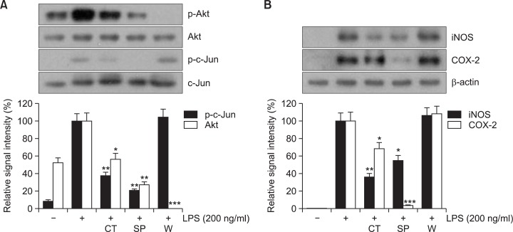
The present data demonstrated that CT significantly suppresses LPS-induced activation of JNK/c-jun (Fig. 5) and Akt (Fig. 6) signaling pathways. In order to delineate the exact signaling pathway by which CT exerts its anti-inflammatory responses, blockade of each signaling pathway was achieved with specific inhibitors and involvement of each signaling pathway in LPS-induced inflammatory response was examined. Inhibition of JNK/c-Jun signaling pathway with SP600125 showed significant suppression of c-jun and Akt phosphorylation, which is quite similar with CT (Fig. 7A). However, inhibition of Akt signaling pathway with wortmannin, a PI3K inhibitor, exhibited only Akt phosphorylation but not c-jun phosphorylation (Fig. 7A). Furthermore, inhibition of JNK/c-Jun signaling pathway significantly attenuated LPS-induced iNOS and COX-2 expression (Fig. 7B). However, inhibition of Akt signaling pathway with wortmannin could not inhibit LSP-induced expression of iNOS and COX-2 (Fig. 7B), strongly suggesting that CT exerts its anti-inflammatory action through the suppression JNK/c-Jun signaling pathway and that apparent suppression of Akt phosphorylation by CT might be due to the lessened necessity of compensatory action as CT attenuates the LPS-induced damage, rather than direct inhibition of Akt phosphorylation by CT.
Fig. 7.
Role of JNK and Akt signaling pathways in N-(p-coumaryol) tryptamine-mediated suppression of LPS-induced RAW264.7 cell activation. RAW264.7 cells were pretreated with CT (N-(p-coumaryol) tryptamine), SP (SP600125, JNK inhibitor), or W (wortmannin, Akt inhibitor), and then exposed to LPS (200 ng/ml) for 1 hr. The cell lysates were prepared and subjected to Western blotting analysis. (A) Phosphorylation levels of Akt and c-jun were examined in the presence of CT, SP, or W. CT and SP significantly suppressed LPS-induced phosphorylation of Akt and c-jun. However, W did not inhibit LPS-induced c-jun phosphorylation. (B) The protein levels of iNOS and COX-2 were examined. Suppression of LPS-induced iNOS and COX-2 expressions were observed with N-(p-coumaryol) tryptamine (CT) and also with JNK inhibitor (SP) but not with Akt inhibitor (W). The images on top are representatives of three independent experiments. The data for quantitative analyses on bottom were obtained from three independent experiments and expressed as mean ± SD (n=3). *p<0.05 and **p<0.01 indicate statistically significant differences from treatments with control alone.
DISCUSSION
The present study clearly demonstrated that N-(p-coumaryol) tryptamine (CT) suppresses LPS-induced inflammatory responses through the suppression of JNK/c-Jun signaling pathway in LPS-challenged RAW264.7 macrophage cells. CT significantly inhibited LPS-induced extracellular release of pro-inflammatory mediators and cytokines. In addition, CT significantly attenuated LPS-induced expression of iNOS and COX-2 proteins.
In accordance with previous reports that CT and its derivatives have pharmacological properties such as anti-inflammatory and anti-oxidant actions (Takii et al., 1999; Takii et al., 2003), the present study demonstrated that CT possesses anti-inflammatory properties in LPS-induced RAW 264.7 macrophage cells. However, the underlying mechanism by which CT and its derivatives exert the anti-inflammatory activity has not been clearly demonstrated. Therefore, the present study elucidated that CT exerts the anti-inflammatory action through the suppression of LPS-induced activation JNK/c-Jun signaling pathway.
Macrophages play essential roles in the host defense against bacterial infection (Rehman et al., 2012). However, aberrant activation of macrophages plays detrimental roles in multiple inflammation-related disorders including sepsis by producing a wide range of pro-inflammatory mediators and cytokines (Rietschel and Brade, 1992). Macrophages initiate LPS-induced pro-inflammatory gene transcription when LPS binds to its membrane receptor, TLR4, which leads to the phosphorylation of multiple kinases, which subsequently activates various pro-inflammatory transcription factors including NF-κB and AP-1 (O’Connell et al., 1998; Guha and Mackman, 2001). We previously reported that LPS causes increased expression of pro-inflammatory mediators and the increased degradation IκB (Vo et al., 2012). CT significantly attenuated LPS-induced extracellular release of pro-inflammatory cytokines such as IL-1β and TNF-α. Although pro-inflammatory cytokines such as IL-1β and TNF-α have been reported to play key roles in the development of inflammatory responses, other cytokines and transcription factors might also play a certain role. Therefore, further studies are necessary to clearly explain the effect of CT other cytokines and transcription factors in LPS-mediated inflammatory conditions.
NF-κB is a major transcription factor responsible for the expression of a variety of pro-inflammatory genes such as mediators such as iNOS, COX-2, and cytokines (Siebenlist et al., 1994). Therefore, the aberrant activation of NF-κB has been associated with various pathological conditions including cancers and autoimmune diseases (Li and Verma, 2002). We previously reported that suppression of NF-κB transcription mediated anti-inflammatory properties of natural products against LPS-induced inflammation in RAW264.7 cells (Vo et al., 2014a; Vo et al., 2014b). The present data showed that CT significantly suppressed the expression of iNOS and COX-2, presumably through the suppression of NF-κB transcription. However, further studies are necessary to clearly delineate the exact mechanism by which CT leads to the suppression of iNOS and COX-2 expression.
Many studies have shown that LPS activates all three MAP kinases such as ERK, JNK, and p38 in macrophages (Sweet and Hume, 1996) and that many of the downstream targets of MAPK pathways are transcription factors, which regulate various genes encoding inflammatory mediators (Zhang et al., 2006; Lee et al., 2009; Zhang et al., 2011). In the present study, LPS treatment exhibited increased phosphorylation of all three MAPKs. However, CT selectively attenuated LPS-induced JNK/c-Jun activation in a concentration-dependent manner with concurrent significant attenuation of LPS-induced pro-inflammatory responses. The JNK/c-Jun signaling pathway has been implicated in the development of inflammatory responses in various conditions leading to cellular dysfunction and organ failures (Ip and Davis, 1998; Supinski et al., 2009). It has been also reported that inhibition of JNK activation significantly attenuates lipopolysaccharide (LPS)-induced inflammatory responses in RAW264.7 cells (Lee et al., 2013). No noticeable changes were observed in phosphorylation of ERK and p38 with CT. Furthermore, inhibition of JNK signaling pathway with specific JNK inhibitor mimicked the action of CT, suggesting that selective suppression of JNK/c-Jun signaling pathway might be sufficient to exert anti-inflammatory effects of CT in RAW 264.7 cells and that JNK/c-Jun pathway rather than ERK and p38 plays a key role in pro-inflammatory responses in RAW264.7 cells.
It has been reported that LPS-induced activation of PI3K/Akt signaling pathway contributes to the development of inflammatory responses including the activation of NF-κB transcription (Madrid et al., 2001; Joh and Kim, 2011). Many reports have shown that suppression of Akt signaling attenuates LPS-induced inflammatory responses in various models (Shao and Lin, 2008; Dilshara et al., 2013; Liu et al., 2013). In the present study, LSP treatment resulted in the phosphorylation of Akt and CT appeared to significantly suppress Akt phosphorylation in a concentration-dependent manner in RAW 264.7 cells. To the contrary, it has been also reported that activation of PI3K/Akt signaling pathway reduces LPS-induced inflammation in various models (Kidd et al., 2008; Xu et al., 2010; Zong et al., 2012). Activation of Akt/PI3K signaling pathway has been reported to exert significant anti-inflammatory effects through the suppression of NF-κB-mediated transcription (Ha et al., 2011; Vo et al., 2014a). Therefore, the role of PI3K/Akt signaling cascades in LPS-induced inflammatory response still remains controversial (Takeshima et al., 2009). In the present study, inhibition of PI3K/Akt signaling pathway failed to suppress LPS-induced expression of iNOS and COX-2, which was shown with JNK inhibition and CT treatment. The data strongly suggest that the PI3K/Akt pathway might not be responsible for the LPS-induced inflammatory responses. Apparent attenuation of Akt phosphorylation with CT might be considered due to the lessened mobilization of compensatory intracellular components as CT attenuates the LPS-induced inflammation, rather than direct inhibition of Akt phosphorylation by CT. However, further studies are necessary to clearly delineate the phenomenon.
In conclusion, the present data clearly demonstrates that CT exerts anti-inflammatory property through the inhibition of JNK/c-Jun signaling pathway in LPS-challenged RAW264.7 macrophage cells. The present study strongly suggests that CT might be a valuable therapeutic agent for the treatment of inflammation-related pathogenic conditions.
REFERENCES
- Andrianaivoravelona JO, Terreaux C, Sahpaz S, Rasolondramanitra J, Hostettmann K. A phenolic glycoside and N-(p-coumaroyl)-tryptamine from Ravensara anisata. Phytochemistry. 1999;52:1145–1148. doi: 10.1016/s0031-9422(99)00158-2. [DOI] [PubMed] [Google Scholar]
- Cho SG, Choi EJ. Apoptotic signaling pathways: caspases and stress-activated protein kinases. J Biochem Mol Biol. 2002;35:24–27. doi: 10.5483/bmbrep.2002.35.1.024. [DOI] [PubMed] [Google Scholar]
- Dilshara MG, Jayasooriya RG, Lee S, Jeong JB, Seo YT, Choi YH, Jeong JW, Jang YP, Jeong YK, Kim GY. Water extract of processed Hydrangea macrophylla (Thunb.) Ser. leaf attenuates the expression of pro-inflammatory mediators by suppressing Akt-mediated NF-kappaB activation. Environ Toxicol Pharmacol. 2013;35:311–319. doi: 10.1016/j.etap.2012.12.012. [DOI] [PubMed] [Google Scholar]
- Guha M, Mackman N. LPS induction of gene expression in human monocytes. Cell Signal. 2001;13:85–94. doi: 10.1016/s0898-6568(00)00149-2. [DOI] [PubMed] [Google Scholar]
- Ha YM, Ham SA, Kim YM, Lee YS, Kim HJ, Seo HG, Lee JH, Park MK, Chang KC. beta(1)-adrenergic receptor-mediated HO-1 induction, via PI3K and p38 MAPK, by isoproterenol in RAW 264.7 cells leads to inhibition of HMGB1 release in LPS-activated RAW 264.7 cells and increases in survival rate of CLP-induced septic mice. Biochem Pharmacol. 2011;82:769–777. doi: 10.1016/j.bcp.2011.06.041. [DOI] [PubMed] [Google Scholar]
- Ip YT, Davis RJ. Signal transduction by the c-Jun N-terminal kinase (JNK)--from inflammation to development. Curr Opin Cell Biol. 1998;10:205–219. doi: 10.1016/s0955-0674(98)80143-9. [DOI] [PubMed] [Google Scholar]
- Itharat A, Hiransai P. Dioscoreanone suppresses LPS-induced nitric oxide production and inflammatory cytokine expression in RAW 264.7 macrophages by NF-kappaB and ERK1/2 signaling transduction. J Cell Biochem. 2012;113:3427–3435. doi: 10.1002/jcb.24219. [DOI] [PubMed] [Google Scholar]
- Joh EH, Kim DH. Kalopanaxsaponin A ameliorates experimental colitis in mice by inhibiting IRAK-1 activation in the NF-kappaB and MAPK pathways. Br J Pharmacol. 2011;162:1731–1742. doi: 10.1111/j.1476-5381.2010.01195.x. [DOI] [PMC free article] [PubMed] [Google Scholar]
- Kidd LB, Schabbauer GA, Luyendyk JP, Holscher TD, Tilley RE, Tencati M, Mackman N. Insulin activation of the phosphatidylinositol 3-kinase/protein kinase B (Akt) pathway reduces lipopolysaccharide-induced inflammation in mice. J Pharmacol Exp Ther. 2008;326:348–353. doi: 10.1124/jpet.108.138891. [DOI] [PMC free article] [PubMed] [Google Scholar]
- Kim YJ, Shin Y, Lee KH, Kim TJ. Anethum graveloens flower extracts inhibited a lipopolysaccharide-induced inflammatory response by blocking iNOS expression and NF-kappaB activity in macrophages. Biosci Biotechnol Biochem. 2012;76:1122–1127. doi: 10.1271/bbb.110950. [DOI] [PubMed] [Google Scholar]
- Lee JW, Bae CJ, Choi YJ, Kim SI, Kim NH, Lee HJ, Kim SS, Kwon YS, Chun W. 3,4,5-Trihydroxycinnamic acid inhibits LPS-induced iNOS expression by suppressing NF-kappaB activation in BV2 microglial cells. Korean J Physiol Pharmacol. 2012;16:107–112. doi: 10.4196/kjpp.2012.16.2.107. [DOI] [PMC free article] [PubMed] [Google Scholar]
- Lee JW, Kim NH, Kim JY, Park JH, Shin SY, Kwon YS, Lee HJ, Kim SS, Chun W. Aromadendrin inhibits lipopolysaccharide-induced nuclear translocation of NF-kappaB and phosphorylation of JNK in RAW 264.7 macrophage cells. Biomol Ther. 2013;21:216–221. doi: 10.4062/biomolther.2013.023. [DOI] [PMC free article] [PubMed] [Google Scholar]
- Lee YJ, Kim S, Lee SJ, Ham I, Whang WK. Anti-oxidant activities of new flavonoids from Cudrania tricuspidata root bark. Arch Pharm Res. 2009;32:195–200. doi: 10.1007/s12272-009-1135-z. [DOI] [PubMed] [Google Scholar]
- Li Q, Verma IM. NF-kappaB regulation in the immune system. Nat Rev Immunol. 2002;2:725–734. doi: 10.1038/nri910. [DOI] [PubMed] [Google Scholar]
- Liu XH, Pan LL, Jia YL, Wu D, Xiong QH, Wang Y, Zhu YZ. A novel compound DSC suppresses lipopolysaccharide-induced inflammatory responses by inhibition of Akt/NF-kappaB signalling in macrophages. Eur J Pharmacol. 2013;708:8–13. doi: 10.1016/j.ejphar.2013.01.013. [DOI] [PubMed] [Google Scholar]
- Madrid LV, Mayo MW, Reuther JY, Baldwin AS., Jr Akt stimulates the transactivation potential of the RelA/p65 Subunit of NF-kappa B through utilization of the Ikappa B kinase and activation of the mitogen-activated protein kinase p38. J Biol Chem. 2001;276:18934–18940. doi: 10.1074/jbc.M101103200. [DOI] [PubMed] [Google Scholar]
- O’Connell MA, Bennett BL, Mercurio F, Manning AM, Mackman N. Role of IKK1 and IKK2 in lipopolysaccharide signaling in human monocytic cells. J Biol Chem. 1998;273:30410–30414. doi: 10.1074/jbc.273.46.30410. [DOI] [PubMed] [Google Scholar]
- Ock J, Kim S, Suk K. Anti-inflammatory effects of a fluorovinyloxyacetamide compound KT-15087 in microglia cells. Pharmacol Res. 2009;59:414–422. doi: 10.1016/j.phrs.2009.02.008. [DOI] [PubMed] [Google Scholar]
- Rehman MU, Yoshihisa Y, Miyamoto Y, Shimizu T. The anti-inflammatory effects of platinum nanoparticles on the lipopolysaccharide-induced inflammatory response in RAW 264.7 macrophages. Inflamm Res. 2012;61:1177–1185. doi: 10.1007/s00011-012-0512-0. [DOI] [PubMed] [Google Scholar]
- Rietschel ET, Brade H. Bacterial endotoxins. Sci Am. 1992;267:54–61. doi: 10.1038/scientificamerican0892-54. [DOI] [PubMed] [Google Scholar]
- Rushworth SA, Chen XL, Mackman N, Ogborne RM, O’Co nnell MA. Lipopolysaccharide-induced heme oxygenase-1 expression in human monocytic cells is mediated via Nrf2 and protein kinase C. J Immunol. 2005;175:4408–4415. doi: 10.4049/jimmunol.175.7.4408. [DOI] [PubMed] [Google Scholar]
- Shao DZ, Lin M. Platonin inhibits LPS-induced NF-kappaB by preventing activation of Akt and IKKbeta in human PBMC. Inflamm Res. 2008;57:601–606. doi: 10.1007/s00011-008-8053-2. [DOI] [PubMed] [Google Scholar]
- Siebenlist U, Franzoso G, Brown K. Structure, regulation and function of NF-kappa B. Annu Rev Cell Biol. 1994;10:405–455. doi: 10.1146/annurev.cb.10.110194.002201. [DOI] [PubMed] [Google Scholar]
- Sim JY, Kim MA, Kim MJ, Chun W, Kwon YS. Acetylcholinesterase inhibitors from the stem of Zea Zea mays. Nat Prod Sci. 2014;20:13–16. [Google Scholar]
- Supinski GS, Ji X, Callahan LA. The JNK MAP kinase pathway contributes to the development of endotoxin-induced diaphragm caspase activation. Am J Physiol Regul Integr Comp Physiol. 2009;297:R825–834. doi: 10.1152/ajpregu.90849.2008. [DOI] [PMC free article] [PubMed] [Google Scholar]
- Sweet MJ, Hume DA. Endotoxin signal transduction in macrophages. J Leukoc Biol. 1996;60:8–26. doi: 10.1002/jlb.60.1.8. [DOI] [PubMed] [Google Scholar]
- Takeshima E, Tomimori K, Kawakami H, Ishikawa C, Sawada S, Tomita M, Senba M, Kinjo F, Mimuro H, Sasakawa C, Fujita J, Mori N. NF-kappaB activation by Helicobacter pylori requires Akt-mediated phosphorylation of p65. BMC Microbiol. 2009;9:36. doi: 10.1186/1471-2180-9-36. [DOI] [PMC free article] [PubMed] [Google Scholar] [Retracted]
- Takii T, Hayashi M, Hiroma H, Chiba T, Kawashima S, Zhang HL, Nagatsu A, Sakakibara J, Onozaki K. Serotonin derivative, N-(p-Coumaroyl)serotonin, isolated from safflower (Carthamus tinctorius L.) oil cake augments the proliferation of normal human and mouse fibroblasts in synergy with basic fibro-blast growth factor (bFGF) or epidermal growth factor (EGF) J Biochem. 1999;125:910–915. doi: 10.1093/oxfordjournals.jbchem.a022368. [DOI] [PubMed] [Google Scholar]
- Takii T, Kawashima S, Chiba T, Hayashi H, Hayashi M, Hiroma H, Kimura H, Inukai Y, Shibata Y, Nagatsu A, Sakakibara J, Oomoto Y, Hirose K, Onozaki K. Multiple mechanisms involved in the inhibition of proinflammatory cytokine production from human monocytes by N-(p-coumaroyl)serotonin and its derivatives. Int Immunopharmacol. 2003;3:273–277. doi: 10.1016/s1567-5769(02)00207-2. [DOI] [PubMed] [Google Scholar]
- Vo VA, Lee JW, Chang JE, Kim JY, Kim NH, Lee HJ, Kim SS, Chun W, Kwon YS. Avicularin inhibits lipopolysaccharide-induced inflammatory response by suppressing ERK phosphorylation in RAW 264.7 macrophages. Biomol Ther. 2012;20:532–537. doi: 10.4062/biomolther.2012.20.6.532. [DOI] [PMC free article] [PubMed] [Google Scholar]
- Vo VA, Lee JW, Kim JY, Park JH, Lee HJ, Kim SS, Kwon YS, Chun W. Phosphorylation of Akt mediates anti-inflammatory activity of 1-p-coumaronyl beta-D-glucoside against lipopolysaccharide-induced inflammation in RAW264.7 cells. Korean J Physiol Pharmacol. 2014a;18:79–86. doi: 10.4196/kjpp.2014.18.1.79. [DOI] [PMC free article] [PubMed] [Google Scholar]
- Vo VA, Lee JW, Shin SY, Kwon JH, Lee HJ, Kim SS, Kwon YS, Chun W. Methyl p-hydroxycinnamate suppresses lipopolysaccharide-induced inflammatory responses through Akt phosphorylation in RAW264.7 cells. Biomol Ther. 2014b;22:10–16. doi: 10.4062/biomolther.2013.095. [DOI] [PMC free article] [PubMed] [Google Scholar]
- Wang WH, Gregori G, Hullinger RL, Andrisani OM. Sustained activation of p38 mitogen-activated protein kinase and c-Jun N-terminal kinase pathways by hepatitis B virus X protein mediates apoptosis via induction of Fas/FasL and tumor necrosis factor (TNF) receptor 1/TNF-alpha expression. Mol Cell Biol. 2004;24:10352–10365. doi: 10.1128/MCB.24.23.10352-10365.2004. [DOI] [PMC free article] [PubMed] [Google Scholar]
- Xu CQ, Liu BJ, Wu JF, Xu YC, Duan XH, Cao YX, Dong JC. Icariin attenuates LPS-induced acute inflammatory responses: involvement of PI3K/Akt and NF-kappaB signaling pathway. Eur J Pharmacol. 2010;642:146–153. doi: 10.1016/j.ejphar.2010.05.012. [DOI] [PubMed] [Google Scholar]
- Zhang HL, Nagatsu A, Watanabe T, Sakakibara J, Okuyama H. Antioxidative compounds isolated from safflower (Carthamus tinctorius L.) oil cake. Chem. Pharm. Bull. (Tokyo) 1997;45:1910–1914. doi: 10.1248/cpb.45.1910. [DOI] [PubMed] [Google Scholar]
- Zhang WY, Lee JJ, Kim IS, Kim Y, Myung CS. Stimulation of glucose uptake and improvement of insulin resistance by aromadendrin. Pharmacology. 2011;88:266–274. doi: 10.1159/000331862. [DOI] [PubMed] [Google Scholar]
- Zhang X, Hung TM, Phuong PT, Ngoc TM, Min BS, Song KS, Seong YH, Bae K. Anti-inflammatory activity of flavonoids from Populus davidiana. Arch Pharm Res. 2006;29:1102–1108. doi: 10.1007/BF02969299. [DOI] [PubMed] [Google Scholar]
- Zong Y, Sun L, Liu B, Deng YS, Zhan D, Chen YL, He Y, Liu J, Zhang ZJ, Sun J, Lu D. Resveratrol inhibits LPS-induced MAPKs activation via activation of the phosphatidylinositol 3-kinase pathway in murine RAW 264.7 macrophage cells. PLoS One. 2012;7:e44107. doi: 10.1371/journal.pone.0044107. [DOI] [PMC free article] [PubMed] [Google Scholar]


