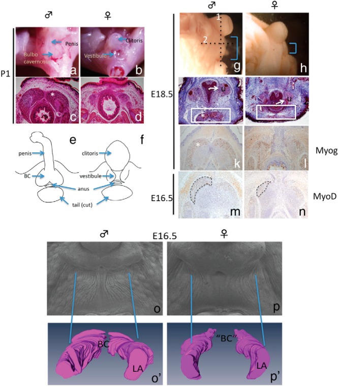
Figure 1.
Characterization of male and female BC muscles. Whole mount (A and B) and transverse sections of BC (C and D) from postnatal samples of the male and female are shown. E and F, A schematic representation of the structures in the perineum. G and H, Embryonic investigations of the perineum with AGD difference observed at E18.5. I and J, Transverse section (dotted line 1 in G) of E18.5 perineum stained with Masson trichrome showing the BC (*) located ventral to the urethra (white box). Horizontal sections (dotted line 2 in G) of E18.5 and E16.5 perineum of the BC region (* and traced line) were stained with anti-Myog (K and L) and MyoD (M and N), respectively. Scanning electron microscope photomicrograph of the perineum (O and P) and a 3D reconstruction (O′ and P′) of the BC or remnant of BC/LA complex based on MyoD-stained sections at E16.5. U, urethra.

