ABSTRACT
Study Design:
Case Report
Background:
Upper extremity deep vein thrombosis (UEDVT) is a rare complication following arthroscopic shoulder surgery. However, it is possible that a patient with an UEDVT will present to physical therapy as the first service to interact with the patient following surgery. As a result, proper screening in the physical therapy setting is essential.
Case Description:
The purpose of this report is to present the case of a 37 year‐old male who developed an upper extremity deep vein thrombosis (UEDVT) following arthroscopic glenohumeral labral repair and arthroscopically assisted biceps tenodesis. This patient presented with disproportionate pain and swelling of his involved upper extremity at his initial evaluation in physical therapy (8 days post‐operatively), which raised the index of suspicion for an UEDVT.
Outcome:
The patient was referred to the emergency department for immediate diagnostic testing and treatment. A Doppler scan provided a definitive diagnosis of UEDVT. Following successful medical treatment with anti‐coagulant therapy, the patient went on to complete an otherwise uneventful course of rehabilitation.
Discussion:
UEDVT events following arthroscopy are rare, and are often attributed to a systemic secondary stimulus. UEDVT following shoulder arthroscopy is a complication that occurs in the orthopaedic setting, but may present primarily to the physical therapist, and as such requires awareness of its clinical presentation and treatment. Care of UEDVT requires a systems‐based approach when considering clinical manifestation, best treatment, and future research
Keywords: Arthroscopy, Shoulder, SLAP Tear, Upper Extremity Deep Venous Thrombosis
INTRODUCTION AND BACKGROUND
Upper extremity deep venous thrombosis (UEDVT) is a rare but serious complication of arthroscopic shoulder surgery.1 UEDVTs are of principal concern as they carry the potential to migrate to the lung resulting in a pulmonary embolism (PE).2
It has been reported that 1–10% of all DVTs occur in the UE, and 9–14% of these cases are complicated by PE.3‐5 In addition, long term comorbidities associated with UEDVT such as post‐thrombotic syndrome (PTS) has been reported to have a 7–44% frequency of occurrence status‐post UEDVT depending on the criteria used for diagnosis.6 The 3‐month UEDVT associated mortality rate without comorbidities, such as cancer or central venous catheter placement, has been reported to be 2.6%.4
Signs and symptoms of UEDVT are extremely variable, just as they are for lower extremity DVTs.4 However, the generally accepted clinical picture of a patient with an UEDVT includes: +1 pitting edema, ipsilateral pain to palpation, erythema, and warmth with palpation of ipsilateral extremity.7 As the popularity of orthopaedic surgical procedures is on the rise, post‐operative complications such as DVTs may become more prevalent in the PT clinic, requiring rigorous screening and a higher index of suspicion.8 As noted by Hannon et al. who recently described a lower extremity DVT (LEDVT) in an otherwise healthy young man, after undergoing ulnar collateral ligament reconstruction.9
The risk of mortality, morbidity, and resulting decreased quality of life are a direct result of this potential post‐operative complication, and warrants vigilance in clinical practice for early detection and appropriate management. The purpose of this paper is to (1) present a case report of a patient who presented in physical therapy with an UEDVT following an arthroscopic glenohumeral labral repair and (2) discuss the incidence of UEDVT associated with arthroscopic shoulder surgery.
CASE DESCRIPTION
A 37 year‐old Caucasian male presented to the primary orthopaedic surgeon (ADM) complaining of significant right shoulder pain sustained during a bench‐press injury. The patient reported that this pain had persisted for five months since the injury, and he had already failed conservative treatment including a course of physical therapy. The patient reported no history of steroid, illicit drug or tobacco use. The patient reported no significant past medical history, which of note was unremarkable for clotting disorders. Family history was negative for clotting disorders.
Preoperative diagnosis was a massive labral tear with evidence of a labral cyst. Conservative and operative treatment options were discussed at length with the patient and the patient chose operative treatment.
The patient subsequently underwent an arthroscopic labral repair, excision of labral cyst, and an arthroscopic assisted open subpectoral biceps tenodesis. The patient was operated on in the lateral position with a bean bag and an axillary roll, with seven pounds of abduction and five pounds of longitudinal traction applied to the operative upper extremity. From incision to closure, the elapsed time was 119 minutes, and time under anesthesia was a total of 125 minutes. There were no obvious complications during the procedure, and there was minimal blood loss. The patient was placed in an external rotation sling and issued a cryo‐cuff to reduce swelling and inflammation post‐operatively.
Eight days post‐operatively the patient presented to physical therapy with the primary therapist (TJSD). During the initial history, the patient reportedly kept his affected UE completely immobilized in the external rotation sling from 0‐8 days post‐op, as per instructions given to him by a post‐operative care nurse. Patient reports not removing the sling, except for showering, and only used the exercise stress ball included with the sling.
During the initial interview, the patient complained of pain at rest, which was increased in the dependent position, and was without relief with post‐operative medication. The patient also complained of increased swelling, and a sensation of pins and needles in the affected UE distal to the elbow while at rest. The patient denied fever or chills.
Clinical Impression #1
Due to the post‐operative nature of this case the differential was focused on pathologies which are congruent with this etiology. Clinical decision‐making was required to determine if proceeding with post‐operative rehabilitation was appropriate for this patient, or if a referral to the medical team may be needed to ensure patient safety before progressing. The differential at this point included three main considerations, which were UEDVT, post‐operative infection of joint or wound site, or normal variant of post‐operative edema. Given that the patient was 8 days from surgery, all three of these diagnoses were equally probable with regards to time course, and due to the non‐specific symptoms, a focused physical examination was required.
Examination
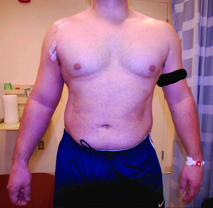
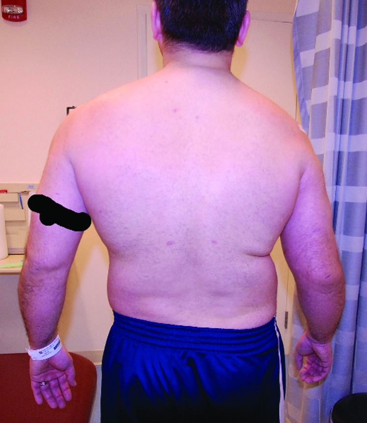
On physical exam the right UE was warm, erythematous, edematous, and very painful to palpation (Figure 1 and Figure 2). The erythema was described with non‐demarcated borders, extending distally along the medial side of the affected UE, without extension laterally, or proximally. There were no foul smelling odors noted from the wound site. Initial passive range of motion (PROM) revealed 10 degrees of external rotation (ER) and 90 degrees of forward flexion (FF). There were no palpable cords indicating superficial thrombophlebitis.10 +1 pitting edema was noted to extend from the axilla to approximately 15 cm distal to the elbow on the affected UE (Table 1). Passive extension of the elbow on the affected UE reproduced pain over the medial aspect of the subaxillary region. Pain to palpation was also noted over the medial aspect of the ipsilateral UE proximal to the elbow, extending to the axilla.
Figure 2.
Figure 1.
Table 1.
Circumferential measurements of the upper extremities 8 days s/p surgery
| Unaffected UE (cm) | Affected UE (cm) | Affected vs. Unaffected (cm) | |
|---|---|---|---|
| 15 cm proximal to elbow | 38.5 | 38.5 | 0 |
| 10 cm proximal to elbow | 36 | 38 | +2 |
| 5 cm proximal to elbow | 33.5 | 37 | +3.5 |
| Elbow (at joint line) | 31 | 34.5 | +3.5 |
| 5 cm distal to elbow | 31.5 | 37 | +5.5 |
| 10 cm distal to elbow | 30 | 35 | +5 |
| 15 cm distal to elbow | 27 | 32 | +5 |
Clinical Impression #2
Following physical exam, the aforementioned differential was considered. Due to the erythematous nature of the UE, pain to palpation away from surgical site and edema distal to the elbow, normal variant post‐operative edema was considered unlikely. Due to the patient's afebrile status, post‐operative infection was also considered unlikely. Due to pain to localized to palpation along the course of the brachial vein, and unilateral pitting edema distal to the elbow, this combination of signs and symptoms raised suspicion of UEDVT over infection.
Intervention
Due to suspected UEDVT the patient's surgeon was notified, and the patient was sent to the emergency department (ED) for immediate assessment of his condition. A Doppler ultrasound study performed in the ED revealed the presence of a DVT in both the axillary and brachial veins of the ipsilateral UE.
The patient was immediately started on Coumadin and Lovenoux to induce a therapeutic International Normalized Ratio (INR) above 2.0. Once the patient's INR of 2.0 was established, the Lovenoux was discontinued, and the patient was continued on long‐term Coumadin therapy for six months under the supervision of his primary care physician (PCP).
CASE OUTCOME
The patient was successfully treated with Coumadin therapy for an UEDVT. The patient did not experience any short or long‐term comorbidities from this thrombotic event. The overall rehabilitation protocol for this patient was unaltered due to this adverse event of his surgery. The patient resumed physical therapy 1 week after starting anticoagulant therapy, and progressed appropriately through expected stages of recovery, without restrictions on PT due to Coumadin therapy. When patient resumed physical therapy after achieving a stable therapeutic INR, PROM measurements included 100 degrees FF and 20 degrees ER. Final clinical outcome was appropriate for this patient, including full ROM and 5/5 strength throughout all planes of glenohumeral motion at 6 months status‐post surgery. Patient reported return to recreational activities including bench press at the 12‐month follow up.
The patient experienced no complications following treatment for the UEDVT. Following a 6 month course of Coumadin, the patient underwent extensive hypercoagulability testing through his PCP. These tests, including thorough screening for occult cancer all returned negative.
DISCUSSION
The purpose of this paper was to report a case of UEDVT following arthroscopic shoulder surgery, and discuss the perceived incidence rate of this rare but potentially serious complication. Current and past literature indicates that UEDVT carries the potential for serious complications and morbidity, and the awareness of this complication in the clinic during post‐operative visits, regardless of provider discipline or the apparently benign nature of the operative procedure, is important to consider for patient safety.11
The clinical decision to send this patient to the ED was based on an 8 day history of immobilization, recent surgical procedure to affected UE, and physical examination revealing +1 pitting edema throughout the UE, erythema and warmth, and ipsilateral tenderness to superficial palpation. Infection was still considered a possibility, but was considered less likely at the time due to a lack of fever.
The UEDVT prediction rule for clinical diagnosis, as developed by Constans et al,12 identified four key points for consideration, scoring one point for venous access to the jugular or subclavian veins/pace maker placement, unilateral pitting edema, localized pain, and ‐1 for the presence of a diagnosis at least equally plausible. Subjects presenting with > 2 positive findings had a 70% probability of having an UEDVT. These criteria were considered for this patient in our clinical decision to refer the patient to the ED, however, and it should be noted that this patient presented to the physical therapy clinic with 2 (localized pain and unilateral pitting edema) of these parameters.
It is important to note that the clinical presentation of DVT is known to be variable in nature, and while the criteria set by Constans et al7 may be useful in supplementing a clinicians findings on history and physical examination, they should not be used alone to rule in or rule out the likelihood of DVT as their validity have not been reported.6
The medical records belonging to the affected patient were ordered and reviewed by a single investigator. Pre, intra, and postoperative information was obtained and reviewed for intraoperative complications and risk factors as defined by current literature. Medical history and subsequent screening for thrombophillic risk factors on an outpatient basis revealed no conditions associated with increased risk for a vascular thromboembolic event () in this patient. Therefore, it seems that the most plausible etiology of this VTE event is the complete disuse of the affected UE status‐post surgery. Due to a lack of muscular assistance promoting venous drainage, the resulting increase in venous stasis is believed to occur in the patient's affected UE. Ultimately, this mimicked the immobilization associated with casting of the lower extremity, which is a known risk factor for ipsilateral limb VTE.13
While there are multiple reports in the literature suggesting that lateral positioning, duration of surgery, use of UE traction, and axillary rolls may play a role in UEDVT formation, the data is speculative, inconclusive and limited to level IV evidence.2,6,11 The arthroscopically assisted mini‐open biceps tenodesis, as described by Wiley et al14, was performed on this patient. Currently, there is no literature that demonstrates any causality between this technique and venous endothelial damage and subsequent UEDVT development.15‐17 To the authors' knowledge, UEDVT is not a complication that is more highly associated with this type of procedure.
Current literature provides inconsistent estimates of UEDVT incidence rate following shoulder arthroscopy, as it is limited to case reports and chart reviews.18‐20 Bongiovanni et al18 performed a retrospective chart review of 1,082 arthroscopic shoulder surgeries performed over the course of three years. Three cases of VTE events were identified with an incidence rate of 0.3%. Randelli et al11 performed a multicenter study investigating the incidence of DVT events following shoulder arthroscopy. Of 9385 surgeries, 6 DVTs were identified for an incidence of 0.06%. Linneman et al21 recently reported on a cohort of 75 patients with UEDVT events unrelated to central venous catheter placement, 39.2% had at least one thrombophilic disorder. Additionally, Blom et al22 reported an 18‐fold increased risk for UEDVT in patients with active malignancy. Accordingly, an idiopathic UEDVT incident may warrant additional screening for genetic hypercoaguability disorder, occult malignancy, and appropriate follow‐up for the risk of developing post‐thrombotic syndrome.11,21,22
In an effort to determine the frequency of UEDVT following shoulder arthroscopy, the number of arthroscopic labral tears repaired by the primary orthopaedic surgeon was queried in the institutional database via the CPT code 29807. The total numbers of labral repairs were calculated between the year 2005 and 2010 to determine a 5‐year incidence rate. The number of DVTs presenting in clinic were queried from the ICD‐9 code 453.82 in the same database, within the same time frame.
The primary orthopaedic surgeon in this case has performed 139 arthroscopic labral repairs and 1653 arthroscopic shoulder procedures in the last 5 years, with incidence rates of UEDVT at 0.7% and 0.06% respectively attributable to this case of UEDVT alone. In the last 5 years (ADM) has performed 207 biceps tenodesis procedures with an UEDVT incidence rate of 0.4%, also attributable to this case alone.
It is important to note that these values represent the known incidence. This value is in line with the published literature, which ranges from 0.06% to 0.3%.11,18 True incidence values are unobtainable due to a lack of a screening policy for this complication. UEDVT commonly presents in a subclinical fashion, and therefore there is a high likelihood that the majority of VTE events go undetected in clinical practice. In this regard, retrospective chart reviews yield a crude estimation however prospective data with rigorous screening may help to elicit true incidence values of this potentially fatal post‐surgical complication.
CONCLUSION
UEDVT following arthroscopic shoulder surgery is an exceedingly rare complication, potentially underdiagnosed, that carries potentially lethal or devastating effects.4 In the physical therapy setting, the index of suspicion for this complication should be raised during the evaluation of post‐operative arthroscopic shoulder patients, as this complication may be underdiagnosed and under reported. Should an UEDVT occur, attention should be directed toward immediate treatment due to the inherent risk of the development of PE.4
REFERENCES
- 1.Spencer FA Emery C Lessard D Goldberg RJ Upper extremity deep vein thrombosis: a community‐based perspective. Am J Med. Aug 2007;120(8):678‐684. [DOI] [PMC free article] [PubMed] [Google Scholar]
- 2.Monreal M Raventos A Lerma R, et al. Pulmonary embolism in patients with upper extremity DVT associated to venous central lines‐‐a prospective study. Thromb Haemost. Oct 1994;72(4):548‐550. [PubMed] [Google Scholar]
- 3.Bernardi E Pesavento R Prandoni P Upper extremity deep venous thrombosis. Semin Thromb Hemost. Oct 2006;32(7):729‐736. [DOI] [PubMed] [Google Scholar]
- 4.Munoz FJ Mismetti P Poggio R, et al. Clinical outcome of patients with upper‐extremity deep vein thrombosis: results from the RIETE Registry. Chest. Jan 2008;133(1):143‐148. [DOI] [PubMed] [Google Scholar]
- 5.Sawyer GA Hayda R Upper‐extremity deep venous thrombosis following humeral shaft fracture. Orthopedics. Feb 2011;34(2):141. [DOI] [PubMed] [Google Scholar]
- 6.Elman EE Kahn SR The post‐thrombotic syndrome after upper extremity deep venous thrombosis in adults: a systematic review. Thromb Res. 2006;117(6):609‐614. [DOI] [PubMed] [Google Scholar]
- 7.Constans J Salmi LR Sevestre‐Pietri MA, et al. A clinical prediction score for upper extremity deep venous thrombosis. Thromb Haemost. Jan 2008;99(1):202‐207. [DOI] [PubMed] [Google Scholar]
- 8.Arciero RA Spang JT Complications in arthroscopic anterior shoulder stabilization: pearls and pitfalls. Instr Course Lect. 2008;57:113‐124. [PubMed] [Google Scholar]
- 9.Hannon J Garrison C Conway J Residents case report: deep vein thrombosis in a high school baseball pitcher following ulnar collateral ligament (ucl) reconstruction. Int J Sports Phys Ther. Aug 2013;8(4):472‐481. [PMC free article] [PubMed] [Google Scholar]
- 10.Chengelis DL Bendick PJ Glover JL Brown OW Ranval TJ Progression of superficial venous thrombosis to deep vein thrombosis. J Vasc Surg. Nov 1996;24(5):745‐749. [DOI] [PubMed] [Google Scholar]
- 11.Randelli P Castagna A Cabitza F Cabitza P Arrigoni P Denti M Infectious and thromboembolic complications of arthroscopic shoulder surgery. J Shoulder Elbow Surg. Jan 2010;19(1):97‐101. [DOI] [PubMed] [Google Scholar]
- 12.Constans J SL Sevestre‐Pietri MA Perusa S Nguon M Degeilh M, et al. A clinical prediction score for upper extremity deep venous thrombosis. Thromb Haemost. 2008;99(202‐207). [DOI] [PubMed] [Google Scholar]
- 13.Healy B Beasley R Weatherall M Venous thromboembolism following prolonged cast immobilisation for injury to the tendo Achillis. J Bone Joint Surg Br. May 2010;92(5):646‐650. [DOI] [PubMed] [Google Scholar]
- 14.Wiley WB Meyers JF Weber SC Pearson SE Arthroscopic assisted mini‐open biceps tenodesis: surgical technique. Arthroscopy. Apr 2004;20(4):444‐446. [DOI] [PubMed] [Google Scholar]
- 15.Becker DA Cofield RH Tenodesis of the long head of the biceps brachii for chronic bicipital tendinitis. Long‐term results. J Bone Joint Surg Am. Mar 1989;71(3):376‐381. [PubMed] [Google Scholar]
- 16.Boileau P Baque F Valerio L Ahrens P Chuinard C Trojani C Isolated arthroscopic biceps tenotomy or tenodesis improves symptoms in patients with massive irreparable rotator cuff tears. J Bone Joint Surg Am. Apr 2007;89(4):747‐757. [DOI] [PubMed] [Google Scholar]
- 17.Ma H Van Heest A Glisson C Patel S Musculocutaneous nerve entrapment: an unusual complication after biceps tenodesis. Am J Sports Med. Dec 2009;37(12):2467‐2469. [DOI] [PubMed] [Google Scholar]
- 18.Bongiovanni SL Ranalletta M Guala A Maignon GD Case reports: heritable thrombophilia associated with deep venous thrombosis after shoulder arthroscopy. Clin Orthop Relat Res. Aug 2009;467(8):2196‐2199. [DOI] [PMC free article] [PubMed] [Google Scholar]
- 19.Burkhart SS Deep venous thrombosis after shoulder arthroscopy. Arthroscopy. 1990;6(1):61‐63. [DOI] [PubMed] [Google Scholar]
- 20.Creighton RA Cole BJ Upper extremity deep venous thrombosis after shoulder arthroscopy: a case report. J Shoulder Elbow Surg. Jan‐Feb 2007;16(1):e20‐22. [DOI] [PubMed] [Google Scholar]
- 21.Linnemann B Meister F Schwonberg J Schindewolf M Zgouras D Lindhoff‐Last E Hereditary and acquired thrombophilia in patients with upper extremity deep‐vein thrombosis. Results from the MAISTHRO registry. Thromb Haemost. Sep 2008;100(3):440‐446. [PubMed] [Google Scholar]
- 22.Blom JW Doggen CJ Osanto S Rosendaal FR Old and new risk factors for upper extremity deep venous thrombosis. J Thromb Haemost. Nov 2005;3(11):2471‐2478. [DOI] [PubMed] [Google Scholar]


