Abstract
Purpose:
To evaluate the outcome of strabismus surgery for congenital superior oblique palsy (SOP) in relation to correction of head tilt and hypertropia. The cohort of patients mainly involved very young children. This is the first study to use a standardized instrument to objectively measure torticollis before and after surgery.
Materials and Methods:
A non-comparative interventional case series of 13 cases of congenital superior oblique palsy with head tilt, who underwent simultaneous superior oblique tuck and inferior oblique recession between Jan 2000 and Dec 2008, were studied.
Results:
The mean duration of SOP until surgery was 36.8 months. Of the 12 unilateral cases, 8 were right-sided. Mean follow-up period was 17 months (range 7-36). The outcome was determined at the last follow-up. Mean pre-and post-operative hypertropia (p.d.) in forced primary position was 19 ± 7 and 2 ± 6, respectively (P < 0.0001). The head tilt reduced from mean of 17 ± 9 to 2 ± 2 degrees (P < 0.0001). Success, defined as hypertropia <5 PD and head tilt less than 5 degrees, was achieved in 69% (9/13. C.I. 42-88%) and 85% (11/13. C.I. 56-96%), respectively. The success rate for achieving both criteria was 61.5% (C.I. 35-88%). Five patients required additional surgery; usually a contralateral inferior rectus muscle recession, which was successful in all cases. One case developed asymptomatic Brown syndrome (7.69% - C.I. 6.7-22.2).
Conclusions:
Simultaneous superior oblique tuck and inferior oblique muscle recession can successfully treat selected cases of congenital superior oblique palsy. About one-third required an additional procedure, which led to total normalization of the head position.
Keywords: Ocular torticollis, superior oblique palsy, surgery
Childhood torticollis has an estimated prevalence of 1.3%.[1] Multiple causative factors have been documented,[2] including orthopedic and non-orthopedic causes. Among the non-orthopedic causes, 22.6% have an ocular etiology.[3] Kushner showed that cyclovertical strabismus with incomitance was the most frequent ocular etiology (62.7%) followed by nystagmus (20.2%).[4]
Superior oblique palsy (SOP) is the single most common cause of ocular torticollis.[4] In the Helveston et al. series[5] of SOP, more than 75% of the cases were congenital. Clinical features included ipsilateral hypertropia, contralateral head tilt, and facial asymmetry. In children, a constant head tilt can cause permanent contracture of the sternocleidomastoid muscle (SCM).[6]
Once congenital superior oblique palsy is diagnosed in a child, one should consider the advisability of early surgical intervention and the type of surgical treatment. The benefits of early surgical intervention are the elimination of ocular torticollis, which will prevent further musculoskeletal changes from occurring in the face and neck and the elimination of hypertropia. Various surgical options include inferior oblique (IO) weakening (myectomy, recession, disinsertion or denervation), superior oblique (SO) tuck or simultaneous IO weakening and SO tuck.[7] Previously reported outcome measures of SOP surgery were either the change in the hypertropia or inferior oblique overaction.[8,9] Kraft et al. looked at the elimination of abnormal head posture as a measure of success of the surgery,[10] while Lau et al. analyzed only the residual torticollis after strabismus surgery for congenital SOP.[6]
We evaluated the outcome of strabismus surgery for congenital SOP in relation to correction of both head tilt and hypertropia. The cohort of patients mainly involved very young children. This is the first study to use a standardized method to objectively measure head tilt before and after surgery.
Materials and Methods
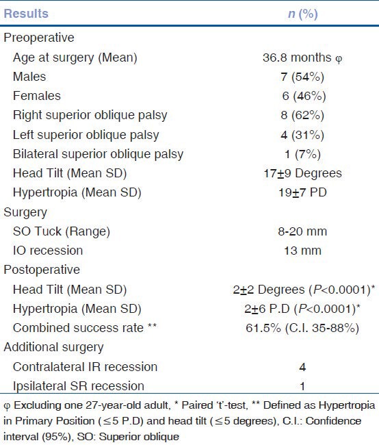
After approval from the Institutional Review Board, patients who underwent strabismus surgery for congenital SOP between January 2000 and December 2008 operated by a single surgeon were identified from a computerized patient database. This non-comparative interventional case series included 13 consecutive patients with congenital superior oblique palsy with torticollis, who underwent simultaneous superior oblique tuck and inferior oblique recession [Table 1]. Congenital SOP was diagnosed based on a history of ocular torticollis and/or vertical misalignment since early childhood (before 6 months of age) and fulfilment of the Park's 3-step test. Exclusion criteria included prior history of trauma to the eye, head, neck or shoulder area, craniosynostosis, and other ocular diseases. The goal of the surgery was to correct the hypertropia and head tilt. All patients had simultaneous SO tuck and IO recession, irrespective of the amount of hypertropia or head tilt.
Table 1.
Demographic and surgical information for 13 patients with congenital superior oblique palsy
The details of history including onset and duration of the signs and symptoms were noted. The initial ophthalmologic examination included, visual acuity, ductions, versions, ocular alignment with prism and cover test in all cardinal gaze positions and Bielschowsky's head tilt test. Presence of any facial asymmetry was also recorded. The SCM muscle was palpated for any tightness. The ocular motility assessment consisted of subjective grading (0-4) of underaction (-) or overaction (+) of the cyclovertical muscles. Measurement of the deviation was performed in primary gaze and the cardinal positions of gaze (whenever possible) at both 33 cm and 6 m. Head tilt was objectively measured using a goniometer. A cycloplegic refraction and evaluation of anterior and posterior segments were performed in all cases. Postoperatively, the above orthoptic assessment was repeated at each follow-up visit.
A goniometer (By Baseline products, White plains, NEW YORK) is an instrument with graded markings, commonly used to measure the range of joint movement and deformity. During measurement, the patient is fixated on a distant target at eye level. One arm of the goniometer was placed parallel to the axis of the face, and the second arm perpendicular to the floor [Fig. 1]. It is helpful to align the second arm with a vertical object, such as a door post.
Figure 1.
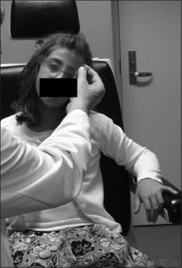
Measurement of head tilt using a goniometer: One arm is lined up with the vertical edge of the door while the other is aligned across the medial and lateral canthus of each eye
The surgical procedures performed were a SO tuck and IO recession of 13 mm since in all the cases, the SO tendon was found to be lax. After performing the exaggerated traction test[11] for oblique muscles to assess tendon laxity, the SO tendon was isolated nasal to the superior rectus through a superotemporal conjunctival fornix incision. The desired amount of tucking was performed nasal to the superior rectus muscle by imbricating the anterior and posterior portions of tendon with temporary interrupted non-absorbable 6-0 Mersilene sutures (Ethicon, Inc, Somerville, NJ). We used Bishop tendon tucker (BAUSCH and LOMB STORZ OPHTHALMICS) for tucking SO muscle. The endpoint of tucking was then decided by performing a traction test by grasping the globe at the limbus in the inferotemporal quadrant and elevating the eye in adduction.[12] A SO tuck was considered adequate, when resistance was felt when the inferior limbus reached an imaginary line drawn between the medial and lateral canthi. A significant resistance detected with the inferior limbus below this line suggested excessive tightness. When no resistance was felt when the inferior limbus moved above this imaginary line, the tuck was considered inadequate. The sutures were tied permanently after ensuring the adequate end point. By using a tendon tucker in this manner, the SO was tucked in this series from 8 to 20 mm. Simultaneously, a standard fornix approach IO recession of 13 mm was performed in all cases. For the bilateral case, a similar procedure was performed on both sides. In addition to the other muscles, in one case, a contralateral inferior rectus (IR) recession of 4.5 mm was performed.
Postoperative examination was performed at 1-week, 1-month, then at 6 - monthly intervals. At every follow-up visit, a complete evaluation of ocular alignment, goniometry, and any postoperative complications were noted. Assessment of the postoperative limitation to elevation in adduction (Iatrogenic Brown's syndrome) was charted on a-1 to-4 scale according to the severity.
Main outcome measures were the effect on hypertropia in primary position and head tilt. Criteria for surgical success was defined as postoperative hypertropia <5PD and head tilt <5 degrees. We analyzed the success rate with regards to hypertropia as well as head tilt i.e., combined success rate.
For a significant residual hypertropia (>5 PD) and head tilt (>5 Degrees), further extraocular muscle surgeries were performed. The surgeries performed were contralateral IR recession or ipsilateral superior rectus (SR) recession. The IR was recessed if the residual hypertropia was worse to the side contralateral to the paresis. The SR was recessed if the residual hypertropia was worse to the side ipsilateral to the paresis. The second surgery was conducted at least 3 months after the first surgery. No patient required horizontal rectus muscle surgery.
SPSS 11.5 for Windows statistical software (Chicago, Illinois) was used in the analyzes. Paired t-tests were used to assess possible association between the variables observed. A P value of less than 0.05 was considered statistically significant.
Results
Thirteen cases (7 males, 6 females) of congenital SOP underwent simultaneous SO tuck (range 8-20 mm) and IO recession (13 mm). Of these, 8 (62%) had right-sided SOP and 4 (31%) were left-sided. One (7.5%) case was bilateral. All had a contralateral head tilt, except the one bilateral case who had chin elevation and right head tilt. The amount of pre-operative head tilt and hypertropia ranged from 10-40 degrees (mean 17) and 6-30 P.D (mean 19), respectively. Age of the patients at first intervention ranged from 7 months to 7 years, except one who was 27 years old. Five of them had a small (less than 8 PD) exotropia (4) or esotropia (1). None of these required any horizontal strabismus surgery. Inferior oblique overaction ranged from +2 to +3, while superior oblique underaction was – 3 to - 1. The mean duration of SOP until surgery was 36.8 months. The mean follow-up period was 17 months (range 7-36). Mild amblyopia was found in one case, which was treated pre-operatively with part time occlusion therapy.
The outcome was determined at the last follow-up. The mean pre- and post-operative hypertropia (P.D.) in forced primary position was 19 ± 7 and 2 ± 6, respectively (P < 0.0001). The head tilt reduced from mean of 17 ± 9 to 2 ± 2 degrees (P < 0.0001). Success, defined as hypertropia <5 PD and head tilt less than 5 degrees, was achieved in 69% (9/13 – C.I. 42-88%) and 85% (11/13 - C.I. 56-96%) of cases, respectively. When both criteria were met, the success rate was 61.5% (C.I. 35 to 88% - Table 1). There was no persistent overaction of the inferior oblique muscle in any of the cases. Subjectively, there was modest improvement in the superior oblique function. There was no correlation between the amount of head tilt, hypertropia, and the length of SO tendon tuck both pre-and post-operatively [Figs. 2 and 3].
Figure 2.
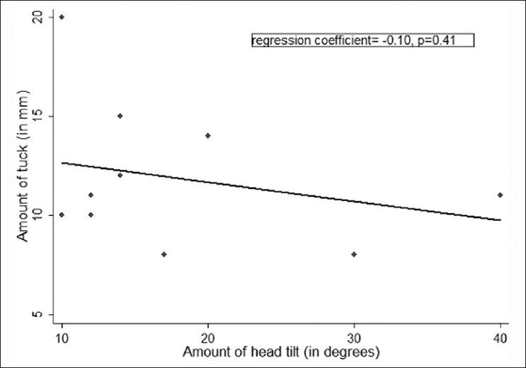
Scatter plot showing that there were no significant correlations between the amount of SO tuck and Head tilt
Figure 3.
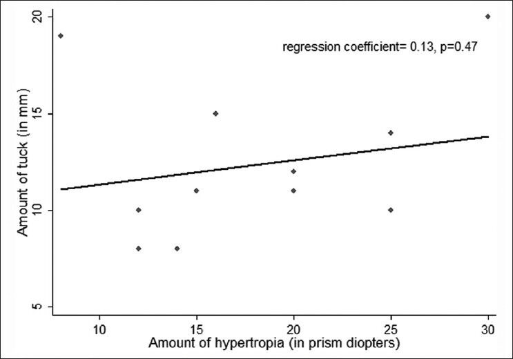
Scatter plot showing that there were no significant correlations between the amount of SO tuck and Hypertropia
Five patients (38%) required additional surgery; contralateral inferior rectus muscle recession (4) and ipsilateral superior rectus recession (1). This decision was based on the amount of maximum hypertropia on side gazes. In the case that developed Brown syndrome (7.69% - C.I. 6.7 to 22.2), there was no hypotropia in primary position, and the patient was asymptomatic. Therefore, in this case, no additional surgery was performed. In 1 (8%) case, there was unmasking of bilateral SOP that required a similar surgical procedure on the newly affected side i.e., simultaneous SO tuck and IO recession as a second procedure. The final outcome was excellent alignment with no significant abnormal head posture (i.e., less than 5 degrees) in all cases.
Discussion
The most notable sign of congenital SOP in children is torticollis. This is due to the induced excyclotorsion or hypertropia effect. To negate this, children adopt a compensatory head posture (contralateral head tilt), which allows them to place the eyes in a particular field of gaze so as to regain binocular single vision. Appropriate surgery should eliminate the compensatory head posture in a high percentage of cases. [Figs. 4 and 5].
Figure 4.
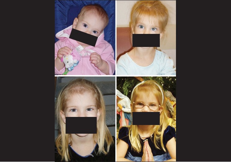
Pre-and post-operative head and eye profile picture of case number 2 showing the elimination of head tilt
Figure 5.
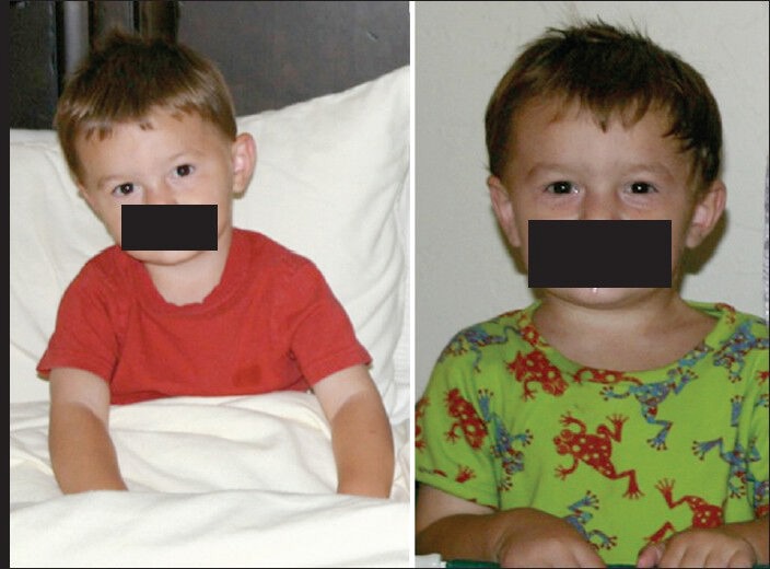
Pre- and post-operative head and eye profile picture of case number 6 showing the elimination of head tilt
Most of the recent literature favors IO weakening and/or SO tuck, in isolation or combination, and additionally, contralateral IR recession or ipsilateral SR recession as a surgical option for the treatment of congenital SOP.[5,7,8,13] In our study, we performed simultaneous SO tuck and IO recession, irrespective of the amount of vertical deviation in primary position. This is due to the consistent findings of a lax tendon in each case in this series. In other series, other anatomic abnormalities of the SO tendon have been reported including malposition of the SO tendon insertion and complete or partial absence of the SO.
In most of the earlier studies, the outcome measures considered were change in vertical deviation and inferior oblique overaction or disappearance of head tilt.[7,8,9] Lau et al. studied only the residual torticollis using a goniometer.[6] In their cohort of 32 patients with congenital SOP, they did not assess the pre-operative head tilt objectively. Post-operatively, they defined 10 degrees of torticollis as significant AHP, which was seen in 32% of their cases. The AHP they used for analysis was the summation of degrees of face turn, head tilt, and chin elevation and depression. Their overall success rate of strabismus surgery in eliminating the significant torticollis was 68%. The surgery they performed was a recession of the IO muscle occasionally combined with a recession of the IR muscle.
Kraft et al. evaluated the effect of strabismus surgery in eliminating AHP in 32 patients.[10] The head tilt alone was seen only in 31% of their cases. The rest had chin up, chin down, face turn in isolation or in combination. Their surgical procedure mainly included inferior oblique weakening and one vertical rectus muscle recession. They measured the head tilt, whenever possible, by using an arc perimeter or surgical protractor with graded markings, and in children, angles were estimated by using known reference angles on horizontal or vertical structures within the examining lane. The criteria for success of elimination of AHP was less than or equal to 10 degrees. This was achieved in 76% of their cases. Although our study subjects were mainly children,[12] we were able to measure the head tilt both pre-and post-operatively using a goniometer. Therefore, we could also study the effect of surgery on the torticollis change using one standard for measurement in addition to the change in vertical deviation.
In our study, surgical success was more stringently defined as hypertropia <5PD and head tilt <5 degrees (as opposed to 10 degrees in other studies.[6,10] Combination of IO recession and SO tendon tuck surgery was unique to our study group as in most other studies, SO tuck was rarely performed. Helveston's large series[5] of 190 cases had both congenital and acquired cases. One hundred and seventy one had IO weakening as part of the initial procedure. Twenty-six of them had SO tuck as a primary surgery. A cure, defined as relief of symptoms or elimination of strabismus and head tilt, was achieved in 92% of the patients. Our combined success rate was 61.5% (C.I. 35% to 88%) while, hypertropia <5 PD and head tilt less than 5 degrees individually was achieved in 69% and 85% of cases, respectively.
Brown syndrome is one of the known complications of SO tendon tuck. In Helveston's series,[14] all developed a transient Brown syndrome. Persistent Brown syndrome was seen in 17% of their cases at an average of 9 months after the surgery, and they required a takedown procedure to reverse this complication. One case in our series developed Brown syndrome, but did not require a take-down of the tuck since he was asymptomatic.
One third of our patients had residual torticollis. After the additional surgery i.e., contralateral IR recession (4) and ipsilateral SR recession (1), none had significant torticollis (i.e., more than 5 degrees).
A goniometer is a useful tool to measure the head tilt, even in children. Further prospective studies with a larger sample size and examining other parameters such as the effect on facial asymmetry and SCM tightness are warranted.
In a recent study by Kaeser et al., it is showed that inferior oblique recession alone is a suitable procedure for most of the congenital superior oblique palsies with a moderate to large deviation in adduction.[15] Post-operative residual vertical deviations were not different in the primary position or in downgaze. However, significantly better alignment was achieved in the inferior oblique recession and superior oblique tuck group in adduction and downgaze in adduction. Consecutive Brown pattern occurred in 18 of 20 patients who underwent inferior oblique recession and superior oblique tuck versus 5 of 20 who underwent inferior oblique recession.
Durnian et al. looked at the success rate of superior oblique tuck alone as a single muscle treatment for selected cases of superior oblique palsy.[16] In their series of 75 cases, 33 were congenital superior oblique palsy. Since they did not find any correlation between tuck size and correction obtained, they suggest that superior oblique tuck should be considered as “one tightness fits all” rather than “one size fits all.”
Our study has limitations of its own in the form of retrospective chart review of patients from one institution, one surgeon performing all measurements, data includes a bilateral case, few cases required additional surgery to achieve desired result. In addition, it is cautioned that simultaneous superior oblique tuck and inferior oblique recession may not be the standard of care in all the cases. This form of surgical treatment needs to be chosen on a case-to-case basis.
Simultaneous superior oblique tuck and inferior oblique muscle recession can successfully treat selected cases of congenital superior oblique palsy. In this series, one-third required an additional procedure, which led to total normalization of the head position. This may guide the physicians in decision-making regarding the surgery and counseling the parents.
Footnotes
Source of Support: Nil.
Conflict of Interest: None declared.
References
- 1.Cheng JC, Au AW. Infantile torticollis: A review of 624 cases. J Pediatr Orthop. 1994;14:802–8. [PubMed] [Google Scholar]
- 2.Kiwak KJ. Establishing an etiology for torticollis. Postgrad Med. 1984;75:126–34. doi: 10.1080/00325481.1984.11698622. [DOI] [PubMed] [Google Scholar]
- 3.Ballock RT, Song KM. The prevalence of nonmuscular causes of torticollis in children. J Pediatr Orthop. 1996;16:500–4. doi: 10.1097/00004694-199607000-00016. [DOI] [PubMed] [Google Scholar]
- 4.Kushner BJ. Ocular causes of abnormal head postures. Ophthalmology. 1979;86:2115–25. doi: 10.1016/s0161-6420(79)35301-5. [DOI] [PubMed] [Google Scholar]
- 5.Helveston EM, Mora JS, Lipsky SN, Plager DA, Ellis FD, Sprunger DT, et al. Surgical treatment of superior oblique palsy. Trans Am Ophthalmol Soc. 1996;94:315–34. [PMC free article] [PubMed] [Google Scholar]
- 6.Lau FH, Fan DS, Sun KK, Yu CB, Wong CY, Lam DS. Residual torticollis in patients after strabismus surgery for congenital superior oblique palsy. Br J Ophthalmol. 2009;93:1616–9. doi: 10.1136/bjo.2008.156687. [DOI] [PubMed] [Google Scholar]
- 7.Reynolds JD, Biglan AW, Hiles DA. Congenital superior oblique palsy in infants. Arch Ophthalmol. 1984;102:1503–5. doi: 10.1001/archopht.1984.01040031223022. [DOI] [PubMed] [Google Scholar]
- 8.Simons BD, Saunders TG, Siatkowski RM, Feuer WJ, Lavina AM, Capó H, et al. Outcome of surgical management of superior oblique palsy: A study of 123 cases. Binocul Vis Strabismus Q. 1998;13:273–82. [PubMed] [Google Scholar]
- 9.Davis AR, Dawson E, Lee JP. Residual symptomatic superior oblique palsy. Strabismus. 2007;15:69–77. doi: 10.1080/09273970701404993. [DOI] [PubMed] [Google Scholar]
- 10.Kraft SP, Donoghue EP, Roarty JD. Improvement of compensatory head posture after strabismus surgery. Ophthalmology. 1992;99:1301–8. doi: 10.1016/s0161-6420(92)31811-1. [DOI] [PubMed] [Google Scholar]
- 11.Guyton DL. Exaggerated traction test for the oblique muscles. Ophthalmology. 1981;88:1035–40. doi: 10.1016/s0161-6420(81)80033-4. [DOI] [PubMed] [Google Scholar]
- 12.Saunders RA. Quantitated superior oblique tendon tuck in the treatment of superior oblique palsy. Am J Orthop. 1985;35:81–9. [Google Scholar]
- 13.Saunders RA. Treatment of superior oblique palsy with superior oblique tendon tuck and inferior oblique muscle myectomy. Ophthalmology. 1986;93:1023–7. doi: 10.1016/s0161-6420(86)33627-3. [DOI] [PubMed] [Google Scholar]
- 14.Helveston EM, Ellis FD. Superior oblique tuck for superior oblique palsy. Aust J Ophthalmol. 1983;11:215–20. [PubMed] [Google Scholar]
- 15.Kaeser PF, Klainguti G, Kolling GH. Inferior oblique muscle recession with and without superior oblique tendon tuck for treatment of unilateral congenital superior oblique palsy. J AAPOS. 2012;16:26–31. doi: 10.1016/j.jaapos.2011.08.012. [DOI] [PubMed] [Google Scholar]
- 16.Durnian JM, Marsh IB. Superior oblique tuck: Its success as a single muscle treatment for selected cases of superior oblique palsy. Strabismus. 2011;19:133–7. doi: 10.3109/09273972.2011.620058. [DOI] [PubMed] [Google Scholar]


