

Porcelain gall bladder or calcified gall bladder is an interesting entity both from the point of view of its unique and eye catching image as well as its controversial association with gall bladder cancer. In a majority of patients, the porcelain gall bladder is associated with gall bladder stones.1,2 In this letter, we describe a patient in whom we detected porcelain gall bladder on endoscopic ultrasound (EUS).
A 65-year-old female with no comorbidities presented to us with upper abdominal pain. There was no history of anorexia and weight loss. Clinical examinations did not reveal any significant findings. Blood investigations were normal. Ultrasound (US) of the abdomen showed calcification in the gall bladder wall and there was no gall bladder mass or stones and similar findings were observed on a contrast-enhanced computed tomography (CECT) of the abdomen (Fig. 1). The upper gastrointestinal endoscopy was normal. EUS was done to evaluate the gall bladder and pancreas. EUS examination revealed that the gall bladder was encircled by a fine ring of calcification suggestive of porcelain gall bladder (Fig. 2) and there were no gall bladder stones. The pancreas was normal. She has been thereafter referred to surgery services for cholecystectomy.
Figure 1.
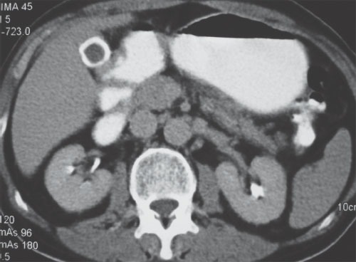
Computed tomography: calcification in the gall bladder wall.
Figure 2.
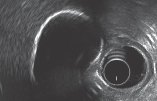
Endoscopic ultrasound: porcelain gall bladder.
Porcelain gallbladder is a rare chronic disease of gallbladder seen more commonly in females and is due to mural calcification of the gallbladder.1 Its cause is unknown. However, as in a majority of the patients it is associated with gall bladder stones, and it is thought to be an unusual presentation of chronic cholecystitis.3,4 Although it has been strongly associated with gall bladder cancer, recent studies suggest that the association between these two entities is less than previously thought.3,4,5 Still the relationship between these two entities is controversial, and given the poor prognosis of gall bladder cancer, most of the surgeons would prefer prophylactic cholecystectomy. Recently, it has been suggested that the risk of gall bladder cancer in porcelain gall bladder depends upon the pattern of calcification.6 The calcification pattern has been described as diffuse intramural calcification and selective mucosal calcification, and it has been shown that patients with selective mucosal calcification have a higher risk of malignancy as compared to diffuse intramural calcification; the pattern that was seen in the index case also. Shimizu et al. also reported that incomplete gallbladder wall calcification, as determined by diagnostic ultrasound, was much more likely to be associated with gallbladder carcinoma as compared to complete wall calcification.7
REFERENCES
- 1.Gupta V, Rao C, Rana SS, et al. Porcelain gallbladder and thalassaemia intermedia: association or coincidence? Dig Liver Dis. 2012;44:e22. doi: 10.1016/j.dld.2012.04.006. [DOI] [PubMed] [Google Scholar]
- 2.Palermo M, Núñez M, Duza GE, et al. Porcelain gallbladder: a clinical case and a review of the literature. Cir Esp. 2011;89:213–7. doi: 10.1016/j.ciresp.2010.09.012. [DOI] [PubMed] [Google Scholar]
- 3.Polk HC. Carcinoma and the calcified gallbladder. Gastroenterology. 1966;50:582–5. [PubMed] [Google Scholar]
- 4.Opatrny L. Porcelain gallbladder. CMAJ. 2002;166:933. [PMC free article] [PubMed] [Google Scholar]
- 5.Towfigh S, McFadden DW, Cortina GR, et al. Porcelain gallbladder is not associated with gallbladder carcinoma. Am Surg. 2001;67:7–10. [PubMed] [Google Scholar]
- 6.Stephen AE, Berger DL. Carcinoma in the porcelain gallbladder: a relationship revisited. Surgery. 2001;129:699–703. doi: 10.1067/msy.2001.113888. [DOI] [PubMed] [Google Scholar]
- 7.Shimizu M, Minura J, Tanaka T, et al. Porcelain gallbladder: relation between its type by ultrasound and incidence of cancer. J Clin Gastroenterol. 1989;11:471–6. [PubMed] [Google Scholar]