Figure 1.
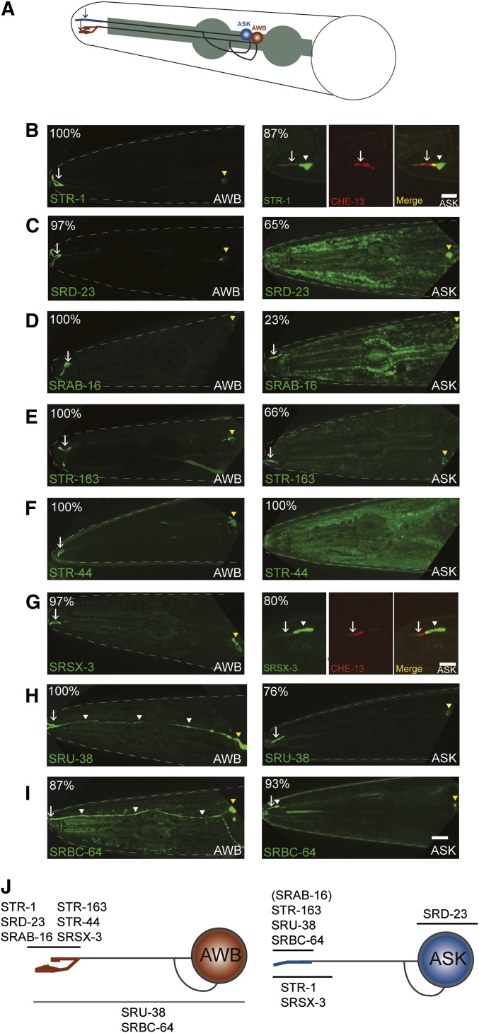
Localization of GPCR::GFP fusion proteins in the AWB and ASK chemosensory neurons. (A) Cartoon of AWB and ASK sensory neurons in the head of C. elegans. Cell bodies are indicated in blue (ASK) or red (AWB). Shaded structure indicates the pharynx. Arrows indicate cilia. Only one of each bilateral pair of neurons is seen in this lateral view. Anterior is at left. (B–I) Representative examples of the localization pattern of indicated GPCR fusion proteins in AWB (left panels) or ASK (right panels) in adult hermaphrodites. Numbers in top left corners indicate the percentage of examined animals exhibiting the phenotype (see Table 1). White arrows indicate cilia; white arrowheads indicate dendrites and dendritic domains proximal to cilia; yellow arrowheads indicate cell bodies. Expression was driven in AWB and ASK under the str-1 and srbc-66 promoters, respectively. (B and G, right panels) Cilia were visualized via cell-specific expression of CHE-13::TagRFP. Lateral views. Bars, 10 μm (B–I, left panels and C–F, H, and I, right panels); 5 μm (B and G right panels). (J) Summary of GPCR localization patterns in AWB and ASK. Horizontal lines indicate subcellular localization patterns of indicated fusion proteins.

