INTRODUCTION
Endoscopic retrograde cholangiopancreatography (ERCP) is the standard procedure for biliary drainage (BD) in patients with benign or malignant biliary obstruction.[1] Therapeutic ERCP requires deep cannulation into the common bile duct (CBD), the success rate of deep cannulation is high, but still not perfect, even with the use of advanced cannulation techniques such as precut sphincterotomy.[2] In particular, periampullary diverticula,[3] tumor infiltration,[4] or altered surgical anatomy,[5] occasionally complicates biliary cannulation. Alternatives if deep biliary cannulation fails include percutaneous transhepatic BD (PTBD)[6] or surgical intervention.[7] However, both PTBD and surgical intervention are associated with considerable morbidity and occasional mortality.[7] We present the first case in Egypt with a malignant pancreatic head mass causing biliary obstruction that was successfully treated with endoscopic ultrasound (EUS)-guided choledochoduodenostomy (EUS-CDS).
CASE AND IMAGE
An 80-year-old female with a history of malaise, weight loss, abnormal liver function tests and biliary obstruction was referred to our facility for an ERCP. She was previously diagnosed with pancreatic head tumor invading superior mesenteric artery, vein and distal CBD. An attempted ERCP failed to cannulate the CBD due to tumor infiltration. An EUS showed large isoechoic head mass (5.5 cm × 4 cm) [Figure 1] associated with dilation of the extra-hepatic bile duct and severe stenosis of the distal CBD [Figure 2]. Using the therapeutic linear echoendoscope (Pentax, Tokyo, Japan), a 22 gauge fine-needle aspiration (FNA) (Wilson-Cook Corporation, North Carolina, USA) was advanced through the wall of the duodenal bulb to the mass [Figure 3] and multiple passes were done using fanning technique to take aspirate which was, later on, diagnosed as neuroendocrine tumor. A 19 gauge FNA (Wilson-Cook Corporation, North Carolina, USA) was advanced through the wall of the duodenal bulb into the CBD [Figure 4] and a cholangiogram was performed by injecting contrast through the needle. A 0.035 inch guide wire (Boston Scientific, Natick, Massachusetts, USA) was then introduced through the needle into the CBD and advanced up to the intra-hepatic biliary tree [Figure 5]. The needle was then removed and a standard cannula (Boston Scientific, Natick, Massachusetts, USA) was advanced over the guide wire into the CBD to dilate the choledochoduodenal fistula tract [Figure 6]. The choledochoduodenal fistula was then created after further dilating the fistula tract with an 8 mm dilating balloon (Wilson-Cook Corporation, North Carolina, USA) [Figure 7]. A plastic stent (10 Fr diameter and 9 cm in length, Wilson-Cook Corporation, North Carolina, USA) was placed over a guiding catheter [Figure 8] within the fistula tract [Figure 9]. However, the stent slightly migrated within the fistula tract during insertion. The patient had abdominal pain and fever for 1 day postendoscopy. Over 3 weeks follow-up, there was no complication, patient bilirubin decreased and she was transferred to oncology center for neoadjuvant therapy.
Figure 1.
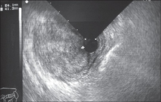
Radial EUS image of isoechoic mass measuring 5.5 × 4 cm at the pancreatic head
Figure 2.
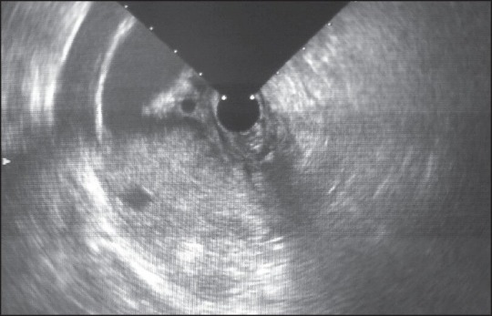
Radial EUS image of the mass invading distal common bile duct, portal vein and superior mesenteric vein
Figure 3.
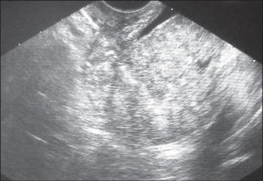
Linear EUS image of isoechoic mass with peripheral hypoechoic rim at the pancreatic head invading distal common bile duct with needle inside during EUS-FNA
Figure 4.
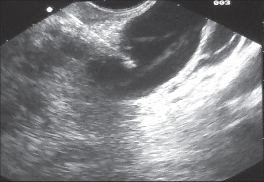
EUS guided puncture of the common bile duct above the mass
Figure 5.
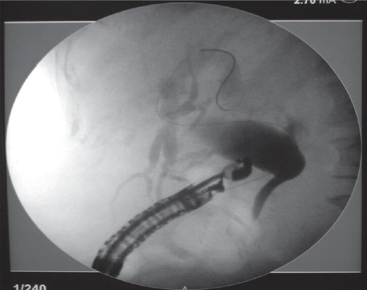
Flouroscopic image of the biliary tree with guide wire inside
Figure 6.
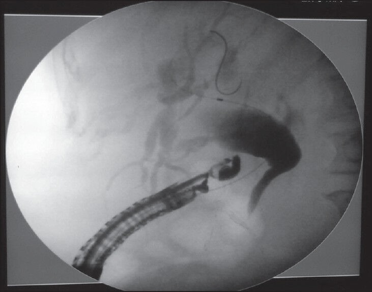
Flouroscopic image of the choledochoduodenal tract dilation using standard cannula
Figure 7.
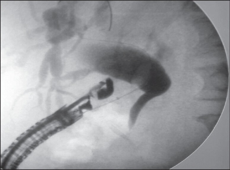
Flouroscopic image of the choledochoduodenal tract dilation using balloon dilator
Figure 8.
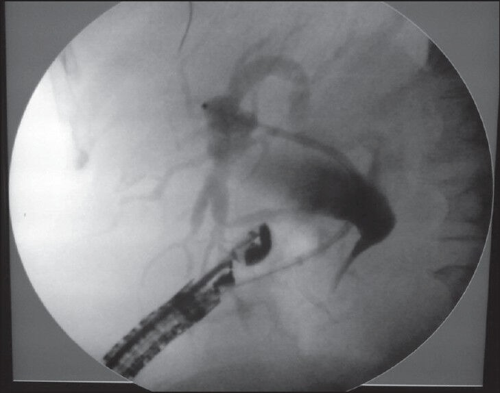
Flouroscopic image of plastic stent (10Fr, 9cm) during insertion over guiding catheter
Figure 9.
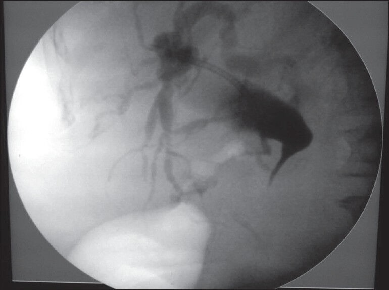
Flouroscopic image of the stent after insertion
DISCUSSION
Since EUS-guided bile duct puncture was first described in 1996,[8] sporadic case reports of EUS-guided BD (EUS-BD) have suggested it as an alternative to PTBD after failed ERCP.[9,10,11,12,13,14,15,16,17,18,19,20,21,22] The potential benefits of EUS-BD include that it is a one-stage procedure, as with ERCP, and internal drainage, avoiding long term external drainage; this can significantly improve the quality of life of terminally ill patients and possibly result in lower morbidity than PTBD or surgery.[23] EUS guided extra-hepatic BD through EUS-CDS has been reported in literature for biliary decompression in advanced pancreatic carcinomas in the context of a failed ERCP.[20] Its major advantage is provision for internal and long lasting BD by virtue of its anatomic location away from the tumor.[24] Notable complications from the procedure are local peritonitis, cholangitis and stent migration.[25] However, to the best of our knowledge, no cases of EUS-BD have been reported in the Arabic world.
Footnotes
Source of Support: Nil.
Conflict of Interest: None declared.
REFERENCES
- 1.Park do H, Jang JW, Lee SS, et al. EUS-guided biliary drainage with transluminal stenting after failed ERCP: Predictors of adverse events and long-term results. Gastrointest Endosc. 2011;74:1276–84. doi: 10.1016/j.gie.2011.07.054. [DOI] [PubMed] [Google Scholar]
- 2.Bailey AA, Bourke MJ, Williams SJ, et al. A prospective randomized trial of cannulation technique in ERCP: Effects on technical success and post-ERCP pancreatitis. Endoscopy. 2008;40:296–301. doi: 10.1055/s-2007-995566. [DOI] [PubMed] [Google Scholar]
- 3.Huang CH, Tsou YK, Lin CH, et al. Endoscopic retrograde cholangiopancreatography (ERCP) for intradiverticular papilla: Endoclip-assisted biliary cannulation. Endoscopy. 2010;42(Suppl 2):E223–4. doi: 10.1055/s-0029-1215008. [DOI] [PubMed] [Google Scholar]
- 4.Iwashita T, Lee JG, Shinoura S, et al. Endoscopic ultrasound-guided rendezvous for biliary access after failed cannulation. Endoscopy. 2012;44:60–5. doi: 10.1055/s-0030-1256871. [DOI] [PubMed] [Google Scholar]
- 5.Koornstra JJ, Fry L, Mönkemüller K. ERCP with the balloon-assisted enteroscopy technique: A systematic review. Dig Dis. 2008;26:324–9. doi: 10.1159/000177017. [DOI] [PubMed] [Google Scholar]
- 6.Beissert M, Wittenberg G, Sandstede J, et al. Metallic stents and plastic endoprostheses in percutaneous treatment of biliary obstruction. Z Gastroenterol. 2002;40:503–10. doi: 10.1055/s-2002-32806. [DOI] [PubMed] [Google Scholar]
- 7.Smith AC, Dowsett JF, Russell RC, et al. Randomised trial of endoscopic stenting versus surgical bypass in malignant low bileduct obstruction. Lancet. 1994;344:1655–60. doi: 10.1016/s0140-6736(94)90455-3. [DOI] [PubMed] [Google Scholar]
- 8.Wiersema MJ, Sandusky D, Carr R, et al. Endosonography-guided cholangiopancreatography. Gastrointest Endosc. 1996;43:102–6. doi: 10.1016/s0016-5107(06)80108-2. [DOI] [PubMed] [Google Scholar]
- 9.Artifon EL, Okawa L, Takada J, et al. EUS-guided choledochoantrostomy: An alternative for biliary drainage in unresectable pancreatic cancer with duodenal invasion. Gastrointest Endosc. 2011;73:1317–20. doi: 10.1016/j.gie.2010.10.041. [DOI] [PubMed] [Google Scholar]
- 10.Bories E, Pesenti C, Caillol F, et al. Transgastric endoscopic ultrasonography-guided biliary drainage: Results of a pilot study. Endoscopy. 2007;39:287–91. doi: 10.1055/s-2007-966212. [DOI] [PubMed] [Google Scholar]
- 11.Burmester E, Niehaus J, Leineweber T, et al. EUS-cholangio-drainage of the bile duct: Report of 4 cases. Gastrointest Endosc. 2003;57:246–51. doi: 10.1067/mge.2003.85. [DOI] [PubMed] [Google Scholar]
- 12.Fabbri C, Luigiano C, Fuccio L, et al. EUS-guided biliary drainage with placement of a new partially covered biliary stent for palliation of malignant biliary obstruction: A case series. Endoscopy. 2011;43:438–41. doi: 10.1055/s-0030-1256097. [DOI] [PubMed] [Google Scholar]
- 13.Giovannini M, Moutardier V, Pesenti C, et al. Endoscopic ultrasound-guided bilioduodenal anastomosis: A new technique for biliary drainage. Endoscopy. 2001;33:898–900. doi: 10.1055/s-2001-17324. [DOI] [PubMed] [Google Scholar]
- 14.Itoi T, Itokawa F, Sofuni A, et al. Endoscopic ultrasound-guided choledochoduodenostomy in patients with failed endoscopic retrograde cholangiopancreatography. World J Gastroenterol. 2008;14:6078–82. doi: 10.3748/wjg.14.6078. [DOI] [PMC free article] [PubMed] [Google Scholar]
- 15.Kahaleh M, Hernandez AJ, Tokar J, et al. Interventional EUS-guided cholangiography: Evaluation of a technique in evolution. Gastrointest Endosc. 2006;64:52–9. doi: 10.1016/j.gie.2006.01.063. [DOI] [PubMed] [Google Scholar]
- 16.Kim YS, Gupta K, Mallery S, et al. Endoscopic ultrasound rendezvous for bile duct access using a transduodenal approach: Cumulative experience at a single center. A case series. Endoscopy. 2010;42:496–502. doi: 10.1055/s-0029-1244082. [DOI] [PubMed] [Google Scholar]
- 17.Siddiqui AA, Sreenarasimhaiah J, Lara LF, et al. Endoscopic ultrasound-guided transduodenal placement of a fully covered metal stent for palliative biliary drainage in patients with malignant biliary obstruction. Surg Endosc. 2011;25:549–55. doi: 10.1007/s00464-010-1216-6. [DOI] [PubMed] [Google Scholar]
- 18.Will U, Meyer F, Schmitt W, et al. Endoscopic ultrasound-guided transesophageal cholangiodrainage and consecutive endoscopic transhepatic Wallstent insertion into a jejunal stenosis. Scand J Gastroenterol. 2007;42:412–5. doi: 10.1080/00365520600881136. [DOI] [PubMed] [Google Scholar]
- 19.Will U, Thieme A, Fueldner F, et al. Treatment of biliary obstruction in selected patients by endoscopic ultrasonography (EUS)-guided transluminal biliary drainage. Endoscopy. 2007;39:292–5. doi: 10.1055/s-2007-966215. [DOI] [PubMed] [Google Scholar]
- 20.Yamao K, Sawaki A, Takahashi K, et al. EUS-guided choledochoduodenostomy for palliative biliary drainage in case of papillary obstruction: Report of 2 cases. Gastrointest Endosc. 2006;64:663–7. doi: 10.1016/j.gie.2006.07.003. [DOI] [PubMed] [Google Scholar]
- 21.Park do H, Song TJ, Eum J, et al. EUS-guided hepaticogastrostomy with a fully covered metal stent as the biliary diversion technique for an occluded biliary metal stent after a failed ERCP (with videos) Gastrointest Endosc. 2010;71:413–9. doi: 10.1016/j.gie.2009.10.015. [DOI] [PubMed] [Google Scholar]
- 22.Brauer BC, Chen YK, Fukami N, et al. Single-operator EUS-guided cholangiopancreatography for difficult pancreaticobiliary access (with video) Gastrointest Endosc. 2009;70:471–9. doi: 10.1016/j.gie.2008.12.233. [DOI] [PubMed] [Google Scholar]
- 23.Ang TL. Current status of endosonography-guided biliary drainage. Singapore Med J. 2010;51:762–6. [PubMed] [Google Scholar]
- 24.Yamao K, Bhatia V, Mizuno N, et al. EUS-guided choledochoduodenostomy for palliative biliary drainage in patients with malignant biliary obstruction: Results of long-term follow-up. Endoscopy. 2008;40:340–2. doi: 10.1055/s-2007-995485. [DOI] [PubMed] [Google Scholar]
- 25.Hara K, Yamao K, Mizuno N, et al. Interventional endoscopic ultrasonography for pancreatic cancer. World J Clin Oncol. 2011;2:108–14. doi: 10.5306/wjco.v2.i2.108. [DOI] [PMC free article] [PubMed] [Google Scholar]


