


Abstract
INTRODUCTION
Desmoid tumor (DT) is a common manifestation of Gardner's Syndrome (GS), although it is a rare condition in the general population. DT in patients with GS is usually located in the abdominal wall and/or intra-abdominal cavity.
PRESENTATION OF CASE
We report a case of a 32 years-old female patient with familial adenomatous polyposis (FAP), who was already submitted to total colectomy and developed multiple DT, located in the abdominal wall and in the left breast. The patient underwent several surgical procedures, with a multidisciplinary team of surgeons. Wide surgical resections of the left breast and the abdominal wall tumors were performed in separate steps. Polypropylene mesh reconstruction and muscle flaps were needed to cover the defects of the thoracic and abdominal walls. After partial necrosis of the adipose-cutaneous flap in the abdomen that required a new skin graft, she had a satisfactory outcome with complete healing of the surgical incisions.
DISCUSSION
DT is frequent in GS, however, breast localization is very rare, with few cases reported in the literature. Recurrence of DT is not negligible, even after a wide surgical resection. GS patients must be followed up closely, and clinical examination, associated with imaging studies, should be performed to detect any signs of tumor.
CONCLUSION
DT represents one of the most significant causes of the morbidity and mortality that affects FAP patients following colectomy. In general, the surgical procedures to excise DT are highly complex, requiring a multidisciplinary team.
Keywords: Desmoid tumor, Gardner's syndrome, Familial adenomatous polyposis, Surgical approach
1. Introduction
Familial Adenomatous Polyposis (FAP) is an autosomal dominant disease caused by mutations in the adenomatous polyposis coli (APC) gene. This genetic disorder causes widespread polyps of the colon and rectum with high potential for colon cancer development in patients who are not submitted to a prophylactic colectomy. The symptoms frequently start between 20 and 40 years of age and the disease has similar gender distribution.1–3
Gardner's syndrome (GS) is better defined as a variant of FAP, which affects one individual in 8300–16,000 live births. Its intestinal lesions are specially prominent, but extracolonic manifestations may also occur, such as osteomas, skin tumors, supernumerary teeth, congenital hypertrophy of the retinal pigment epithelium and desmoid tumor.2,4–6 The desmoid tumors (DT) are locally aggressive mesenchymal neoplasms originated from fibroblasts and they are the second cause of mortality in FAP patients after colorectal carcinoma. DT occurs in 3.5–5.7% of GS and can affect many locations, being most common in the abdominal wall and/or in the intra-abdominal cavity, as well as in the mesentery.3 We present a case of a young patient with GS, who underwent to a total colectomy with ileorectal anastomosis, and developed multiple DT, particularly in the breast, which is an unusual location of this tumor.
2. Presentation of the case
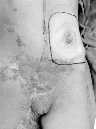
A 32 years-old female patient has been followed at our Coloproctology Outpatient Clinic at the University of Campinas for the past 2 years. She was sent to our clinic, reporting progressive growth of a large exophytic lesion with central ulceration in the lower abdominal wall, extending to the right inguinal area. She has the antecedent of GS and her surgical history includes removal of scalp fibrolipoma 9 years ago; resection of nodules (DT) in the abdominal wall 5 years ago; complete removal of a left breast nodule that showed low-grade pattern with aggressive fibromatosis in the following year; and total colectomy with ileorectal anastomosis 3 years ago. Histopathological sections showed multiple adenomas and moderately differentiated adenocarcinoma of the cecum T2N0M0. Nine months after total colectomy, she presented herself at the emergency room reporting severe abdominal pain with nauseas and vomiting, which was diagnosed as a small bowel obstruction that required surgery and a diversion ileostomy. No family history of intestinal polyps or tumors was reported.
Afterwards, the patient was followed at the Mastology Outpatient Clinic in the same hospital, due to recurrence of the left breast tumor. Mamography examination revealed a lesion classified as BI-RADS (Breast Imaging, Reporting and Data System7) grade IV and biopsy showed a DT. The first approach was conservative with tamoxifen and non-steroidal anti-inflammatory. At this time, she was referred to our Unit to evaluate the lower abdominal wall lesion. The patient underwent a chest and abdomen computed tomography (CT) scan, which revealed an expansive left breast lesion involving the anterior chest wall, and others lesions in the subcutaneous abdominal wall, which were located in the hypogastric region. The two largest abdominal wall lesions extended to the adjacent skin and muscle layer, measuring 100 mm × 53 mm × 73 mm one of them, and 65 mm × 38 mm × 53 mm the other (Figs. 1 and 2).
Fig. 1.
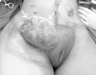
Lower abdominal wall desmoid tumor, extending to right inguinal area.
Fig. 2.
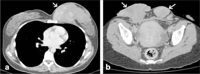
CT scan images show desmoid tumors: (a) left breast (b) lower abdominal wall.
Surgeries were scheduled: the patient first underwent a left radical mastectomy associated with pectoralis muscles resection, sternum partial resection and removal of medial portions of the 3rd to 5th ribs with monofilament polypropylene mesh reconstruction and latissimus dorsi muscle flap. This surgery was performed by teams of Mastology, Thoracic Surgery and Plastic Surgery of the Clinical Hospital at University of Campinas. The patient had a satisfactory outcome and she was discharged from the hospital in the 8th postoperative day. Histopathological sections showed DT with free margins. She subsequently underwent resection of the abdominal wall tumor two months later. Reconstruction after tumor excision was done with polypropylene mesh and a thigh adipose-cutaneous flap. The flap donor site was covered with a partial skin graft. The patient developed partial necrosis of the adipose-cutaneous flap being submitted to debridement and muscle flap, followed by a new partial skin graft. She was finally discharged from hospital in the 47th postoperative day. Histopathological sections also showed DT (Figs. 3 and 4).
Fig. 3.
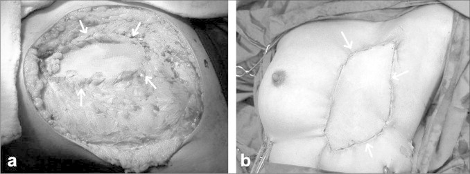
Surgical aspects after breast tumor removal: (a) mesh reconstruction; (b) latissimus dorsi muscle flap.
Fig. 4.
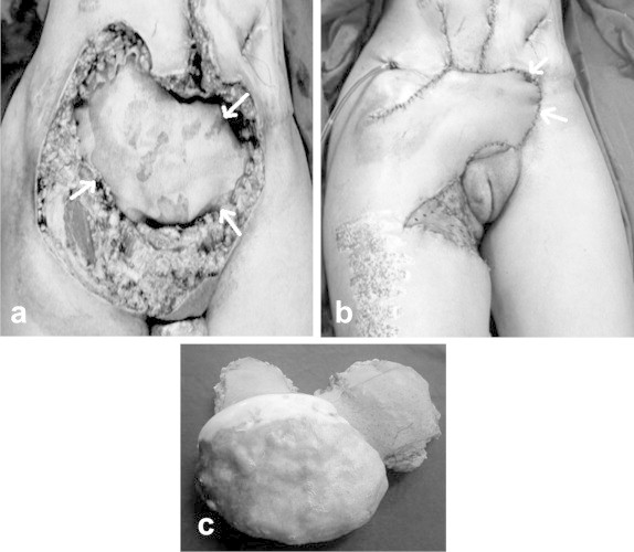
Surgical aspects after abdominal wall tumor resection: (a) mesh reconstruction; (b) adipose-cutaneous flap; (c) surgical specimen.
After 30 days of hospital discharge, the patient was submitted to a new surgical procedure. The preoperative colonoscopy performed before the two major procedures described above had shown a polypoid lesion of 30 mm × 40 mm in the anterior wall of the rectum, 4 cm from the anal verge. The aspect of the ileorectal anastomosis was normal. An endoanal resection of the lesion was done. The biopsy revealed tubulovillous adenoma with a focus of high-grade dysplasia and free margins. The complication after this procedure was the development of rectovaginal fistula of low discharge.
In the outcome, the patient developed two small DT nodes in the abdominal wall. Concomitant excision of the nodes and rectovaginal fistula repair were performed 6 months after the endoanal surgery. The patient developed fistula recurrence, and needed a new surgical procedure. After eventful postoperative courses, the patient has been currently on tamoxifen and celecoxib with no evidence of DT and no evidence of rectovaginal fistula. She is waiting for ileostomy closure (Fig. 5).
Fig. 5.
Final aspect of the abdomen after healed surgical incisions.
3. Discussion
GS is an autosomal dominant genetic disorder with almost complete penetrance (80%) and variable expression.8–10 Most patients have a positive family history of the disease, whereas 20% of cases have spontaneous mutation, without a family history, as described in the present report.2 Restorative proctocolectomy is the procedure of choice to treat the colonic manifestation of the GS, which presents usually more than one hundred colonic adenomatous polyps. The rectum preservation may be an option when there are less than 20 rectal polyps and absence of epithelial high grade dysplasia; but careful monitoring of rectal segment is required in the follow-up. Endoscopic polypectomy or even polyp endoanal resection is performed if lesions are detected.
Diagnosis of GS may present difficulties due to the variety of its clinical manifestations. While some may present with all the classical manifestations of the syndrome, others may show only one or two of them. In general, skin and bone lesions develop approximately 10 years before the appearance of intestinal polyps.4 Osteomas are the most frequent bone lesions, while epidermal or sebaceous cysts are the most common cutaneous manifestations of GS.5,11
Although DTs are not very frequent as bone and cutaneous lesions, they constitute a major cause of morbidity and mortality for patients with GS, and determine their outcomes.12 The most common sites of DT are surgical incisions, abdominal cavity (especially the mesentery) and the retroperitoneum.13,14 Usually, DT appears in the first 3 years after colon surgery. In the present case, the first extracolonic manifestations appeared before colectomy, including the left breast DT. This site of DT is quite rare and there are few reports in the literature.15 Breast DT is a rare condition, even in patients without GS.12
The treatment of choice for DT is surgical resection with clear margins. However, its recurrence can reach 80%; incomplete resection is associated with high risk of recurrence. The surgical and/or clinical management will depend on the location and extent of the tumor. Frequently complex surgical procedures must be employed, as described in the present case. DT in the abdominal wall should be treated surgically since they are easier to be removed and recurrence rates are lower when compared to mesenteric or retroperitoneal DT.3 Considering unresectable tumors, therapeutic options are chemotherapy drugs, non-steroidal anti-inflammatory, antiestrogen therapy, interferon and imatinib associated with radiotherapy.16,17
Considering our case report; the two large tumors in the left breast were successfully removed without recurrence. Although the patient presented abdominal wall DT recurrence, it was possible to excise it by means of a new surgical approach, without morbidity. In those procedures, reconstruction with polypropylene meshes was necessary, as described by Xu et al.18 In addition, flap repair for the defects of the thoracic and abdominal walls was needed.
4. Conclusion
DT represents one of the most significant causes of morbidity and mortality that affects FAP patients following colectomy. For this reason, patients with GS must be followed up closely and abdominal examination should be performed to detect any signs or symptoms of tumors. In addition, a general clinical examination to identify mesenchymal tumors located in uncommon sites, as in the breast, should be made. Recurrence of DT is not negligible, even after wide surgical resection. Imaging techniques such as CT scan and/or ultrasound can help in the surveillance for its early detection. In general, the surgical procedures to excise DT are highly complex, requiring a multidisciplinary team, which is only possible in a tertiary center. Moreover, rectal endoscopic monitoring in the cases with ileorectal anastomosis is very important to avoid cancer development in the remaining rectum.
5. Informed consent
The case report was performed in accordance with the Clinical Hospital Ethical Committee of the University of Campinas. Written informed consent was obtained from the patient for publication of this case report and accompanying images. A copy of the written consent is available for review by the Editor-in-Chief of this journal on request.
Conflict of interest statement
Lílian V. Pinheiro and other co-authors have no conflict of interest.
Funding
There was no funding for this research.
Ethical approval
The case report was performed in accordance with the Clinical Hospital Ethical Committee of the University of Campinas and the patient assigned the informed consent.
Author contributions
Lílian Vital Pinheiro contributed with data collection and wrote the paper.
João José Fagundes contributed to the data collection and participated in the surgeries.
Cláudio Saddy Rodrigues Coy contributed to the study design and participated in the surgeries.
Cesar Cabello dos Santos, Ivan Toro, Marcelo Michellino, Paulo Henrique Fachina participated in the gynecologic, thoracic and plastic surgeries.
Marc Ward reviewed and helped to write the paper.
Raquel Franco Leal contributed to the study design, wrote the paper.
Maria de Lourdes Setsuko Ayrizono contributed to the study design and revision of the manuscript.
Acknowledgement
We thank Prof. T. Torriani of University of Campinas for English review.
References
- 1.Lee B.D., Lee W., Oh S.H. A case report of Gardner syndrome with hereditary widespread osteomatous jaw lesions. Oral Surg Oral Med Oral Pathol Oral Radiol Endodontol. 2009;107(3):68–72. doi: 10.1016/j.tripleo.2008.10.018. [DOI] [PubMed] [Google Scholar]
- 2.Jonathan B., Claire H., Mary T. Gardner syndrome – Review and report of a case. Oral Oncol Extra. 2005;41:89–92. [Google Scholar]
- 3.Fotiadis C., Tsekouras D.K., Antonakis P. Gardner's syndrome: a case report and review of the literature. World J Gastroenterol. 2005;11(34):5408–5411. doi: 10.3748/wjg.v11.i34.5408. [DOI] [PMC free article] [PubMed] [Google Scholar]
- 4.Gómez García E.B., Knoers N.V. Gardner's syndrome (familial adenomatous polyposis): a cilia-related disorder. Lancet Oncol. 2009;10(7):727–735. doi: 10.1016/S1470-2045(09)70167-6. [DOI] [PubMed] [Google Scholar]
- 5.Cristofaro M.G., Giudice A., Amantea M. Gardner's syndrome: a clinical and genetic study of a family. Oral Surg Oral Med Oral Pathol Oral Radiol. 2013;115(3):1–6. doi: 10.1016/j.oooo.2011.10.020. [DOI] [PubMed] [Google Scholar]
- 6.Gu G.L., Wang S.L., Wei X.M. Diagnosis and treatment of Gardner syndrome with gastric polyposis: a case report and review of the literature. World J Gastroenterol. 2008;14(13):2121–2123. doi: 10.3748/wjg.14.2121. [DOI] [PMC free article] [PubMed] [Google Scholar]
- 7.American College of Radiology . 4th ed. American College of Radiology; Reston, VA: 2003. Breast imaging reporting and data system, Breast Imaging Atlas. [Google Scholar]
- 8.Merg A., Lynch H.T., Lynch J.F. Hereditary colon cancer-Part I. Curr Probl Surg. 2005;42(4):195–256. doi: 10.1067/j.cpsurg.2005.01.004. [DOI] [PubMed] [Google Scholar]
- 9.Mao C., Huang Y., Howard J.M. Carcinoma of the ampulla of Vater and mesenteric fibromatosis (desmoid tumor) associated with Gardner's syndrome: problems in management. Pancreas. 1995;10(3):239–245. doi: 10.1097/00006676-199504000-00004. [DOI] [PubMed] [Google Scholar]
- 10.Cruz-Correa M., Giardiello F.M. Familial adenomatous polyposis. Gastrointest Endosc. 2003;58(6):885–894. doi: 10.1016/s0016-5107(03)02336-8. [DOI] [PubMed] [Google Scholar]
- 11.Juhn E., Khachemoune A. Gardner syndrome: skin manifestations, differential diagnosis and management. Am J Clin Drematol. 2010;11(2):117–122. doi: 10.2165/11311180-000000000-00000. [DOI] [PubMed] [Google Scholar]
- 12.Turina M., Pavlik C.M., Heinimann K. Recurrent desmoids determine outcome in patients with Gardner syndrome: a cohort study of three generations of an APC mutation-positive family across 30 years. Int J Colorectal Dis. 2013;28(6):865–872. doi: 10.1007/s00384-012-1600-x. [DOI] [PubMed] [Google Scholar]
- 13.Brown C.S., Jeffrey B., Korentager R. Desmoid tumors of the bilateral breasts in a patient without Gardner syndrome: a case report and review of literature. Ann Plast Surg. 2012;69(2):220–222. doi: 10.1097/SAP.0b013e31821e8faf. [DOI] [PubMed] [Google Scholar]
- 14.Leal R.F., Silva P.V.V.T., Ayrizono M.L.S. Desmoid tumor in patients with familial adenomatous polyposis. Arq Gastroenterol. 2010;47:373–378. doi: 10.1590/s0004-28032010000400010. [DOI] [PubMed] [Google Scholar]
- 15.Rammohan A., Wood J.J. Desmoid tumour of the breast as a manifestation of Gardner's syndrome. Int J Surg Case Rep. 2012;3(5):139–142. doi: 10.1016/j.ijscr.2012.01.004. [DOI] [PMC free article] [PubMed] [Google Scholar]
- 16.Escobar C., Munker R., Thomas J.O. Update on desmoid tumors. Ann Oncol. 2012;23(3):562–569. doi: 10.1093/annonc/mdr386. [DOI] [PubMed] [Google Scholar]
- 17.Camargo V.P., Keohan M.L., D’Adamo D.R. Clinical outcomes of systemic therapy for patients with deep fibromatosis (desmoid tumor) Cancer. 2010;116(9):2258–2265. doi: 10.1002/cncr.25089. [DOI] [PMC free article] [PubMed] [Google Scholar]
- 18.Xu H.M., Han J.G., Ma S.Z. Related citations treatment of massive desmoid tumour and abdominal wall reconstructed with meshes in Gardner's Syndrome. J Plast Recontr Aesthet Surg. 2010;63(6):1058–1060. doi: 10.1016/j.bjps.2009.04.013. [DOI] [PubMed] [Google Scholar]