Abstract
As a disease commonly encountered in daily practice, acute appendicitis is usually diagnosed and managed easily with a low mortality and morbidity rate. However, acute appendicitis may occasionally become extraordinarily complicated and life threatening. A 56-year-old man, healthy prior to this admission, was brought to the hospital due to spiking high fever, poor appetite, dysuria, progressive right flank and painful swelling of the thigh for 3 d. Significant inflammatory change of soft tissue was noted, involving the entire right trunk from the subcostal margin to the knee joint. Painful disability of the right lower extremity and apparent signs of peritonitis at the right lower abdomen were disclosed. Laboratory results revealed leukocytosis and an elevated C-reactive protein level. Abdominal CT revealed several communicated gas-containing abscesses at the right retroperitoneal region with mass effect, pushing the duodenum and the pancreatic head upward, compressing and encasing inferior vena cava, destroying psoas muscle and dissecting downward into the right thigh. Laparotomy and right thigh exploration were performed immediately and about 500 mL of frank pus was drained. A ruptured retrocecal appendix was the cause of the abscess. The patient fully recovered at the end of the third post-operation week. This case reminds us that acute appendicitis should be treated carefully on an emergency basis to avoid serious complications. CT scan is the diagnostic tool of choice, with rapid evaluation followed by adequate drainage as the key to the survival of the patient.
Keywords: Acute appendicitis, Retrocecal appendicitis, Complication, Retroperitoneal abscess, Thigh abscess
INTRODUCTION
Acute appendicitis is a disease commonly encountered in daily practice, and a very low morbidity and mortality rate can be achieved with proper diagnosis and management at present[1,2]. Generally, a non-perforated acute appendicitis can be managed by urgent appendectomy, while perforated appendicitis which may be associated with the formation of localized abscess in the right iliac fossa or in the pelvic cavity, can be managed depending on their symptoms either by early appendectomy or by interval appendectomy following percutaneous drainage[3]. Even with a more severe form of ruptured appendicitises such as those complicated with diffuse peritonitis as commonly encountered in patients of pre-school age, the post-operative recovery is usually smooth[4]. However, acute appendicitises such as those forming appendiceal masses and extensive abscesses or resulting in intestinal obstruction may sometimes become more complicated and require a prolonged treatment period[3,4]. These complications should not be overlooked in order to avoid further sequelae. Hence, we present here a rare and critical case of ruptured retrocecal acute appendicitis with extensive formation of retroperitoneal and thigh abscess, re-emphasizing the importance of early diagnosis and prompt management for this common disease.
CASE REPORT
A 56-year-old man was generally in good health when he first experienced right flank and right thigh pain with daily spiking fever of up to 39 °C for 3 d. He also experienced progressive loss of appetite, epigastric fullness and painful disability of the right thigh. There was minimal abdominal pain initially, but this became more prominent over the right lower quadrant a few days later. He was brought to our hospital 3 d after the onset of the symptoms.
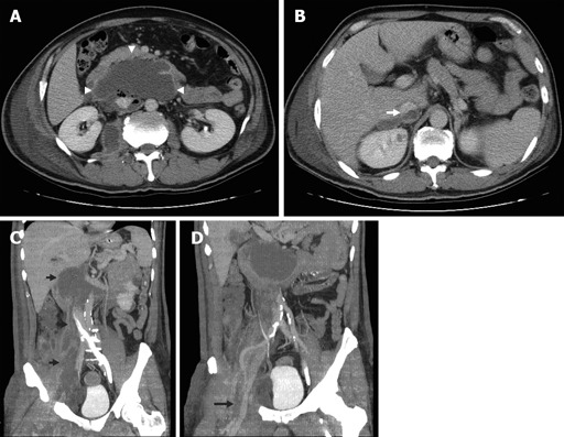
Physical examination on admission revealed an acute ill-looking man with a body temperature of 39.6 °C. He was slightly anemic in appearance and breathed both shallowly and rapidly at a rate of 28/min. He was lying in supine position with his right knee joint mildly flexed and hip joint externally rotated, being reluctant to move his right leg because of severe tenderness. A positive psoas stretch test was performed and the results indicated that there were significant inflammatory signs such as local heat, swelling, edema, and tenderness disclosed at areas involving the entire right trunk from right subcostal region to right knee joint, but no subcutaneous emphysema or crepitation was noted. Palpation of the abdomen revealed tenderness at right lower quadrant without muscle rigidity and no mass-like lesion was palpable. Laboratory data indicated leukocytosis with a WBC count of 16.4×10⁶/mL, of which 80% were mature neutrophils and 3% were immature neutrophils. The hemoglobin level was slightly decreased to the level of 12.8 g/dL and his platelet count was also decreased to 617×106/mL. The C-reactive protein level was 26.1 mg/dL. All the other blood chemistry data were within the normal range. X-ray of the abdomen revealed an indistinct shadow of right psoas muscle. CT scan of the abdomen revealed formation of multiple gas-containing abscesses involving the entire right retroperitoneum with mass effect. At the upper abdominal region, the duodenum and pancreatic head were pushed upward by the abscess, the right perinephric space was filled with the abscess and the inferior vena cava at the same level was encased and compressed. There was also a suspicious septic thrombus inside the inferior vena cava. In addition to destroying the right psoas muscle, the abscess also dissected downward to the right thigh through the femoral canal, forming an abscess between muscle groups (Figure 1).
Figure 1.

Retroperitoneal and right thigh abscess in a patient with ruptured retrocecal appendicitis. A: Retroperitoneal abscess with mass effect, demonstrating the upward-pushed duodenum and pancreatic head as well as compressed and encased inferior vena cava (white arrowheads); B: suspicious septic thrombus inside inferior vena cava (white arrow); C: reconstructed coronal image demonstrating extensive involvement of the abscess (short black arrows); D: dissection of the abscess in the right thigh via the femoral canal just beneath the femoral vessels (long black arrow).
The patient underwent a laparotomy immediately for retroperitoneal exploration, which revealed more than 500 mL of feculent fluid collection in the above-mentioned locations. However, the intra-abdominal cavity was clear without any contamination. A gangrenous ruptured appendix was identified with its entire length embedded in the retroperitoneum, draining fecal content into it and forming an abscess. Appendectomy was done cautiously making sure that no necrotic residual appendix remained in the retroperitoneal cavity. The abscess was communicated with those at the thigh as expected, through the femoral canal just behind the inguinal ligament, even though a separate incision was made at the right thigh to ensure complete drainage of the abscess. Fortunately, the muscle groups of the right thigh were still viable and therefore no debridement of the muscle was performed. Multiple sump drainages were inserted at the end of the operation. The bacterial culture revealed an Escherichia coli infection.
The patient recovered smoothly with his fever subsiding 2 d after surgery and oral intake resumed in 4 d. Painful disability of his right thigh improved immediately after surgery and he was able to walk 12 d later. The patient was discharged uneventfully 3 wk after the surgery.
DISCUSSION
Acute appendicitis is the most common abdominal emergency worldwide and can usually be managed smoothly even if it is perforated. However, formation of retroperitoneal abscesses remains one of the most serious but rare complications of acute appendicitis and is always associated with perforation of a retrocecal appendix due to delayed diagnosis and treatment[5-7]. Knowing that the anatomical position of the appendix is variable and 65% of the appendix has been reported to be at the retrocecal[8], the importance of early management for acute appendicitis cannot be over-emphasized.
There are quite a few case reports discussing related problems similar to the present case, such as psoas and thigh abscesses[9,10], lower extremity subcutaneous emphysema of abdominal origin[11-15] and rare complications of acute appendicitis[16,17]. Among these reports, descriptions are similar regarding patients’ presentation and their management. In short, the onset of symptoms is usually insidious and atypical, initial medical treatment is usually unsuccessful. The causes of abscess formation are usually unclear before surgery and patients are usually critical on presentation. Surgical management is mandatory but may or may not be effective and mortality is not an uncommon result. In addition to the above-mentioned experiences, there are still some important issues that can be addressed based on our present case and a literature review.
First of all, it is still difficult to distinguish primary from secondary psoas abscesses. While the primary psoas abscess is defined as an abscess of unknown origin, its diagnostic symptoms and signs are almost identical to those of the secondary psoas abscess[9,10]. For example, the insidious onset of abscess formation is not responsive to medical treatment, the classical triads of psoas abscess such as fever, flank pain, and limitation of hip movement, can all present in both primary and secondary psoas abscesses[6-8,11]. After reviewing the information from the literature, we found that only the results of bacterial culture, if could be obtained before surgery, could indicate the nature of the abscess being primary or secondary. The most common causative pathogen of primary psoas abscess is Staphylococcus aureus, while that of secondary psoas abscess is usually mixed intestinal floras[9,10,12,18].
Second, as CT scan is used as a modality in addition to the physical signs for definite diagnosis in most of the reported cases, there is no doubt that CT scan of the abdomen with contrast is the most widely-used imaging study with the highest accuracy and efficiency[5-10,19]. CT scan of the abdomen not only helps in the establishment of the diagnosis, but also in the evaluation of the extension of involvement and in its treatment. For example, application of advanced high speed helical CT (which was the case in this report and has never been demonstrated before) can demonstrate a reconstructed coronal image that is of great help for surgical planning because it simulates the operation field. In addition, the drainage of abscess can be achieved by percutaneous and retroperitoneal approach or by laparotomy based on CT findings.
Third, whether the abscess is managed surgically or non-surgically should be carefully evaluated. With the improving technique and the accumulating experiences of interventional radiology, there are several reports demonstrating substantial results by percutaneous drainage of the abscess and then by surgery only if percutaneous drainage fails or is contraindicated. For example, Benoist et al[20] demonstrated that percutaneous drainage can drain 81% postoperative abdominal abscesses in patients without sepsis at presentation. Cantasdemir et al[21] showed that primary and secondary iliopsoas abscesses can be successfully treated with percutaneous drainage in patients without secondary abdominal pathology. Percutaneous drainage of the abscesses is less invasive. Gerzof et al[22] have recommended that percutaneous drainage should be done for most complex abscesses, but we are still uncertain if the percutaneous approach is adequate for our patient, because our patient was in a critical condition requiring prompt drainage of the abscess which cannot be achieved by percutaneous method and the infection could not be controlled if there was a persistent existing focus as previously reported by Ushiyama et al[15]. In addition, according to the results reported by Benoist et al[20] and Cantasdemir et al[21], a combined therapy (that is drainage in combination with surgery) might be contraindicated in our patient because he was septic and had a persistent existing intra-abdominal focus. Hence, it is our experience that surgery is advantageous over percutaneous drainage in patients under critical conditions such as perforated appendicitis, diverticulitis or malignancy[14].
Fourth, though abscesses inside the thigh is due to direct extension from retroperitoneum, it might be wise to create a separate incision at the thigh to drain the abscess rather than from the trunk. Draining thigh abscess by an incision at the thigh has two advantages. First, the abscess can be more easily and directly approached. Second, the viability of muscle and fascia of the thigh as well as the need for further debridement can be adequately evaluated. This is supported by the fact that some of thigh abscesses can be cured by drainage alone[6,7], while others with extensive myonecrosis require debridement or amputation[5,13].
In conclusion, formation of huge retroperitoneal abscesses with thigh involvement is a serious complication of perforated acute appendicitis. To improve the treatment outcome, patients with retroperitoneal infection should undergo CT scan in order to find the origin of the infection and to choose the best way to drain the abscess.
Footnotes
S- Editor Wang XL and Guo SY L- Editor Elsevier HK E- Editor Kong LH
References
- 1.Blomqvist PG, Andersson RE, Granath F, Lambe MP, Ekbom AR. Mortality after appendectomy in Sweden, 1987-1996. Ann Surg. 2001;233:455–460. doi: 10.1097/00000658-200104000-00001. [DOI] [PMC free article] [PubMed] [Google Scholar]
- 2.Hale DA, Molloy M, Pearl RH, Schutt DC, Jaques DP. Appendectomy: a contemporary appraisal. Ann Surg. 1997;225:252–261. doi: 10.1097/00000658-199703000-00003. [DOI] [PMC free article] [PubMed] [Google Scholar]
- 3.Lally KP, Cox CS, Andrassy RJ. Appendix. In: Townsend CM, Beauchamp RD, Evers BM, Mattox KL: Sabiston textbook of surgery, editors. 17th ed. Philadelphia: Elsevier Saunders; 2004. pp. 1381–1399. [Google Scholar]
- 4.Ellis H, Natbanson LK. Appendix and appendectomy. In: Zinner MJ, Schwartz SI, Ellis H, editors. Maingot’s abdominal operations. 10th ed. Connecticut: Appleton & Lange; 1997. pp. 1191–1227. [Google Scholar]
- 5.Edwards JD, Eckhauser FE. Retroperitoneal perforation of the appendix presenting as subcutaneous emphysema of the thigh. Dis Colon Rectum. 1986;29:456–458. doi: 10.1007/BF02561584. [DOI] [PubMed] [Google Scholar]
- 6.Gutknecht DR. Retroperitoneal abscess presenting as emphysema of the thigh. J Clin Gastroenterol. 1997;25:685–687. doi: 10.1097/00004836-199712000-00027. [DOI] [PubMed] [Google Scholar]
- 7.Sharma SB, Gupta V, Sharma SC. Acute appendicitis presenting as thigh abscess in a child: a case report. Pediatr Surg Int. 2005;21:298–300. doi: 10.1007/s00383-004-1356-7. [DOI] [PubMed] [Google Scholar]
- 8.El-Masry NS, Theodorou NA. Retroperitoneal perforation of the appendix presenting as right thigh abscess. Int Surg. 2002;87:61–64. [PubMed] [Google Scholar]
- 9.Agrawal SN, Dwivedi AJ, Khan M. Primary psoas abscess. Dig Dis Sci. 2002;47:2103–2105. doi: 10.1023/a:1019693400742. [DOI] [PubMed] [Google Scholar]
- 10.Kleiner O, Cohen Z, Barki Y, Mares AJ. Unusual presentation of psoas abscess in a child. J Pediatr Surg. 2001;36:1859–1860. doi: 10.1053/jpsu.2001.28870. [DOI] [PubMed] [Google Scholar]
- 11.Nicell P, Tabrisky J, Lindstrom R, Peter M. Thigh emphysema and hip pain secondary to gastrointestinal perforation. Surgery. 1975;78:555–559. [PubMed] [Google Scholar]
- 12.Rotstein OD, Pruett TL, Simmons RL. Thigh abscess. An uncommon presentation of intraabdominal sepsis. Am J Surg. 1986;151:414–418. doi: 10.1016/0002-9610(86)90481-2. [DOI] [PubMed] [Google Scholar]
- 13.Jager GJ, Rijssen HV, Lamers JJ. Subcutaneous emphysema of the lower extremity of abdominal origin. Gastrointest Radiol. 1990;15:253–258. doi: 10.1007/BF01888788. [DOI] [PubMed] [Google Scholar]
- 14.Kobayashi H, Sakurai Y, Shoji M, Nakamura Y, Suganuma M, Imazu H, Hasegawa S, Matsubara T, Ochiai M, Funabiki T. Psoas abscess and cellulitis of the right gluteal region resulting from carcinoma of the cecum. J Gastroenterol. 2001;36:623–628. doi: 10.1007/s005350170047. [DOI] [PubMed] [Google Scholar]
- 15.Ushiyama T, Nakajima R, Maeda T, Kawasaki T, Matsusue Y. Perforated appendicitis causing thigh emphysema: a case report. J Orthop Surg (Hong Kong) 2005;13:93–95. doi: 10.1177/230949900501300118. [DOI] [PubMed] [Google Scholar]
- 16.Kao CT, Tsai JD, Lee HC, Wang NL, Shih SL, Lin CC, Huang FY. Right perinephric abscess: a rare presentation of ruptured retrocecal appendicitis. Pediatr Nephrol. 2002;17:177–180. doi: 10.1007/s00467-001-0794-x. [DOI] [PubMed] [Google Scholar]
- 17.Chang TN, Tang L, Keller K, Harrison MR, Farmer DL, Albanese CT. Pylephlebitis, portal-mesenteric thrombosis, and multiple liver abscesses owing to perforated appendicitis. J Pediatr Surg. 2001;36:E19. doi: 10.1053/jpsu.2001.26401. [DOI] [PubMed] [Google Scholar]
- 18.Ricci MA, Rose FB, Meyer KK. Pyogenic psoas abscess: worldwide variations in etiology. World J Surg. 1986;10:834–843. doi: 10.1007/BF01655254. [DOI] [PubMed] [Google Scholar]
- 19.Haaga JR. Imaging intraabdominal abscesses and nonoperative drainage procedures. World J Surg. 1990;14:204–209. doi: 10.1007/BF01664874. [DOI] [PubMed] [Google Scholar]
- 20.Benoist S, Panis Y, Pannegeon V, Soyer P, Watrin T, Boudiaf M, Valleur P. Can failure of percutaneous drainage of postoperative abdominal abscesses be predicted. Am J Surg. 2002;184:148–153. doi: 10.1016/s0002-9610(02)00912-1. [DOI] [PubMed] [Google Scholar]
- 21.Cantasdemir M, Kara B, Cebi D, Selcuk ND, Numan F. Computed tomography-guided percutaneous catheter drainage of primary and secondary iliopsoas abscesses. Clin Radiol. 2003;58:811–815. doi: 10.1016/s0009-9260(03)00274-5. [DOI] [PubMed] [Google Scholar]
- 22.Gerzof SG, Johnson WC, Robbins AH, Nabseth DC. Expanded criteria for percutaneous abscess drainage. Arch Surg. 1985;120:227–232. doi: 10.1001/archsurg.1985.01390260085012. [DOI] [PubMed] [Google Scholar]


