Figure 3.
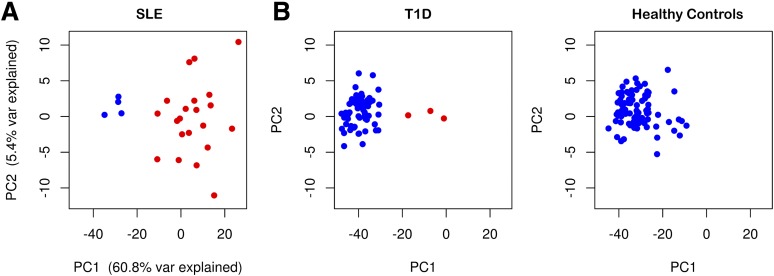
An IFN signature can be detected in peripheral blood of patients with SLE and T1D. A: Plot depicting the two first PCs obtained from the expression of the 1,111 IFN-inducible probe sets defined in patients with SLE (n = 25) in this study. The percentage of variance explained by each PC is shown on the respective axis. B: Plots depicting the first two PCs obtained from the projection of the expression of the IFN-inducible probe sets in subjects from cross-sectional cohorts of patients with T1D (n = 64; left panel) and adult healthy controls (n = 87; right panel) onto the PC axes defined in the analysis of the patients with SLE (see Research Design and Methods for details). The cross-sectional cohorts of patients with T1D include 15 adult patients with long-standing T1D (median 13 years since diagnosis; median age 31 years) enrolled from the Cambridge BioResource and 49 recently diagnosed patients (median 1.4 years since diagnosis; median age 12 years) recruited from the Diabetes-Genes, Autoimmunity and Prevention study. Samples that show clear evidence of upregulation of IFN-inducible genes by hierarchical clustering are depicted in red.

