

Abstract
A 30-year-old unmarried, heterosexual male presented with an 8-month history of tense blistering skin lesions over the hands. Physical examination revealed facial hypertrichosis and multiple erosions with crusts and scars over the dorsum of both hands. Woods lamp examination of the urine, histopathology and urinary porphyrin levels were suggestive of porphyria cutanea tarda (PCT). The patient responded well to hydroxychloroquine and antiretroviral drugs. This case report calls for a detailed evaluation and HIV testing in every patient with PCT.
Keywords: HIV, hydroxychloroquine, porphyria cutanea tarda
INTRODUCTION
Porphyria cutanea tarda (PCT), a relatively uncommon metabolic disease, is the most common cutaneous porphyria and has a higher incidence rate in HIV-infected patients when compared with the general population. Several reports have shown HIV infection to be an independent risk factor for PCT in contrast to the earlier belief of it being a coincidental association with PCT.[1,2,3] We report this association due to its rarity in the Indian scenario and to alert the treating consultant regarding the need to consider testing for HIV in a patient with PCT.
CASE REPORT
A 30-year-old unmarried male presented with an 8-month history of tense fluid-filled lesions over the back of both hands leaving behind erosions and scars. There was history of photosensitivity, weight loss and high-risk behavior over the past 1 year. He reported moderate alcohol intake in the past, but denied drinking during the previous 1 year. There was no history suggestive of liver disease or of having similar complaints in the family. Physical examination revealed multiple erosions with crusts, scars over the dorsum of both hands [Figure 1] and face. Minimal hypertrichosis of the face was present [Figure 2].
Figure 1.

Clinical photograph showing multiple erosions with crusts and scars over the dorsum of both hands
Figure 2.
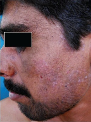
Minimal hypertrichosis and erosions with crusts over the left side of the face
Examination of oral mucosa and genitals were within the normal limits.
Blood investigations revealed: Hemoglobin level 13.4 g/L, total count 4,200 cells/cumm, iron 146 mcg/mL, ferritin 429.2 ng/mL. Except for the elevated aspartate aminotransferase (AST) levels of 128 U/L, the remaining parameters of liver function tests were within normal limits. On serological testing, patient was found to be HIV positive with a CD4 count of 32 cells/mm3. Serological screening for hepatitis-B and C were negative. No abnormality was detected on abdominal ultrasonography.
Examination of urine under Wood's light revealed a bright pink fluorescence consistent with the presence of porphyrins [Figure 3]. Histopathological examination revealed a subepidermal bulla with scanty lymphocytic infiltrate and hemorrhage. There was deposition of periodic acid–Schiff (PAS) stained material around the dermal capillary blood vessels [Figure 4].
Figure 3.
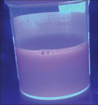
Bright pink fluorescence on woods lamp examination
Figure 4.

A pauci-inflammatory subepidermal bulla with hemorrhage and periodic acid–Schiff (PAS) positive material around dermal capillaries. The floor of the bulla shows preserved dermal papillae (PAS magnification, ×10)
Direct immunofluorescence study showed linear IgG deposition along the basement membrane. Urinary porphyrin levels were elevated.
Based on the clinical presentation and laboratory findings, a diagnosis of PCT was made. The patient was treated with low-dose hydroxychloroquine 200 mg twice weekly, antiretroviral drugs and was advised photo protective measures. Three months following the initiation of treatment, there was clinical remission of the skin lesions [Figure 5] with a CD4 count of 232 cells/mm3.
Figure 5.
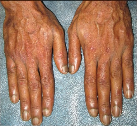
Resolution of the erosions over the dorsum of both hands
DISCUSSION
Porphyria cutanea tarda is a disorder of heme synthesis characterized by reduced activity of the enzyme uroporphyrin decarboxylase in the hepatocytes (acquired form) or in both erythrocytes and hepatocytes (familial form).
Mansourati et al. initially reported the association of PCT with HIV in 1987. They reviewed 75 reported cases of HIV infection with PCT; almost all (96%) of the patients were male. At the time of diagnosis, the mean age was 36.5 years and the mean CD4 count of 166 cells/mm3. Forty-fivepercent of the patients had hepatitis C virus infection, 55% consumed alcohol heavily, and 89% had abnormal liver function test results. The diagnosis of PCT preceded the detection of HIV infection in 40% of cases.[2] Clinical features of PCT appeared prior to the detection of HIV infection in our patient with no evidence of polycythemia and late cutaneous manifestations of PCT.
The mechanism of HIV infection that causes PCT is not fully understood.
Several hypotheses have been postulated to explain the association between PCT and HIV. The common factor in all the different hypotheses of PCT is an aberration of hepatic function of significant severity that normal porphyrin metabolism is no longer maintained. Our patient had deranged liver function in the form of raised AST. HIV could impair cytochrome P-450 dependent mixed-function oxidase system, the rate limiting mitochondrial enzyme delta-aminolevulinic acid synthase and heme oxygenase.[1,4] A disruption in the heme synthesis occurring secondary to the altered steroid hormone levels leading to elevated estrogen levels has been observed in HIV-infected men.[5,6] Ineffective hematopoiesis associated with HIV infection can result in a subsequent increase in hepatic iron. Direct hepatic damage by HIV has also been suggested.[2]
Cutaneous photosensitivity resulting in the development of subepidermal blisters is a major feature of PCT. Histopathological examination of the involved skin in PCT may show linear globules of eosinophilic PAS positive material termed caterpillar bodies, which may be present in the epidermis overlying the bullae with festooning of dermal papillae. This finding was not observed in our case.
Direct immunofluorescence study in PCT is characterized by IgG, IgA ± IgM, C3 in the walls of the papillary dermal blood vessels and along the dermoepidermal junction. Our patient had deposition of IgG along the dermoepidermal junction without the deposition of complement or involvement of the blood vessels. This finding has been reported in 2 out of the 28 patients in a study.[7]
For persons with PCT, avoiding sun exposure and other porphyrinogenic precipitants is important. Phlebotomy and low-dose chloroquine or hydroxychloroquine are specific forms of treatment for PCT. Hydroxychloroquine has been described in both short-term high-dose and longer-term low-dose regimens. Though high-dose treatment (250 mg tds for 3 days) results in rapid clinical and biochemical remission, it is associated with flu-like symptoms and hepatotoxicity. Low-dose hydroxychloroquine regimens of 200 mg twice weekly have been shown to be effective in producing clinical remission and reducing porphyrin levels.[8] Our patient was treated with low-dose hydroxychloroquine and was advised photo protection.
With an increased incidence in HIV-infected patients and a clinical presentation which can precede the detection of HIV infection, the role of a dermatologist in detecting PCT cannot be overemphasized.[5] The coexistence of PCT and HIV infection has been rarely reported from Indian literature, being described in one case previously to the best of our knowledge.[9] Thus, the diagnosis of PCT should prompt a comprehensive workup for HIV infection as earlier diagnosis would result in earlier treatment, which could prevent further immunodeficiency.
Footnotes
Source of Support: Nil.
Conflict of Interest: None declared.
REFERENCES
- 1.Wissel PS, Sordillo P, Anderson KE, Sassa S, Savillo RL, Kappas A. Porphyria cutanea tarda associated with the acquired immune deficiency syndrome. Am J Hematol. 1987;25:107–13. doi: 10.1002/ajh.2830250112. [DOI] [PubMed] [Google Scholar]
- 2.Mansourati FF, Stone VE, Mayer KH. Porphyria cutanea tarda and HIV/AIDS: A review of pathogenesis, clinical manifestations and management. Int J STD AIDS. 1999;10:51–6. doi: 10.1258/0956462991912944. [DOI] [PubMed] [Google Scholar]
- 3.Drobacheff C, Derancourt C, Van Landuyt H, Devred D, de Wazieres B, Cribier B, et al. Porphyria cutanea tarda associated with human immunodeficiency virus infection. Eur J Dermatol. 1998;8:492–6. [PubMed] [Google Scholar]
- 4.McAlister F, McClean K, Hamilton PG, Houston S. Human immunodeficiency virus infection and porphyria cutanea tarda: Coexistence of risk factors or causative association? Clin Infect Dis. 1995;20:348–51. doi: 10.1093/clinids/20.2.348. [DOI] [PubMed] [Google Scholar]
- 5.Reynaud P, Goodfellow K, Svec F. Porphyria cutanea tarda as initial presentation of the acquired immunodeficiency syndrome in two patients. J Infect Dis. 1990;161:1032–3. doi: 10.1093/infdis/161.5.1032. [DOI] [PubMed] [Google Scholar]
- 6.Blauvelt A, Harris HR, Hogan DJ, Jimenez-Acosta F, Ponce I, Pardo RJ. Porphyria cutanea tarda and human immunodeficiency virus infection. Int J Dermatol. 1992;31:474–9. doi: 10.1111/j.1365-4362.1992.tb02693.x. [DOI] [PubMed] [Google Scholar]
- 7.Vieira FM, Aoki V, Oliveira ZN, Martins JE. Study of direct immunofluorescence, immunofluorescence mapping and light microscopy in porphyria cutanea tarda. An Bras Dermatol. 2010;85:827–37. doi: 10.1590/s0365-05962010000600008. [DOI] [PubMed] [Google Scholar]
- 8.Malkinson FD, Levitt L. Hydroxychloroquine treatment of porphyria cutanea tarda. Arch Dermatol. 1980;116:1147–50. [PubMed] [Google Scholar]
- 9.Sharma YK, Virmani NC, Dash KN, Deo KS, Mave V, Gupta A, et al. Photo quiz. A 23-year-old man presenting with fluid-filled skin lesions, patchy pigmentation, and skin breakage after trivial trauma. Clin Infect Dis. 2013;56:851. doi: 10.1093/cid/cis992. [DOI] [PubMed] [Google Scholar]