Figure 1.

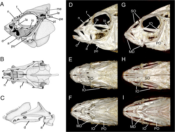
Lateral line canal system in Amatitlania nigrofasciata (A-C), Tramitichromis sp. (D-F), and Aulonocara stuartgranti (G-I). Drawings based on a cleared and stained skull of an adult convict cichlid, A. nigrofasciata(A-C) illustrate the different dermatocranial bones that contain the pored lateral line canals. The supraorbital canal (SO) is contained within the nasal and frontal bones and is visible in both lateral and dorsal views (A, B). The mandibular canal (MD) is contained within the dentary and anguloarticular bones, which are visible in lateral view (C). The infraorbital canal (IO) is contained within the lacrimal and infraorbital bones, and the preopercular canal (PO) is contained within the preopercular bone, and is visible in lateral view (A). 3-D reconstructions of μCT data from a Tramitichromis sp. (adult, 79 mm SL) in lateral, dorsal and ventral views (D, E, F), and A. stuartgranti (adult, 78 mm SL) in lateral, dorsal, and ventral views (G, H, I) clearly show larger canal pores in Aulonocara (widened lateral line canals), and in the mandibular canal in particular (for example, I vs. F), when compared to Tramitichromis (narrow lateral line canals). a, anguloarticular; d, dentary; f, frontal; io, infraorbital bones; la, lacrimal; le, lateral extrascapular; me, medial extrascapular; n, nasal; pe, posttemporal; po, preoperculum; pt, pterotic. A from [28], reprinted with permission of Academic Press/Elsevier, Inc.; B, C from [29], reprinted with permission of Wiley and Sons, Inc.
