Abstract
PURPOSE
To develop an optical coherence tomography (OCT) pachymetry map based keratoconus risk scoring system.
SETTING
This multi-center study was conducted in Doheny Eye Institute, University of Southern California (Los Angeles, CA, USA), Department of Ophthalmology, Affiliated Eye Hospital of Wenzhou Medical College (Wenzhou, China), and Brass Eye Center (New York, NY, USA).
DESIGN
Prospective cross-sectional observational study.
METHODS
A Fourier-domain OCT was used to acquire corneal pachymetry map in normal and keratoconus subjects. Pachymetric variables were: minimum, minimum-median, superior - inferior (S-I), superonasal - inferotemporal (SN-IT), and the vertical location of the thinnest cornea (Ymin). A logistic regression formula and a scoring system were developed based on these variables. Keratoconus diagnostic accuracy was measured by the area under the receiver operating characteristic curve (AROC).
RESULTS
One hundred thirty-three eyes from 67 normal subjects, 84 eyes from 52 keratoconus subjects were recruited. The keratoconus logistic regression formula = 0.543 × minimum + 0.541 × (S-I) − 0.886 × (SN-IT) + 0.886 × (minimum-median) + 0.0198 × Ymin. The formula gave better diagnostic power with AROC than the best single variable (formula = 0.975, minimum = 0.942, P < 0.01). The diagnostic power with AROC of the keratoconus risk score (0.949) was similar to that of the formula (P = 0.08).
CONCLUSION
The OCT corneal pachymetry map based logistic regression formula and the keratoconus risk scoring system provided high accuracy in keratoconus detection. These normal methods may be useful in keratoconus screening.
INTRODUCTION
Keratoconus is a progressive ectatic corneal disease characterized by noninflammatory thinning and protrusion of the cornea.1 Early stage keratoconus is the primary risk factor for post-LASIK ectasia,2, 3 a serious complication of laser refractive surgeries.
Currently, Placido-disk-based corneal topography remains the predominant method for detecting keratoconus.4–7 However, topography screening methods have shortcomings. First, topography may not detect all patients at risks. Randleman et al. reported 27% of 93 post-refractive surgery ectasia cases had normal preoperative topography and 29% of these had borderline preoperative topography.8 Second, satisfactory topography maps may not be available owing to cornea irregularity or tear film breakup. Third, normal eyes could be diagnosed as keratoconus eyes due to corneal distortion like contact lens-induced warpage and misalignment of corneal apex.9, 10 Keratoconus is also characterized by focal thinning and posterior topographic steepening.11 Pachymetric mapping may be able to identify keratoconus cases with normal or borderline topography.12 Previous studies have shown that using pachymetric variables is feasible in keratoconus screening.13–16 Slit scanning imaging, Scheimpflug imaging and optical coherence tomography (OCT) are different approaches in pachymetric mapping. Slit-scanning imaging has a tendency to underestimate corneal thickness in presence of scar.17 Pachymetric measurements acquired with OCT were more repeatable than those obtained with Scheimpflug imaging in keratoconic eyes in one study.18
Optical coherence tomography (OCT) is capable of noncontact imaging of the cornea with micron-level high resolution. It can accurately map the corneal thickness of normal and keratoconic eyes.19, 20 Our previous study introduced a sensitive and specific pachymetry-based method to help screening keratoconus using a time-domain OCT system.20 Fourier-domain OCT, a newer generation of OCT, is capable of acquiring scans 10–100 times faster than time-domain OCT systems.21–23 In this study, we used an 840 nm wavelength Fourier-domain OCT system with higher resolution and scanning speed to investigate an improved keratoconus screening strategy.
METHODS
Subjects
The subjects of this cross-sectional observational study were recruited at 3 study sites: Doheny Eye Institute (DEI) at University of Southern California (Los Angeles, CA, USA), Wenzhou Medical College (WMC, Wenzhou, China), and Brass Eye Center (BEC, New York, NY, USA). This study was approved by the institutional review board of the universities. It followed the tenets of the Declaration of Helsinki and was in accord with the Health Insurance Portability Act of 1996.
All eyes were classified into 2 groups: normal and keratoconus. The inclusion criteria for normal subjects were normal topography, normal slit-lamp biomicroscopy, and best spectacle-corrected visual acuity (BSCVA) of 20/20 or better in both eyes. Keratoconic eyes included in this study were diagnosed clinically. Inclusion criteria were topography characteristic of keratoconus (skewed asymmetric bow-tie, inferior steep spot, etc.)24 and more than one clinical sign including slit lamp findings of Munson’s sign, Vogt’s striae, Fleischer’s ring, apical thinning, Rizutti’s sign, etc. Eyes with late keratoconic changes such as corneal scars or hydrops were excluded as they do not pose any diagnostic challenge and have anomalous pachymetric findings. Exclusion criteria of all eyes included history of other corneal diseases, and eyes with previous ocular surgeries.
Optical Coherence Tomography Imaging
The OCT pachymetry map scans were acquired with a Fourier-domain OCT system (RTVue, Optovue, Inc., Fremont, CA, USA). The instrument used an 840 nm wavelength light source. It has a scan speed of 26,000 A-scans per second. It has a transverse resolution of 15 μm and an axial resolution of 5 μm (in tissue). A corneal adapter module lens (CAM) was mounted for anterior segment imaging. The “pachymetry” scan pattern was used to map the cornea. The pattern consists of 8 high-definition meridional scans (1024 axial scans per meridian) that were acquired in 0.32 seconds. The scan diameter of the pachymetry map was 6 mm. The maps (Figure 1.) were divided into octant zones: superior (S), superotemporal (ST), temporal (T), inferotemporal (IT), inferior (I), inferonasal (IN), nasal (N), superonasal (SN), central (C) and annular rings (2, 5, and 6mm diameters). The location of the minimum corneal thickness was marked as “※”.
Figure 1.

Optical coherence tomography corneal pachymetry map of a keratoconus subject showing pachymetric variables. The five individual pachymetric variables are 1. minimum = location of minimum corneal thickness (marked as ※), 2. minimum-median = the minimum corneal thickness minus the mean corneal thickness averaged from central 5 mm diameter, 3. The S-I: The average thickness of the superior (S) octant minus that of the inferior (I) octant, 4. The SN-IT: The average thickness of the SN octant minus that of the IT octant (variables 3 and 4 are measured within 2–5mm diameters, marked as red circles on the right), 5. Ymin = vertical location of the minimum (marked as red double arrows on the left).
Each eye was scanned 3 times during a single visit with the subject sitting. The subject’s head was stabilized with a chin/forehead rest. The subject’s gaze was fixed with an internal fixation target. The OCT and video camera images were displayed in real time to aid alignment. Subjects were repositioned after each OCT scan. The pachymetry scans were centered by pupil.
Optical Coherence Tomography Pachymetric Variables
Several diagnostic variables were constructed using the OCT pachymetric map with the aim of capturing the focal and asymmetric nature of keratoconic corneal thinning. The variables were calculated from the central 5 mm diameter of the pachymetry map. The octant values were averaged in the 2 to 5 mm diameter zone. The 5 pachymetric diagnostic variables were as follows:20
Minimum: the minimum corneal thickness.
Minimum-median: the minimum corneal thickness minus the mean corneal thickness averaged from central 5 mm diameter.
The S-I: The average thickness of the superior (S) octant minus that of the inferior (I) octant.
The SN-IT: The average thickness of the SN octant minus that of the IT octant.
Vertical location of the minimum (Ymin): Locations superior to the pupil center had positive values and locations inferior to the vertex had negative values.
General corneal thinning was captured by the minimum variable. Focal thinning was captured by the minimum-median variable. Asymmetric thinning was captured by the S-I and SN-IT variables, and by the vertical location of the minimum.
Statistical Analysis
The repeatability of each OCT pachymetric variable was calculated by pooled standard deviation (SD) of repeated measurements25 and the intraclass correlation coefficient (ICC). The mean value ± SD of each pachymetric variable was calculated for each group. The normality of the OCT diagnostic variables was confirmed by a Kolmogorov–Smirnov test on the dataset, in which 1 eye was randomly selected from each normal subject. The generalized estimating equation was used to account for the inter-eye correlation in the tests to compare means. 26
A formula combining 5 pachymetric variables was derived by logistic regression based on 5 pachymetric variables data from all eyes. Clinical diagnosis of normal or keratoconus was used to classify the eyes in the logistic regression.
A keratoconus risk scoring system table was designed based on the pachymetric measurements of the normal subjects. The score cutoffs of each individual pachymetric variable were 20, 5, 1 percentile of the measurements from the normal eyes. Take minimum for example, this variable will get a score of 1, 2, 3 if its value exceeds 20, 5, 1 percentile thresholds, respectively. A composite keratoconus risk score was calculated for each eye by summing up the scores from all 5 variables. A five variables OR logic method was also evaluated for keratoconus screening. The OR logic value is calculated as the minimum of the standardized (mean will be 0 and standard deviation will be 1 after the standardization) values of the five individual variables. It was equivalent to use the OR logic that an eye was identified as keratoconus if any of its 5 pachymetry based variables exceeded the 5 percentile threshold.
Receiver operating characteristic analyses were performed to compare the diagnostic performance of individual pachymetric variables, five variables OR logic, the keratoconus risk score, and the logistic regression formula. To account for potential correlation between the eyes from the same subject, generalized estimating equation (GEE) method27 was used when applicable. The level of significance was set at P<0.05. All statistic analyses were performed in SAS 9.1 (SAS Institute Inc., Cary, NC, USA).
RESULTS
In normal group, 133 eyes from 67 normal subjects (one eye excluded due to unanalyzable pachymetry map) were recruited at Doheny Laser Vision Center of DEI. In keratoconus group, 20 eyes from 12 subjects were recruited from BEC; 32 eyes from 21 subjects were recruited from WMC, and 30 eyes from 19 subjects were recruited from DEI. The mean age of normal group was 38.3 ± 11.5 years (range 18~61), male = 37; the mean age of keratoconus group was 35.2 ± 13.6 years (range 18~61), male = 32. The mean age was not significantly different between groups. There were no differences in gender distribution between groups.
Table 1 showed refractive error and corneal topographic information of the two groups. Keratoconus group had more refractive errors comparing with normal group (P < 0.001). Keratoconus group had steeper keratometric readings comparing with normal group (P < 0.001). The keratoconic corneas were thinner than normal corneas (P < 0.001).
Table 1.
Refractive Errors and Keratometric Characteristics of Normal and Keratoconic Eyes.
| Groups | Sphere (D) | Cylinder (D) | Flat Sim K (D) | Steep Sim K (D) | CCT(μm) |
|---|---|---|---|---|---|
| Normal | −1.39 ± 2.21 (−5.50~2.00) | −0.43 ± 0.61 (−3.50~0) | 42.89 ± 1.58 (40.60~44.11) | 44.17 ± 1.71 (41.9~45.98) | 533 ± 32 (490~598) |
|
| |||||
| Keratoconus | −3.74 ± 4.62 (−18.00~4.25) | −3.13 ± 2.71 (−11.25~0) | 45.78 ± 5.34 (34.5~70.8) | 50.27 ± 6.38 (41.2~72.6) | 471 ± 50 (237~553) |
| P < 0.001 | P < 0.001 | P < 0.001 | P < 0.001 | P < 0.001 | |
Sim K = simulated K, D = dioptor, FFK = forme fruste keratoconus, CCT = central corneal thickness as measured by optical coherence tomography pachymetry map and averaged within 2mm diameter; mean values ± standard deviation (range) are listed, P values are comparing normal with keratoconus group.
The repeatabilities of the individual OCT pachymetric variables were given in Table 2. The minimum thickness had the best intraclass correlation coefficient (1.00 for all both groups). The ICCs of minimum-median, S-I, and SN-IT ranged from 0.88 to 0.99. Ymin had relatively lower ICCs of 0.85 (normal) and 0.84 (keratoconus) compared with that of other variables.
Table 2.
Repeatability of Optical Coherence Tomography Corneal Pachymetric Variables.
| Groups Unit: μm | Minimum | Minimum-Median | S-I | SN-IT | Ymin | |||||
|---|---|---|---|---|---|---|---|---|---|---|
|
| ||||||||||
| PSD | ICC | PSD | ICC | PSD | ICC | PSD | ICC | PSD | ICC | |
| Normal | 1.6 | 1.00 | 2.2 | 0.89 | 4.2 | 0.94 | 3.9 | 0.93 | 241 | 0.85 |
| Keratoconus | 3.3 | 1.00 | 5.1 | 0.99 | 14.2 | 0.98 | 12.8 | 0.97 | 323 | 0.84 |
S-I = superior-inferior, SN-IT = superonasal-inferotemporal, Ymin = vertical location of the minimum.
PSD = pooled standard deviation, ICC = intraclass correlation coefficient.
The descriptive statistics of OCT pachymetric variables were given in Table 3. Comparing normal group with keratoconus group, all pachymetric variables were statistically significantly different (P <0.001).
Table 3.
Optical Coherence Tomography Corneal Pachymetric Variables.
| GroupsUnit: μm | Minimum | Minimum-Median | S-I | SN-IT | Ymin |
|---|---|---|---|---|---|
| Normal | 523.7 ± 29.8 | −16.5 ± 5.6 | 18.6 ± 13.0 | 22.7 ± 12.0 | −383 ± 417 |
|
| |||||
| Keratoconus | 426.4 ± 68.8 | −59.9 ± 47.7 | 66.2± 65.1 | 73.7 ± 49.7 | −917 ± 549 |
| P<0.001 | P<0.001 | P<0.001 | P<0.001 | P<0.001 | |
S-I = superior-inferior, SN-IT = superonasal-inferotemporal, Ymin = vertical location of the minimum; mean values ± standard deviation are listed, P values are comparing normal with keratoconus group.
The logistic regression formula combining 5 individual pachymetric variables were derived as follows: keratoconus logistic regression formula = 0.543 × minimum + 0.541 × (S-I) − 0.886 × (SN-IT) + 0.886 × (minimum-median) + 0.0198 × Ymin. According to the formula, terms SN-IT and minimum-median had the greatest weight when evaluating the risk of keratoconus.
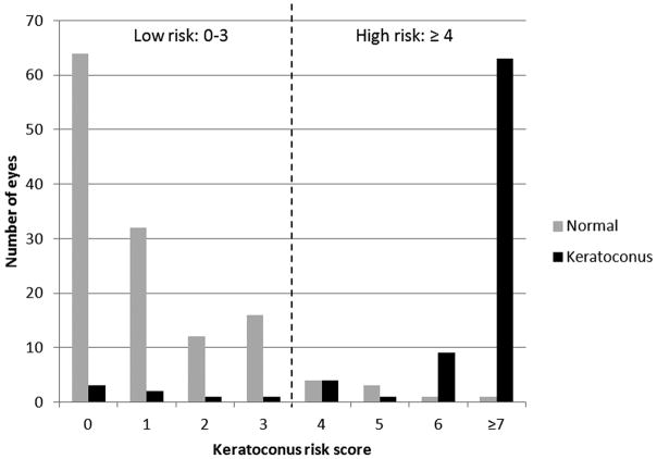
The keratoconus risk scoring system table was presented in Table 4. The histogram of the composite keratoconus risk score was shown in Figure 2. The cutoff of the composite keratoconus risk score was set as follows: low risk: 0 – 3; high risk: ≥ 4. 93.2% of normal eyes and 8.3% of keratoconus eyes were evaluated as having low risk of keratoconus.
Table 4.
Keratoconus Risk Scoring System Table for Clinical Evaluation Based on Optical Coherence Tomography Corneal Pachymetry Map.
| Variables (μm) | 0 | 1 | 2 | 3 | OD | OS |
|---|---|---|---|---|---|---|
| Minimum | >499 | 499 ~ 476 | 475 ~ 455 | <455 | ||
| Minimum-Median | >−21 | −21 ~ −25 | −26 ~ −29 | <−29 | ||
| S-I | <30 | 30~40 | 41~49 | >49 | ||
| SN-IT | <33 | 33~42 | 43~51 | >51 | ||
| Ymin | >−734 | −734 ~ −1069 | −1070 ~ −1353 | <−1353 | ||
| Keratoconus Risk Score | ||||||
Each component variable will be assigned a score of 1, 2, or 3 if it exceeds 20, 5, 1 percentile thresholds; score = 0 if within 20 percentile threshold. The keratoconus risk score of the eye is the summation of all single variables scores. S-I = superior-inferior, SN-IT = superonasal-inferotemporal, Ymin = vertical location of the minimum. Keratoconus risk score 0~3: low risk, ≥4: high risk.
Figure 2.
Distribution of eyes evaluated with the keratoconus risk scoring system table.
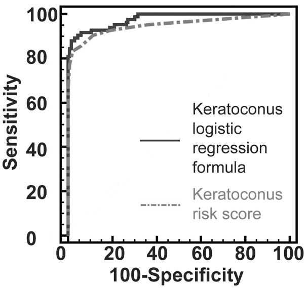
The diagnostic power of the pachymetric variables, keratoconus risk score, and logistic regression formula were listed in Table 5. For keratoconus group, all individual variables had fair to good diagnostic power to detect keratoconus eyes with AROC (0.818–0.942). The logistic regression formula had the best overall diagnostic power with AROC of 0.975 (Figure 3). The second best was the keratoconus risk score with AROC of 0.949 (Figure 3). The five variables OR logic gave an AROC of 0.935.
Table 5.
Diagnostic Accuracy of Optical Coherence Tomography Pachymetric Single Variables and Composite Methods in Keratoconus Detection.
| Minimum | Minimum-Median | S-I | SN-IT | Ymin | Five Variables OR Logic | Keratoconus Risk Score | Logistic Regression Formula | |
|---|---|---|---|---|---|---|---|---|
| Sensitivity at 95% Specificity | 72.6% | 82.1% | 57.1% | 76.1% | 36.9% | 70.2% | 85.7% | 90.5% |
| AROC | 0.942 | 0.925 | 0.839 | 0.896 | 0.818 | 0.935 | 0.949 | 0.975 |
S-I = superior-inferior, SN-IT = superonasal-inferotemporal, Ymin = vertical location of the Minimum, AROC = area under the receiver operative characteristic curve.
Figure 3.
Diagnostic analysis of the keratoconus logistic regression formula and the keratoconus risk score. The keratoconus logistic regression formula had an area under the receiver operating characteristic curve (AROC) value of 0.975. The keratoconus risk score had an AROC value of 0.949 (P = 0.08).
Diagnostic power with AROC were compared between single OCT pachymetric variables, keratoconus risk score, and the logistic regression formula. No statistic significant difference was detected comparing keratoconus risk score with minimum (P = 0.65), minimum-median (P = 0.20) and the logistic regression formula (P = 0.08). The logistic regression formula had statistic significant better diagnostic power with AROC than all single OCT pachymetric variables (P<0.01).
DISCUSSION
The OCT system is capable of imaging both the anterior and posterior surfaces of the cornea. The OCT pachymetric measurements were accurate and highly repeatable. The Fourier-domain OCT system used in this study was capable of a speed of 26,000 axial scans per second, which is more than 10 times faster than the time-domain OCT systems based on original OCT technology. The 840 nm Fourier-domain OCT system also has an axial resolution of 5 μm, which is more than 3 times better than a conventional 1310 nm time-domain OCT system. Faster scan speed can reduce data acquisition time, minimize eye movement during the scan, and improve repeatability of pachymetric measurements. In our study, we measured intra-session repeatability of minimum corneal thickness, which was 1.6 μm in the normal group and 3.3 μm in the keratoconus group. This was better than the intra-session repeatability of central corneal thickness (CCT) reported by Li et al of 4.9 μm and 5.8 μm using 2 types of time-domain OCT.28 Our results were similar to the result of a previous Fourier-domain OCT study which reported an intra-session repeatability of 1.3 μm for CCT.23
The 2 possible landmarks for centering the corneal map scans are the corneal vertex and the pupil. In this study, we used pupil as centering landmarks for 2 reasons. First, the corneal vertex can be altered by shape change of the corneal in keratoconic eyes. In keratoconus, the ectasia is usually inferotemporal; therefore, the corneal vertex is also shifted inferotemporally. Thus, using the pupil as the center might better reveal asymmetric thinning in keratoconus.20 Second, in our previous study, we found that pupil centration gave better repeatability than vertex centration.23
Our group first proposed using OCT pachymetric-based variables for keratoconus screening.20 Corneal thickness had been proposed to be a useful parameter for the clinical identification of keratoconus.29–31 Studies using ultrasound or slit scanning technologies have found that the difference (or ratio) between the peripheral and the thinnest (or central) corneal thickness was significantly greater in eyes with keratoconus than in normal eyes.14, 32–34
In this study, we used 5 pachymetric variables representing different characteristics of keratoconus as follows: general thinning: minimum; focal thinning: minimum-median; asymmetric thinning: S-I, SN-IT, and Ymin. By combining these pachymetric variables into one formula, we were able to improve the diagnostic power with AROC to 0.975. According to literature, AROC values of the most widely studied topography-based KISA% for keratoconus diagnosis is 0.91.20 There were various studies applying pachymetric variables in detecting keratoconus. Li et al. used the diagnostic criteria of any 1 OCT pachymetric parameter below the keratoconus cutoff and yielded an AROC of 0.99.20 Ambrosio et al. introduced various Scheimpflug system based pachymetric variables to differentiate normal and keratoconic corneas and yielded an AROC of 0.987.14 Saad et al. applied indices generated from corneal thickness and curvature measurements and the discriminant function reached an AROC of 0.99.16 Our method gave very high accuracy in keratoconus screening. The AROC of logistic regression formula was better than all five single OCT pachymetric variables, proving that the combined formula improved the diagnostic power of keratoconus detection. In our previous study, we used 1 abnormal and 2 abnormal criteria to diagnose keratoconus.20 The 5 variables OR logic used in this study was a modification of the 1 abnormal criteria of the previous study. In this study, the 5 variables OR logic (AROC = 0.935) worked comparable well to the keratoconus risk scoring system table (AROC = 0.949) but not as good as the logistic regression formula (AROC = 0.975). This is probably because keratoconic corneas may have changes characterized by all 5 pachymetric variables, rather than only 1 or 2 of them.
The best overall single pachymetric variable for detecting keratoconus risks was minimum (AROC = 0.942). The second best variable was minimum-median (AROC = 0.925). This verified that focal thinning and corneal thinning are important characteristics of keratoconus. Variables of asymmetric thinning (S-I 0.839 and SN-IT 0.896) had lower diagnostic accuracy in this study. It may due to the difficulties of accurately centering the OCT scan on keratoconic eyes. A limitation of the OCT pachymetry scan was that the pupil position was not recorded. Simultaneous capture of the pupil position would be a significant improvement since this would allow more accurate centration of the pachymetry map on the pupil center in post-processing, which would in turn improve the accuracy of the asymmetric thinning indices.
Early detection of keratoconus is important for refractive surgery pre-operative screening. In this study we designed a keratoconus risk scoring system table based on corneal pachymetric characteristics of normal subjects for clinical evaluation. The table had similar keratoconus detection power compared with the logistic regression formula according to our data. By using this table, we were able to identify 91.7% of the keratoconus subjects.
Overall, the keratoconus risk scoring table and logistic regression formula both improved sensitivity in diagnosing keratoconus. The disadvantage of the logistic regression formula is that it was optimized for the particular group of keratoconus subjects involved in the study. The performance of the logistic regression formula needs to be further evaluated with another group of keratoconus subjects (evaluation group). In comparison, the keratoconus risk scoring table has the advantage that it was simply based on the normal population average measurements. The pachymetric variations among normal subjects are much less than that of the keratoconus. Therefore, we expect the keratoconus risk scoring table to provide more robust performance than the logistic regression formula.
There were several other limitations for this study. First, the OCT system used in this study provides a limited scan diameter up to 6 mm. Thus, this corneal thickness change outside of 6 mm diameters will be missed. This may limit the sensitivity of the indices in detecting forme fruste keratoconus (FFK). Second, although good accuracy on keratoconus diagnosis was shown, the diagnosis power for FFK still needs to be improved. Not presented in this paper, our results showed that OCT pachymetry map based FFK diagnosis was not sufficient. Other pachymetric and topographic information, such as corneal epithelial thickness and posterior corneal curvature, may be combined with whole corneal thickness information to improve FFK and keratoconus diagnoses. We are conducting other studies on those aspects. By combining various information of the cornea, we might be able to improve the diagnosis power of FFK to a higher level.
In summary, we showed that OCT pachymetry map based analysis could detect abnormal corneal thinning in keratoconus eyes. By combining pachymetric variables into one formula, we were able to improve the diagnosis power for keratoconus. A clinical evaluation table scoring keratoconus risks might facilitate clinicians to screen keratoconus.
WHAT WAS KNOWN
Corneal topography, optical coherence tomography corneal pachymetry map, and several other methods can help diagnosing and screening keratoconus.
WHAT THIS PAPER ADDS
A formula combining various corneal pachymetric variables can further improve keratoconus diagnosis. A keratoconus screening system was developed for potential clinical evaluation of keratoconus risk.
Acknowledgments
Financial support:
This study was supported by NIH grant R01 EY018184 and a research grant from Optovue Inc.
Footnotes
This study was presented at the American Society of Cataract and Refractive Surgery (ASCRS) annual meeting, San Diego, California, USA. March 2011.
Financial and proprietary interest:
Oregon Health and Science University (OHSU), David Huang, Yan Li, and Maolong Tang have a significant financial interest in Optovue, Inc. (Fremont, CA, USA), a company that may have a commercial interest in the results of this research and technology. These potential conflicts of interest have been reviewed and managed by OHSU. Robert Brass receives speaker honoraria from Optovue, Inc. Bing Qin, Shihao Chen, Qinmei Wang, Xinbo Zhang and Xiaoyu Wang have no proprietary interest in the topic of this manuscript.
References
- 1.Rabinowitz YS. Keratoconus. Surv Ophthalmol. 1998;42:297–319. doi: 10.1016/s0039-6257(97)00119-7. [DOI] [PubMed] [Google Scholar]
- 2.Binder PS. Risk factors for ectasia after LASIK. J Cataract Refract Surg. 2008;34:2010–1. doi: 10.1016/j.jcrs.2008.08.035. [DOI] [PubMed] [Google Scholar]
- 3.Randleman JB, Trattler WB, Stulting RD. Validation of the Ectasia Risk Score System for preoperative laser in situ keratomileusis screening. Am J Ophthalmol. 2008;145:813–8. doi: 10.1016/j.ajo.2007.12.033. [DOI] [PMC free article] [PubMed] [Google Scholar]
- 4.Rabinowitz YS. Videokeratographic indices to aid in screening for keratoconus. J Refract Surg. 1995;11:371–9. doi: 10.3928/1081-597X-19950901-14. [DOI] [PubMed] [Google Scholar]
- 5.Varssano D, Kaiserman I, Hazarbassanov R. Topographic patterns in refractive surgery candidates. Cornea. 2004;23:602–7. doi: 10.1097/01.ico.0000121699.74077.f0. [DOI] [PubMed] [Google Scholar]
- 6.Ambrosio R, Jr, Klyce SD, Wilson SE. Corneal topographic and pachymetric screening of keratorefractive patients. J Refract Surg. 2003;19:24–9. doi: 10.3928/1081-597X-20030101-05. [DOI] [PubMed] [Google Scholar]
- 7.Li X, Rabinowitz YS, Rasheed K, Yang H. Longitudinal study of the normal eyes in unilateral keratoconus patients. Ophthalmology. 2004;111:440–6. doi: 10.1016/j.ophtha.2003.06.020. [DOI] [PubMed] [Google Scholar]
- 8.Randleman JB, Woodward M, Lynn MJ, Stulting RD. Risk assessment for ectasia after corneal refractive surgery. Ophthalmology. 2008;115:37–50. doi: 10.1016/j.ophtha.2007.03.073. [DOI] [PubMed] [Google Scholar]
- 9.Cheng HC, Lin KK, Chen YF, Hsiao CH. Pseudokeratoconus in a patient with soft contact lens-induced keratopathy: assessment with Orbscan I. J Cataract Refract Surg. 2004;30:925–8. doi: 10.1016/j.jcrs.2003.08.016. [DOI] [PubMed] [Google Scholar]
- 10.Doyle SJ, Hynes E, Naroo S, Shah S. PRK in patients with a keratoconic topography picture. The concept of a physiological ‘displaced apex syndrome’. Br J Ophthalmol. 1996;80:25–8. doi: 10.1136/bjo.80.1.25. [DOI] [PMC free article] [PubMed] [Google Scholar]
- 11.Lim L, Wei RH, Chan WK, Tan DT. Evaluation of keratoconus in Asians: role of Orbscan II and Tomey TMS-2 corneal topography. Am J Ophthalmol. 2007;143:390–400. doi: 10.1016/j.ajo.2006.11.030. [DOI] [PubMed] [Google Scholar]
- 12.Ambrosio R, Jr, Dawson DG, Salomao M, Guerra FP, Caiado AL, Belin MW. Corneal ectasia after LASIK despite low preoperative risk: tomographic and biomechanical findings in the unoperated, stable, fellow eye. Journal of refractive surgery. 2010;26:906–11. doi: 10.3928/1081597X-20100428-02. [DOI] [PubMed] [Google Scholar]
- 13.Ambrosio R, Jr, Alonso RS, Luz A, Coca Velarde LG. Corneal-thickness spatial profile and corneal-volume distribution: tomographic indices to detect keratoconus. Journal of cataract and refractive surgery. 2006;32:1851–9. doi: 10.1016/j.jcrs.2006.06.025. [DOI] [PubMed] [Google Scholar]
- 14.Ambrosio R, Jr, Caiado AL, Guerra FP, Louzada R, Roy AS, Luz A, Dupps WJ, Belin MW. Novel pachymetric parameters based on corneal tomography for diagnosing keratoconus. Journal of refractive surgery. 2011;27:753–8. doi: 10.3928/1081597X-20110721-01. [DOI] [PubMed] [Google Scholar]
- 15.Buhren J, Kook D, Yoon G, Kohnen T. Detection of subclinical keratoconus by using corneal anterior and posterior surface aberrations and thickness spatial profiles. Investigative ophthalmology & visual science. 2010;51:3424–32. doi: 10.1167/iovs.09-4960. [DOI] [PubMed] [Google Scholar]
- 16.Saad A, Gatinel D. Topographic and tomographic properties of forme fruste keratoconus corneas. Investigative ophthalmology & visual science. 2010;51:5546–55. doi: 10.1167/iovs.10-5369. [DOI] [PubMed] [Google Scholar]
- 17.Khurana RN, Li Y, Tang M, Lai MM, Huang D. High-speed optical coherence tomography of corneal opacities. Ophthalmology. 2007;114:1278–85. doi: 10.1016/j.ophtha.2006.10.033. [DOI] [PubMed] [Google Scholar]
- 18.Szalai E, Berta A, Hassan Z, Modis L., Jr Reliability and repeatability of swept-source Fourier-domain optical coherence tomography and Scheimpflug imaging in keratoconus. Journal of cataract and refractive surgery. 2012;38:485–94. doi: 10.1016/j.jcrs.2011.10.027. [DOI] [PubMed] [Google Scholar]
- 19.Li Y, Shekhar R, Huang D. Corneal pachymetry mapping with high-speed optical coherence tomography. Ophthalmology. 2006;113:792–9. e2. doi: 10.1016/j.ophtha.2006.01.048. [DOI] [PMC free article] [PubMed] [Google Scholar]
- 20.Li Y, Meisler DM, Tang M, Lu AT, Thakrar V, Reiser BJ, Huang D. Keratoconus diagnosis with optical coherence tomography pachymetry mapping. Ophthalmology. 2008;115:2159–66. doi: 10.1016/j.ophtha.2008.08.004. [DOI] [PMC free article] [PubMed] [Google Scholar]
- 21.Christopoulos V, Kagemann L, Wollstein G, Ishikawa H, Gabriele ML, Wojtkowski M, Srinivasan V, Fujimoto JG, Duker JS, Dhaliwal DK, Schuman JS. In vivo corneal high-speed, ultra high-resolution optical coherence tomography. Archives of ophthalmology. 2007;125:1027–35. doi: 10.1001/archopht.125.8.1027. [DOI] [PMC free article] [PubMed] [Google Scholar]
- 22.Sarunic MV, Asrani S, Izatt JA. Imaging the ocular anterior segment with real-time, full-range Fourier-domain optical coherence tomography. Archives of ophthalmology. 2008;126:537–42. doi: 10.1001/archopht.126.4.537. [DOI] [PMC free article] [PubMed] [Google Scholar]
- 23.Li Y, Tang M, Zhang X, Salaroli CH, Ramos JL, Huang D. Pachymetric mapping with Fourier-domain optical coherence tomography. J Cataract Refract Surg. 2010;36:826–31. doi: 10.1016/j.jcrs.2009.11.016. [DOI] [PMC free article] [PubMed] [Google Scholar]
- 24.Binder PS, Lindstrom RL, Stulting RD, Donnenfeld E, Wu H, McDonnell P, Rabinowitz Y. Keratoconus and corneal ectasia after LASIK. Journal of cataract and refractive surgery. 2005;31:2035–8. doi: 10.1016/j.jcrs.2005.12.002. [DOI] [PubMed] [Google Scholar]
- 25.Warmke J, Drysdale R, Ganetzky B. A distinct potassium channel polypeptide encoded by the Drosophila eag locus. Science. 1991;252:1560–2. doi: 10.1126/science.1840699. [DOI] [PubMed] [Google Scholar]
- 26.Zeger SL, Liang KY. Longitudinal data analysis for discrete and continuous outcomes. Biometrics. 1986;42:121–30. [PubMed] [Google Scholar]
- 27.Liang KY, Zeger SL. Longitudinal data analysis using generalized linear models. Biometrika. 1986;73:13–22. [Google Scholar]
- 28.Li H, Leung CK, Wong L, Cheung CY, Pang CP, Weinreb RN, Lam DS. Comparative study of central corneal thickness measurement with slit-lamp optical coherence tomography and visante optical coherence tomography. Ophthalmology. 2008;115:796–801. e2. doi: 10.1016/j.ophtha.2007.07.006. [DOI] [PubMed] [Google Scholar]
- 29.Haque S, Simpson T, Jones L. Corneal and epithelial thickness in keratoconus: a comparison of ultrasonic pachymetry, Orbscan II, and optical coherence tomography. J Refract Surg. 2006;22:486–93. doi: 10.3928/1081-597X-20060501-11. [DOI] [PubMed] [Google Scholar]
- 30.Gherghel D, Hosking SL, Mantry S, Banerjee S, Naroo SA, Shah S. Corneal pachymetry in normal and keratoconic eyes: Orbscan II versus ultrasound. J Cataract Refract Surg. 2004;30:1272–7. doi: 10.1016/j.jcrs.2003.11.049. [DOI] [PubMed] [Google Scholar]
- 31.Ucakhan OO, Ozkan M, Kanpolat A. Corneal thickness measurements in normal and keratoconic eyes: Pentacam comprehensive eye scanner versus noncontact specular microscopy and ultrasound pachymetry. J Cataract Refract Surg. 2006;32:970–7. doi: 10.1016/j.jcrs.2006.02.037. [DOI] [PubMed] [Google Scholar]
- 32.Avitabile T, Marano F, Uva MG, Reibaldi A. Evaluation of central and peripheral corneal thickness with ultrasound biomicroscopy in normal and keratoconic eyes. Cornea. 1997;16:639–44. [PubMed] [Google Scholar]
- 33.Gromacki SJ, Barr JT. Central and peripheral corneal thickness in keratoconus and normal patient groups. Optom Vis Sci. 1994;71:437–41. doi: 10.1097/00006324-199407000-00003. [DOI] [PubMed] [Google Scholar]
- 34.Pflugfelder SC, Liu Z, Feuer W, Verm A. Corneal thickness indices discriminate between keratoconus and contact lens-induced corneal thinning. Ophthalmology. 2002;109:2336–41. doi: 10.1016/s0161-6420(02)01276-9. [DOI] [PubMed] [Google Scholar]


