Abstract
The objective of this study was to evaluate a novel accelerometer-based sensor system, the Walkabout Portable Gait Monitor (WPGM), for use in kinetic gait analysis of dogs. The accelerometer was compared to the common reference standard of force platform analysis. Fifteen client-owned, orthopedically sound dogs of various breeds underwent simultaneous force platform and accelerometer gait trials to measure peak vertical forces (PVFs). The agreement between PVF for the accelerometer and force platform was measured using concordance correlation coefficient (CCC) and was found, overall, to be moderate [CCC = 0.51; 95% confidence interval (CI): 0.46 to 0.56]. The agreement between PVF for the accelerometer and force platform for the forelimbs was positive and substantial (CCC = 0.79; 95% CI: 0.74 to 0.84) and for the hind limbs was positive and low (CCC = 0.34; 95% CI: 0.29 to 0.38). As measured by the accelerometer, PVF was systematically higher than as measured by the force platform (forelimbs 55.3 N, hind limbs 144.3 N). It was also found that, when positioned over the lumbar spine, the WPGM cannot measure PVF of the individual forelimbs and hind limbs, which limits its use as a clinical tool to measure kinetic variables in dogs.
Résumé
L’objectif de la présente étude était d’évaluer un nouveau système d’accéléromètre, le Walkabout Portable Gait Monitor (WPGM), pour utilisation dans l’analyse de la cinétique de l’allure chez des chiens. L’accéléromètre fut comparé au standard de référence habituel qu’est l’analyse par plaque de force. Quinze chiens de races variées appartenant à des clients, et sans évidence d’atteinte orthopédique ont été soumis de manière simultanée à un test de plaque de force et une étude de la cinétique de l’allure afin de mesurer les forces verticales maximales (PVF). L’accord entre la PVF pour l’accéléromètre et la plaque de force fut mesuré en utilisant le coefficient de corrélation de concordance (CCC) et fut trouvé, de manière globale, à être modéré [CCC = 0,51 %; intervalle de confiance (CI) 95 % : 0,46 à 0,56]. L’accord entre la PVF pour l’accéléromètre et la plaque de force pour les membres antérieurs était positif et élevé (CCC = 0,79; 95 % CI : 0,74 à 0,84) et pour les membres postérieurs était positif et faible (CCC = 0,34; 95 % CI : 0,29 à 0,38). Tel que mesuré par l’accéléromètre, la PVF était systématiquement supérieure à celle mesurée par la plaque de force (membres antérieurs 55,3 N, membres postérieurs 144,3 N). Il fut également trouvé que, lorsque positionné par-dessus la colonne lombaire, le WPGM ne peut mesuré la PVF des membres antérieurs pris individuellement et des membres postérieurs, ce qui limite son utilisation comme outil clinique pour mesurer des variables cinétiques chez les chiens.
(Traduit par Docteur Serge Messier)
Introduction
Gait analysis is used for evaluating lameness, monitoring its progression, and assessing response to surgical and medical therapies (1). The most common method used by veterinarians to diagnose canine orthopedic injuries and monitor disease progression is subjective visual observation. However, the human ability to reliably measure the degree of lameness in a patient and to perceive subtle changes during ambulation is limited (2,3). To accurately and reliably assess a dog’s gait, veterinarians benefit from the use of objective and reproducible gait analysis tools.
Objective gait analysis in dogs is broadly split into kinematic gait analysis and kinetic gait analysis (4,5). Kinematic gait analysis quantifies the motion of the gait cycle, while kinetic gait analysis quantifies the force of the gait cycle (1,6). Force platform analysis and pressure walkway system analysis are the most common types of kinetic gait analysis tools, both of which have been assessed for use in dogs (4–9).
Objective gait analysis systems are not often used in a private practice setting due to the cost of specialized equipment, space allocation, and the training required for their use (1,7). The ideal gait analysis system should provide reliable and accurate data, be sensitive enough to identify subtle gait changes, not alter the gait of the patient, provide gait analysis at different speeds of locomotion, be simple and quick to use, have reasonable operating costs and user-friendly software, and produce comprehensive data (10).
Force platform analysis has been studied more extensively than pressure platform analysis. It measures forces in 3 planes: vertical, craniocaudal, and mediolateral. Vertical forces measured by force platform analysis have been shown to be more reliable for characterizing gait and detecting lameness than craniocaudal and mediolateral forces (4,5,11). Furthermore, peak vertical force (PVF) has been shown to be the most accurate single-variable indicator of lameness (2,11).
Accelerometers are small, lightweight devices that are attached to the body to record the position and acceleration of the body in space. These acceleration measurements can be used to assess forces acting on the body, since force is a function of acceleration and body mass. Currently, accelerometry in dogs has been limited to spontaneous activity monitoring (12,13). Activity monitoring has been used to objectively determine responses to medical therapy and to estimate the energy expenditure of dogs (14–17). The use of accelerometry for objective gait analysis in dogs offers great potential in clinical practice and, to the authors’ knowledge, has not been previously investigated.
The Walkabout Portable Gait Monitor (WPGM) is a triaxial accelerometer that has been previously evaluated for use in humans (18–20). In humans, accelerometers are strapped around the lumbar or thoracic spine and require only 20 s for data collection (18,20–25). Trunk accelerometry is an area of gait analysis that is rapidly growing in human medicine (21–26). Previous studies on the WPGM have focused primarily on its kinematic properties, but it is also capable of providing kinetic information (18–20). The WPGM has been used to evaluate outcomes following surgery, such as total knee arthroplasty (18), to quantify the degree of lameness in patients awaiting total hip arthroplasty (20), and to evaluate morbidity associated with procedures such as free fibular grafts (19). The advantages of the WPGM include its mobility, convenient setup, rapid data collection, user-friendly software, and the economical cost of purchase and operation.
The objective of this study was to evaluate the agreement of the kinetic data provided by the WPGM compared to the common reference standard of force platform analysis. We hypothesized that the PVF from the WPGM would be equivalent to the PVF measured by the force platform.
Materials and methods
Participating dogs
Eligible dogs for study inclusion consisted of healthy, orthopedically sound dogs of any breed, weighing more than 18 kg and older than 12 mo of age. Dogs on non-steroidal anti-inflammatory drugs, steroids, or oral nutraceuticals (glucosamine, chondroitin sulfate, omega-3 fatty acids) were not selected. The dogs were screened for orthopedic and neurologic diseases based on subjective gait evaluation, physical examination, and radiographic examination of the elbows, hips, and stifles. Dogs with evidence of orthopedic or neurologic disease were excluded from the study. Dogs that were pacing during the trials, dogs unable to produce valid force platform trials (due to anxiety, pulling on the leash, not stepping on the platform), dogs that had invalid (calibration error) or atypical accelerometer graphs, and dogs with a symmetry index of > 6% were also excluded. The study protocol was approved by the Institutional Animal Care and Use Committee (Protocol #12-004) in compliance with the Canadian Council on Animal Care guidelines. Informed owner consent was obtained.
Peak vertical force measurements
Accelerometer
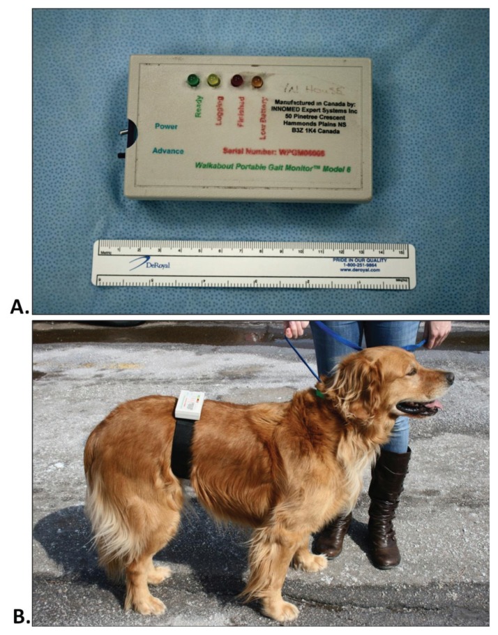
The WPGM (Model 6; INNOMED Expert Systems, Bedford, Nova Scotia) uses a triaxial capacitor-based accelerometer system. The sensors and hardware are mounted in a 67-mm × 115-mm × 25-mm pack weighing 166 g and are powered by a 9-VDC alkaline battery (Figure 1A). The sensor pack is dorsally mounted over the thoracic or lumbar area and secured by a Velcro strap (Figure 1B). The WPGM records acceleration in 3 planes: mediolateral (X), craniocaudal (Y), and vertical (Z). The gait is sampled at a rate of 200 hertz (Hz) with a range of ± 5 g. The data are digitized and temporarily stored on a Secure Digital (SD) memory card. The data are then transferred to a dedicated computer software program (Gaitview 2007, Version 1.0.3.8; INNOMED Expert Systems) for storage and processing.
Figure 1.
Walkabout Portable Gait Monitor (WPGM) alone (A) and positioned over the mid-lumbar spine of a dog (B). This is a triaxial accelerometer system that measures acceleration in the 3 cardinal planes and is used to obtain kinetic and kinematic gait data.
Force platform
The force plate analysis was carried out using a biomechanical platform (OR6-7; Advanced Medical Technology, Watertown, Massachusetts, USA) embedded in, and level with, a 12-m runway. To determine the velocity and acceleration across the force platform, 3 sets of polarized retro-photoelectric cell sensors (MEK92-PAD; Sircon Controls, Mississauga, Ontario) were positioned adjacent to the walkway, 1 m apart, with the middle sensor at the mid-level of the force plate. The data from the platform was processed and stored in real time using a dedicated computer and software program (Acquire, Version 7.35S; Sharon Software, Dewett, Michigan, USA). The dogs were trotted across the force platform at a velocity of 2 ± 0.3 m/s, with a maximum horizontal acceleration of ± 0.5 m/s2. Trials were considered valid when both the ipsilateral forelimbs and hind limbs struck the center of the plate without traction on the leash.
Data collection and processing
The body weight of each dog was recorded in kilograms on the same electronic scale. Force platform and WPGM measurements were conducted in parallel. The same handler (KC) was used for all trials. The WPGM belt was placed snugly around the mid-abdomen and the dogs were allowed to acclimatize to the WPGM, force platform, and handler for 30 min (Figure 1B). All trials were recorded by an analog video camera (Canon HV20; Canon USA, Lake Success, New York, USA). The WPGM was switched on at the start of the runway, the record button was pressed, and the device was sharply tapped (“tap time”) in a vertical direction before the dog moved. The dog was then trotted over the force platform while the accelerometer was recording. The first 5 valid force platform trials were collected for all 4 limbs.
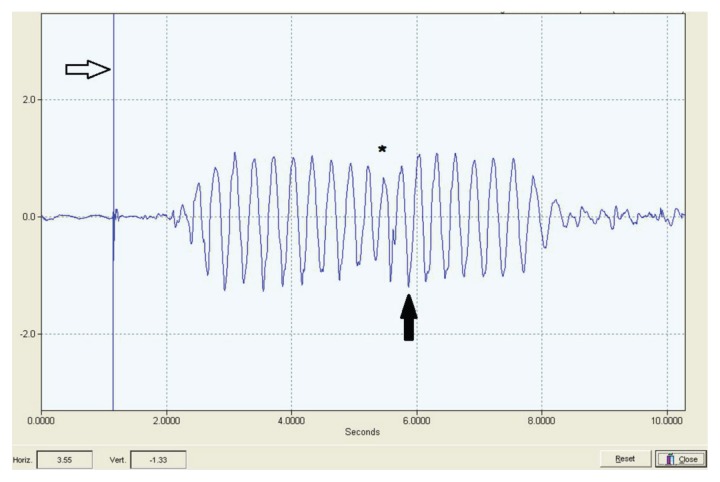
The time of measurement was synchronized with the aid of the video. Briefly, the analog video was digitized and a time-overlay was added to the video (Pinnacle Studio Ultimate 12; Pinnacle Systems, Mountain View, California, USA). A media player (Windows Media Player 2007; Microsoft, Redmond, Washington, USA) was used to record the “tap time” and the “strike time” of the paw on the force plate, and generate a “delta time” or time difference between the 2 events. The “tap time” could be identified on the WPGM acceleration graph by a sharp vertical peak (Figure 2). The subsequent paw-strike on the force platform was then identified on the accelerometer graph by adding the “delta time” to the “tap time”. This method enabled the fore and hind paw-strikes on the force platform to be identified on the accelerometer graph.
Figure 2.
Graph of Dog #10 during the trot, demonstrating the typical waveform of acceleration (m/s2) in the vertical direction (y-axis) over time (x-axis; s). A sharp spike is seen (hollow arrow) indicating the “tap time”, which was used as a reference point to determine which paw-strike occurred on the force platform. Peak vertical acceleration in a downward direction was used to calculate peak vertical force (solid arrow). It can be seen that the dog decelerates as it approaches the force platform (asterisk).
Mean velocity (m/s), acceleration (m/s2), and peak vertical force (N) were determined from the force platform. From the WPGM, the peak acceleration for contralateral forelimbs and hind limbs for the vertical axis was recorded. Vertical forces (N) were then calculated from the peak vertical acceleration and body weight of the dog using the formula (Newton’s second law of motion):
Using the PVF from the force platform analysis, symmetry indices were calculated for the forelimbs and hind limbs (6). Dogs with a symmetry index of ≤ 6% were considered orthopedically sound (6).
Statistical analysis
The agreement between the measured peak vertical forces (PVFs) from the force platform and the WPGM was assessed using conventional descriptive and analytical approaches (27). Descriptively, concordance plots (28) and Bland-Altman limits-of-agreement plots (29) were generated. Concordance plots were used to assess the dispersal and alignment of paired measurements around a 45-degree line representing the reference line of perfect concordance. A regression line for the observed measurements was added to assess potential systematic deviation from the reference line. The dispersal of the paired measurements about the regression line was used to assess the degree of imprecision of the data; data points close to the regression line indicate better precision. The Bland-Altman limits-of-agreement plots were used to assess the evolution of the differences between paired PVF measurements of the 2 methods (y-axis) according to the magnitude of the PVF measured (x-axis). This plot was useful for determining whether the amplitude of the measured force alters the agreement.
Analytically, the agreement between the PVF of the force platform and that of the WPGM was quantified using the Lin’s concordance correlation coefficient (CCC) (28). The CCC ranges from −1 to 1 and a CCC of 1 indicates a perfect agreement. It combines the bias-correction factor, Cb, which reflects the accuracy of the data, and the Pearson correlation coefficient, r, which reflects the precision of the data. Both Cb and r range from −1 to 1 and the closer they are to 1, the better the accuracy and the precision, respectively. The CCC is computed as the product of Cb and r, and a CCC of lower value (away from 1) may indicate a lack of precision, a lack of accuracy, or both. For example, a set of data may show excellent precision, i.e., r estimate very close to 1 and paired observations show little dispersal around a straight regression line, but poor accuracy, i.e., Cb away from 1 and the regression line diverts from the perfect concordance line by its slope and/or intercept, and would result in poor agreement, i.e., CCC away from 1. In addition, 95% limits of agreement were estimated to assess whether the average difference between the 2 methods was significant (29).
The consistency of PVF measurements when measuring the same limb across 5 trials was estimated for the force platform and accelerometer using the repeatability coefficient (RC). The RC is interpreted as the maximum likely differences, i.e., 95%, observed between pairs of measured PVF using the same method for the same limb. The repeatability coefficient was then computed using the following formula (27):
where σi 2 is the variance of PVF across trials for the same limb. The within-limb variance, σi 2, was estimated using a hierarchical model without an explanatory variable and with 3 levels: dog, limbs within a dog, and trials within a limb. All the analyses were conducted using the statistical package Stata 12.0 (StataCorp, College Station, Texas, USA).
Results
Twenty-four dogs met the inclusion criteria and were enrolled in the study. Nine dogs were excluded from data analysis. Of these, 3 dogs were excluded because they were unable to complete valid force platform trials due to pacing (n = 1) and anxiety (n = 2). It was also determined that 1 of these dogs was lame on the day of the trial. Two dogs were excluded for a symmetry index greater than 6%, 2 dogs were excluded due to a calibration error of the accelerometer, and 1 dog was excluded because the trial numbers were incorrect and the force platform trials could not be matched to the accelerometer trials. One dog was excluded because the accelerometric gait pattern was atypical, which was likely due to a slight time delay between the contact time of the forelimb and the hind limb.
In total, force platform and accelerometer data were available for statistical analysis for 15 dogs. The study population included 7 neutered male dogs and 8 spayed female dogs. The median age was 4.3 y (from 1.0 to 8.6 y), the median body weight was 27 kg (from 18 to 47 kg), and the median body condition score was 3.0/5.0 (from 3.0 to 4.0). Ten breeds were represented, including standard poodle (n = 3), greyhound (n = 2), Border collie cross (n = 2), Labrador cross (n = 2), and 1 each of golden retriever, samoyed, Newfoundland, German shepherd cross, boxer cross, and Dogue de Bordeaux cross.
The estimated average difference and concordance correlation coefficient (CCC) between peak vertical forces as measured by the accelerometer (PVFAcc) and the peak vertical forces measured by the force platform (PVFFP) for various subsets of the data are summarized in Table I. On average, PVFAcc were approximately 100 N higher than PVFFP, but this difference did not differ significantly from zero (95% limits of agreement: −24.0 to 134.6).
Table I.
Summary table of the agreement between the peak vertical forces as measured by the force platform and accelerometer for the overall data set, each individual limb, each side (right and left), and forelimbs and hind limbs for 15 dogs. Estimated parameters include the difference average, the 95% limits of agreement, and the concordance correlation coefficient (CCC), which is the product of the Pearson’s correlation coefficient (r) and the bias-correction factor (Cb)
| Data subset | Observations | Difference average (N) | 95% Limits of agreement |
CCC (r × Cb) | 95% CI | r (precision) | Cb (accuracy) |
|---|---|---|---|---|---|---|---|
| Overall | 300 | 99.806 | −30.105–229.717 | 0.509 | 0.455–0.564 | 0.78 | 0.653 |
| Trial | |||||||
| Trial #1 | 60 | 106.524 | −22.879–235.927 | 0.466 | 0.346–0.587 | 0.765 | 0.609 |
| Trial #2 | 60 | 99.578 | −30.042–229.198 | 0.509 | 0.386–0.631 | 0.783 | 0.65 |
| Trial #3 | 60 | 95.441 | −30.044–220.926 | 0.541 | 0.409–0.651 | 0.799 | 0.677 |
| Trial #4 | 60 | 98.052 | −28.384–224.489 | 0.537 | 0.417–0.656 | 0.805 | 0.667 |
| Trial #5 | 60 | 99.435 | −41.939–240.808 | 0.494 | 0.364–0.625 | 0.749 | 0.66 |
| Side | |||||||
| Right | 150 | 101.387 | −41.064–243.837 | 0.472 | 0.390–0.555 | 0.734 | 0.643 |
| Left | 150 | 98.225 | −18.204–214.655 | 0.546 | 0.475–0.618 | 0.825 | 0.663 |
| Location | |||||||
| Front | 150 | 55.285 | −23.999–134.569 | 0.787 | 0.738–0.835 | 0.925 | 0.851 |
| Back | 150 | 144.327 | 33.645–255.009 | 0.336 | 0.292–0.380 | 0.879 | 0.382 |
| Limb | |||||||
| Right forelimb | 75 | 50.009 | −22.608–122.625 | 0.806 | 0.741–0.872 | 0.927 | 0.87 |
| Left forelimb | 75 | 60.561 | −24.097–145.218 | 0.77 | 0.699–0.841 | 0.926 | 0.832 |
| Right hindlimb | 75 | 152.764 | 30.165–275.364 | 0.316 | 0.255–0.376 | 0.888 | 0.356 |
| Left hindlimb | 75 | 135.89 | 40.593–231.187 | 0.361 | 0.299–0.423 | 0.888 | 0.407 |
The overall agreement between PVFAcc and PVFFP was moderate (CCC = 0.51; 95% CI: 0.46 to 0.56), with substantial precision (Pearson’s correlation coefficient: r = 0.78) and moderate accuracy (bias-correction factor, Cb = 0.653). The repeatability coefficient (RC) was more than twice as high for PVFAcc (RC = 77.0; 95% CI: 70.4 to 84.2) than for PVFFP (RC = 31.8; 95% CI: 29.1 to 34.6), indicating that the accelerometer is significantly less repeatable and, therefore, less precise than PVFs measured by the force platform.
The agreement between the 2 methods did not appear to change substantially across trials and when comparing left and right sides of the dog (Table I). However, the agreement between the 2 methods was greater when measuring forelimbs than when measuring hind limbs (CCC = 0.79 and 0.34, respectively). While the precision of PVFACC and PVFFP was high for both the forelimbs (r = 0.93) and the hind limbs (r = 0.88), the accuracy in the forelimbs was substantially higher than in the hind limbs (Cb = 0.85 and 0.38, respectively), which resulted in better agreement in the forelimbs than in the hind limbs. On average, the PVFAcc were 144.3 N higher for the hind limbs and only 55.3 N higher for the forelimbs (Table I).
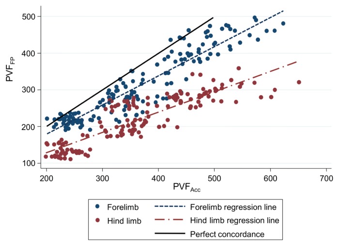
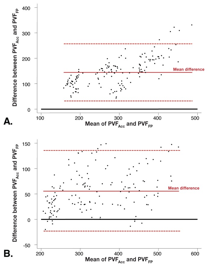
The concordance plot (Figure 3) illustrates the difference in agreement between forelimbs and hind limbs. The regression line (related to the accuracy) for the hind limbs is much diverted (both slope and intercept) relative to the 45-degree reference line of perfect concordance when compared to the regression line of the forelimbs. However, the dispersal of the data (indicator of precision) about the regression lines does not appear to be different between forelimbs and hind limbs, which reflects the similar estimated Pearson’s “r”. The Bland-Altman limits-of-agreement plots revealed that for the hind limbs the disagreement between the 2 methods increases when PVF increases (Figure 4A). This trend was not apparent for the forelimbs (Figure 4B). The average PVFAcc did not differ much between forelimbs and hind limbs (366.8 and 362.8 N, respectively), when PVFFP differed substantially between the forelimbs and hind limbs (311.6 and 218.5 N, respectively). The comparison of the paired PVFs across each individual limb revealed the same discrepancy between the forelimb and hind limb agreement (Table I).
Figure 3.
Concordance plot between the peak vertical forces as measured by the force platform (PVFFP) and accelerometer (PVFAcc) for the forelimbs (blue disks) and hind limbs (red disks) of 15 dogs. The 45° solid line represents perfect concordance.
Figure 4.
Bland-Altman limits-of-agreement plots for the hind limbs (A) and the forelimbs (B). The red dashed lines around the mean difference represent the 95% limits of agreement, while the plain black line represents the line of nil difference.
Discussion
The objective of this study was to investigate whether a novel accelerometer, the WPGM, could provide measures of PVFs generated by dogs during the trot that were comparable to the reference standard of force platform analysis. The versatility of the WPGM would enhance routine gait analysis in a clinical context compared to the force platform, which is almost exclusively used for research purposes.
Overall, the agreement between the PVF measured by the WPGM and that measured by the force platform was moderate. The main factor underlying the moderate agreement was that the accelerometer systematically measured higher PVF than the force platform, which does not support our hypothesis that the values would be equivalent (Table I and Figure 3). This may be explained by the fact that the accelerometer was placed over the lumbar spine of the dog, and therefore measures the whole body as it accelerates and decelerates in a vertical direction. When a dog trots, it places the forelimb and contralateral hind limb on the ground simultaneously (30). Unlike the force platform, which measures forces from 1 individual limb at a time, the accelerometer when placed on the trunk measures the force when 2 contralateral limbs strike the ground simultaneously. Therefore, PVF values for the “forelimb” and “hind limb” as measured by the accelerometer were measuring the same outcome, i.e., simultaneous forelimb and contralateral hind limb strikes.
This also explains why the average PVF as measured by the accelerometer was the same for the forelimbs and hind limbs, while PVFs measured by the force platform were approximately 60% higher for forelimbs than for hind limbs. This difference in the forelimb and hind limb PVFFP is explained by the fact that the forelimbs in the dog typically carry 60% of the body weight, while the hind limbs share 40% of the body weight, independent of gait (4,5). The fact that the accelerometer measures the combination of a forelimb and a hind limb and that the forelimbs carry most of the weight, also explains why the agreement between the 2 methods was stronger for the forelimbs than for the hind limbs (Figure 3).
The moderate agreement between trunk accelerometry and force platform analysis in the current study is in contrast to findings of previous studies conducted in humans where the agreement was high (31,32). The seemingly better precision and accuracy of the accelerometer in human bipeds compared to canine quadrupeds can be explained by the WPGM measuring acceleration of a single limb at a time in humans. The fact that the vertical data from the WPGM does not readily distinguish forelimb and hind limb forces limits its clinical application as a kinetic gait analysis tool for dogs.
A second contributing factor to the imperfect agreement between the 2 devices is attenuation of forces by the limbs. Since trunk acceleration is measuring simultaneous forelimb and hind limb strikes in the dog, one would expect that the PVFAcc would equal the sum of the PVFFP of the forelimb and PVFFP of the hind limb. In contrast, this study found that vertical trunk force was approximately 30% lower than the sum of the PVFFP of the forelimb and hind limb. This attenuation of force at the level of the lumbar spine can be explained by absorption of energy by the body. This finding is similar to another study that compared trunk accelerometry to force platform analysis during a heel-rise test in humans and found that the accelerometer-derived variables were on average 13% lower than the ground reaction forces (31). The percentage of attenuation at the lumbar spine may be lower in humans because of the upright gait, whereas in quadrupeds, force is distributed over 4 limbs.
It was noted in this study that as PVFs of the hind limbs increased, more disagreement was observed between the 2 methods. This could be explained by a different weight repartition in heavier dogs whereby more weight is carried by the forelimbs. A previous study comparing the weight distribution of small and large breed dogs at a walking gait, however, found weight distribution to be independent of weight (9). The reason for this trend is unclear.
In terms of precision, the repeatability coefficients revealed that the inconsistency between 2 measurements from the same limb was twice as high with the accelerometer as with the force platform. This finding is in contrast to previous studies of trunk accelerometry, which found it to be highly reliable in a test-retest model (31,33). One explanation for the larger variability in the accelerometer measurements in this study is the possibility of an error in the estimation of which limb strike on the accelerometer signal correlated with the limb strike on the force platform. If the estimated limb strike was immediately before the “true” limb strike, additional variability may have been introduced by the dog decelerating or accelerating as it approached the force plate (Figure 2). These changes in the accelerometer signal were incidentally identified during the data analysis, and the corresponding behavior was verified by watching the videos of the dogs in slow motion. A second possibility is that the accelerometer is more sensitive than the force platform at detecting changes in vertical movement. It is not known if this level of variability is clinically relevant.
The primary limitation of this study is that the WPGM must be placed over the trunk of the dog. Although this makes assessment of agreement between the 2 devices challenging, it was still possible to evaluate agreement because the forces are similar and related. Advances in technology may allow a smaller device to be attached directly to the limb to overcome this limitation in the future. Alternatively, paired accelerometers placed over the mid-thoracic and mid-lumbar spine may provide additional information. A second limitation is that the body weight range of 29 kg was narrower than the weight range of the normal dog population. This likely resulted in a slight underestimation of the true agreement between the 2 devices.
In conclusion, PVF values derived from the WPGM were consistently higher with greater variability than corresponding PVF values from the force platform, which resulted in moderate agreement between the 2 devices. When mounted on the lumbar spine, the WPGM cannot distinguish PVF isolated to a single limb, which limits its clinical application at this time.
Acknowledgments
This research was supported by a grant from the Atlantic Veterinary College Companion Animal Trust Fund. The authors thank Dr. Shiori Arai for her assistance with the gait trials.
References
- 1.Gillet RL, Angle TC. Recent developments in canine locomotor analysis: A review. Vet J. 2008;178:165–176. doi: 10.1016/j.tvjl.2008.01.009. [DOI] [PubMed] [Google Scholar]
- 2.Evans R, Horstman C, Conzemius M. Accuracy and optimization of force platform gait analysis in Labradors with cranial cruciate ligament disease evaluated at a walking gait. Vet Surg. 2005;34:445–449. doi: 10.1111/j.1532-950X.2005.00067.x. [DOI] [PubMed] [Google Scholar]
- 3.Quinn MM, Keuler NS, Lu Y, Faria ML, Muir P, Markel MD. Evaluation of agreement between numerical rating scales, visual analogue scoring scales, and force plate gait analysis in dogs. Vet Surg. 2007;36:360–367. doi: 10.1111/j.1532-950X.2007.00276.x. [DOI] [PubMed] [Google Scholar]
- 4.Budsberg SC, Verstraete MC, Soutas-Little RW. Force plate analysis of the walking gait in healthy dogs. Am J Vet Res. 1987;48:915–918. [PubMed] [Google Scholar]
- 5.Rumph PF, Lander JE, Kincaid S, Baird DK, Kammermann JR, Visco DM. Ground reaction force profiles from force platform gait analyses of clinically normal mesomorphic dogs at the trot. Am J Vet Res. 1994;55:756–761. [PubMed] [Google Scholar]
- 6.Voss K, Imhof J, Kaestner S, Montavon PM. Force plate gait analysis at the walk and trot in dogs with low-grade hind limb lameness. Vet Comp Orthop Traumatol. 2007;20:299–304. doi: 10.1160/vcot-07-01-0008. [DOI] [PubMed] [Google Scholar]
- 7.Besancon MF, Conzemius MG, Derrick TR, Ritter MJ. Comparison of vertical forces in normal greyhounds between force platform and pressure walkway measurement systems. Vet Comp Orthop Traumatol. 2003;16:153–157. [Google Scholar]
- 8.Lascelles BD, Roe SC, Smith E, et al. Evaluation of a pressure walkway system for measurement of vertical limb forces in clinically normal dogs. Am J Vet Res. 2006;67:277–282. doi: 10.2460/ajvr.67.2.277. [DOI] [PubMed] [Google Scholar]
- 9.Kim J, Kazmierczak KA, Breur GJ. Comparison of temporospatial and kinetic variables of walking in small and large breed dogs on a pressure-sensing walkway. Am J Vet Res. 2011;72:1171–1177. doi: 10.2460/ajvr.72.9.1171. [DOI] [PubMed] [Google Scholar]
- 10.Leach D. Noninvasive technology for assessment of equine locomotion. Compendium. 1987;9:1124–1135. [Google Scholar]
- 11.Fanchon L, Grandjean D. Accuracy of asymmetry indices of ground reaction forces for diagnosis of hind limb lameness in dogs. Am J Vet Res. 2007;68:1089–1094. doi: 10.2460/ajvr.68.10.1089. [DOI] [PubMed] [Google Scholar]
- 12.Chan CB, Spierenburg M, Ihle SL, Tudor-Locke C. Use of pedometers to measure physical activity in dogs. J Am Vet Med Assoc. 2005;226:2010–2015. doi: 10.2460/javma.2005.226.2010. [DOI] [PubMed] [Google Scholar]
- 13.Hansen BD, Lascelles BD, Keene BW, Adams AK, Thomson AE. Evaluation of an accelerometer for at-home monitoring of spontaneous activity in dogs. Am J Vet Res. 2007;68:468–475. doi: 10.2460/ajvr.68.5.468. [DOI] [PubMed] [Google Scholar]
- 14.Michel KE, Brown DC. Determination and application of cut points for accelerometer-based activity counts of activities with differing intensity in pet dogs. Am J Vet Res. 2011;72:866–870. doi: 10.2460/ajvr.72.7.866. [DOI] [PubMed] [Google Scholar]
- 15.Rialland P, Bichot S, Lussier B, et al. Effect of a diet enriched with green-lipped mussel on pain behavior and functioning in dogs with clinical osteoarthritis. Can J Vet Res. 2013;77:66–74. [PMC free article] [PubMed] [Google Scholar]
- 16.Wernham BGJ, Trumpatori B, Hash J, et al. Dose reduction of meloxicam in dogs with osteoarthritis-associated pain and impaired mobility. J Vet Intern Med. 2011;25:1298–1305. doi: 10.1111/j.1939-1676.2011.00825.x. [DOI] [PubMed] [Google Scholar]
- 17.Wrigglesworth DJ, Mort ES, Upton SL, Miller AT. Accuracy of the use of triaxial accelerometry for measuring daily activity level as a predictor of daily maintenance energy requirements in healthy adult Labrador retrievers. Am J Vet Res. 2011;72:1151–1155. doi: 10.2460/ajvr.72.9.1151. [DOI] [PubMed] [Google Scholar]
- 18.Orlik B, Dunbar MJ, Amirault DJ, Hennigar AW, Leahey L. Validity and reliability of accelerometric gait analysis in the assessment of osteoarthritis of the knee. Proc Annu Meet CSB/SCB. 2004:22. [Google Scholar]
- 19.Macdonald KI, Taylor SM, Trites JR, et al. Effect of fibula free flap harvest on the gait of head and neck cancer patients: Preliminary results. J Otolaryngol Head Neck Surg. 2011;40(Suppl 1):S34–40. [PubMed] [Google Scholar]
- 20.Kemp KA, Dunbar MJ, Hennigar A. Examination of radiographic features and lurch: A measure of asymmetric gait among patients awaiting total hip arthroplasty. Proc Annu Meet COA/CORS. 2009. [Last accessed March 22, 2013]. Available from: http://www.coa-aco.org/images/stories/meetings/whistler_09/CORS_3_Mechanics_and_Materials.
- 21.Doi T, Hirata S, Ono R, Tsutsumimoto K, Misu S, Ando H. The harmonic ratio of trunk acceleration predicts falling among older people: Results of a 1-year prospective study. J Neuroeng Rehabil. 2013;10:7. doi: 10.1186/1743-0003-10-7. [DOI] [PMC free article] [PubMed] [Google Scholar]
- 22.Moe-Nilssen R, Helbostad JL. Estimation of gait cycle characteristics by trunk accelerometry. J Biomech. 2004;37:121–126. doi: 10.1016/s0021-9290(03)00233-1. [DOI] [PubMed] [Google Scholar]
- 23.Huisinga J, Mancini M, St George RJ, Horak FB. Accelerometry reveals differences in gait variability between patients with multiple sclerosis and healthy controls. Ann Biomed Eng. 2013;41:1670–1679. doi: 10.1007/s10439-012-0697-y. [DOI] [PMC free article] [PubMed] [Google Scholar]
- 24.Iosa M, Fusco A, Morone G, et al. Assessment of upper-body dynamic stability during walking in patients with subacute stroke. J Rehabil Res Dev. 2012;49:439–450. doi: 10.1682/jrrd.2011.03.0057. [DOI] [PubMed] [Google Scholar]
- 25.Mancini M, Carlson-Kuhta P, Zampieri C, Nutt JG, Chiari L, Horak FB. Postural sway as a marker of progression in Parkinson’s disease: A pilot longitudinal study. Gait Posture. 2012;36:471–476. doi: 10.1016/j.gaitpost.2012.04.010. [DOI] [PMC free article] [PubMed] [Google Scholar]
- 26.Parker K, Hanada E, Adderson J. Gait variability and regularity of people with transtibial amputations. Gait Posture. 2013;37:269–273. doi: 10.1016/j.gaitpost.2012.07.029. [DOI] [PubMed] [Google Scholar]
- 27.Barnhart HX, Haber MJ, Lin LI. An overview on assessing agreement with continuous measurements. J Biopharm Stat. 2007;17:529–569. doi: 10.1080/10543400701376480. [DOI] [PubMed] [Google Scholar]
- 28.Lin LI. A concordance correlation coefficient to evaluate reproducibility. Biometrics. 1989;45:255–268. [PubMed] [Google Scholar]
- 29.Bland JM, Altman DG. Statistical methods for assessing agreement between two methods of clinical measurement. Lancet. 1986;1:307–310. [PubMed] [Google Scholar]
- 30.Bertram JE, Lee DV, Case HN, Todhunter RJ. Comparison of the trotting gaits of Labrador retrievers and Greyhounds. Am J Vet Res. 2000;61:832–838. doi: 10.2460/ajvr.2000.61.832. [DOI] [PubMed] [Google Scholar]
- 31.Schmid S, Hilfiker R, Radlinger L. Reliability and validity of trunk accelerometry-derived performance measurements in a standardized heel-rise test in elderly subjects. J Rehabil Res Dev. 2011;48:1137–1144. doi: 10.1682/jrrd.2011.01.0003. [DOI] [PubMed] [Google Scholar]
- 32.Leahey JL, Deluzio KJ. Validation of a portable accelerometer-based motion tracking system. Proc Annu Meet COA. 2003;45 [Google Scholar]
- 33.Henriksen M, Lund H, Moe-Nilssen R, Bliddal H, Danneskiod-Samsoe B. Test-retest reliability of trunk accelerometric gait analysis. Gait Posture. 2004;19:288–297. doi: 10.1016/S0966-6362(03)00069-9. [DOI] [PubMed] [Google Scholar]


