Abstract
Pneumocystis pneumonia (PcP) is a significant cause of morbidity and mortality in immunocompromised patients. In humans, PcP is caused by the opportunistic fungal species Pneumocystis jirovecii. Progress in Pneumocystis research has been hampered by a lack of viable in vitro culture methods, which limits laboratory access to human-derived organisms for drug testing. Consequently, most basic drug discovery research for P. jirovecii is performed using related surrogate organisms such as Pneumocystis carinii, which is derived from immunosuppressed rodents. While these studies provide useful insights, important questions arise about interspecies variations and the relative utility of identified anti-Pneumocystis agents against human P. jirovecii. Our recent work has identified the histone acetyltransferase (HAT) Rtt109 in P. carinii (i.e., PcRtt109) as a potential therapeutic target for PcP, since Rtt109 HATs are widely conserved in fungi but are absent in humans. To further address the potential utility of this target in human disease, we now demonstrate the presence of a functional Rtt109 orthologue in the clinically relevant fungal pathogen P. jirovecii (i.e., PjRtt109). In a fashion similar to that of Pcrtt109, Pjrtt109 restores H3K56 acetylation and genotoxic resistance in rtt109-null yeast. Recombinant PjRtt109 is an active HAT in vitro, with activity comparable to that of PcRtt109 and yeast Rtt109. PjRtt109 HAT activity is also enhanced by the histone chaperone Asf1 in vitro. PjRtt109 and PcRtt109 showed similar low micromolar sensitivities to two reported small-molecule HAT inhibitors in vitro. Together, these results demonstrate that PjRtt109 is a functional Rtt109 HAT, and they support the development of anti-Pneumocystis agents directed at Rtt109-catalyzed histone acetylation as a novel therapeutic target for human PcP.
INTRODUCTION
Pneumocystis pneumonia (PcP) is a significant cause of morbidity and mortality among patients with HIV infection or other immunosuppressive conditions (1–4). The incidence of PcP has risen significantly among certain non-HIV patients due to the increased use of immunosuppressive therapies related to the management of organ transplantation, autoimmune diseases, and cancer (5–7). The reported mortality rates for PcP range between 10 and 30% for AIDS patients and between 30 and 70% for selected non-HIV-infected patients with immunosuppression (8–13). Several factors contribute to poor PcP outcomes, including delayed diagnosis (14, 15) and complex host-pathogen interactions (16–21). Like other opportunistic fungal pathogens, there is also the emerging threat of Pneumocystis populations developing resistance to the currently available therapeutic agents (22–24). In humans, PcP is caused by the opportunistic fungal species Pneumocystis jirovecii, which specifically infects human hosts and is not viable in other immunosuppressed mammalian hosts. Unfortunately, research progress has been hindered by the lack of continuous in vitro propagation methods for Pneumocystis, which limits ready access to viable organisms for laboratory research and drug discovery studies (25, 26). As a result, most drug discovery for PcP has been performed with other species of Pneumocystis, such as Pneumocystis carinii or Pneumocystis murina generated in rats or mice, respectively (27–29). However, questions remain regarding whether agents identified as having anti-Pneumocystis activity against P. carinii possess critical activity against the causal pathogen in human disease, namely, P. jirovecii. Validation of anti-P. jirovecii drug activity and fundamental target characterization studies represent critical steps in the process of therapeutic target validation.
In this light, we recently characterized the histone acetyltransferase (HAT) Rtt109 in P. carinii (30, 31). Rtt109 HATs were first discovered in Saccharomyces cerevisiae because they catalyze a specific atypical posttranslational histone modification, i.e., histone H3 lysine 56 acetylation (H3K56ac) (32–35). Rtt109-catalyzed H3K56ac occurs during the S phase of the cell cycle and promotes genotoxic resistance, as it is associated with DNA replication and DNA repair (34–37). Rtt109 homologues have been found widely across the fungal kingdom, but no sequence homologies have been found in humans, making these potentially attractive targets for antifungal drug development. In humans and other mammals, H3K56ac is catalyzed by the HATs p300/CREB-binding protein (CBP) or GCN5 (38, 39). Additional evidence indicates that deletion of rtt109 in the opportunistic fungal pathogen Candida albicans reduces fungal infection burdens in mouse models (40, 41). On this basis, we postulate that specific inhibitors of fungal Rtt109-catalyzed histone acetylation may be useful as novel antifungal agents, with minimal mammalian toxicities, and may have activity against recalcitrant organisms such as P. jirovecii (42, 43). Pneumocystis is challenging to treat with standard antifungals, and the use of the available agents can be limited by drug-related toxicities. Accordingly, efficacious anti-Pneumocystis agents with minimal human toxicities still represent an unmet clinical need, despite significant efforts over the course of several decades (44–49).
In the current investigation, we show that P. jirovecii expresses a functional Rtt109 HAT (i.e., P. jirovecii Rtt109 [PjRtt109]). We confirm the location of the Pjrtt109 gene within the recently sequenced P. jirovecii genome. Using heterologous expression, we demonstrate that Pjrtt109 restores H3K56ac levels and genotoxic resistance in rtt109-null yeast. PjRtt109 protein exhibits HAT activity in vitro, and this activity is enhanced by the addition of the histone chaperone Asf1. Finally, we demonstrate that PjRtt109 enzymatic activity can be inhibited by reported small-molecule HAT inhibitors, one of which reduces the viability of Pneumocystis organisms. Both PjRtt109 and P. carinii Rtt109 (PcRtt109) were inhibited by low micromolar concentrations of these two compounds in vitro. Together, these results demonstrate that PjRtt109 is a functional Rtt109 HAT, representing an attractive target for therapeutic development targeting human PcP.
MATERIALS AND METHODS
Generation of full-length Pjrtt109 cDNA and subcloning into expression vectors.
The full-length 1,143-bp Pjrtt109 cDNA was synthesized commercially (GenScript USA) and subcloned into pUC57. This plasmid containing the full-length Pjrtt109 cDNA reading frame was then used as a template in PCRs using Pfu DNA polymerase (Life Technologies). The cDNA was then cloned into the yeast pYES2.1 TOPO or pGEX-4T1 bacterial expression vector. Induction of gene expression in both bacteria and yeast has been described previously (30).
Verification of Pjrtt109 in P. jirovecii genome.
The PCR with the partial Pjrtt109 DNA sequence was conducted using standard protocols. Briefly, total genomic DNA from P. jirovecii was recovered from bronchoalveolar lavage (BAL) fluid specimens from potential positive cases of Pneumocystis pneumonia. We obtained clinical waste BAL fluid samples after all clinical diagnostic testing had been performed. We used the entire residual samples (generally <5 ml) to isolate P. jirovecii organisms, as described previously (30). The entire P. jirovecii isolate was lysed in toto, and nucleic acids were extracted. Freshly isolated P. jirovecii genomic DNA was prepared with the IsoQuick nucleic acid extraction kit (Orca Research). After isolation, approximately 250 ng of DNA was used in a PCR utilizing Pfu DNA polymerase with the following Pjrtt109 gene-specific primers: forward, 5′-TGGTGGGCAAAAGTGTTGG-3′; reverse, 5′-GTGTCTCAAAATCAGAACGC-3′. To verify that these primers would not amplify segments of human genomic DNA, the primer set was also tested against human DNA isolated from healthy lung cells (Amsbio). Using another set of specific primers, the human glyceraldehyde-3-phosphate dehydrogenase (hGAPDH) gene was amplified to verify that this DNA supply was not degraded. The sequences for the hGAPDH primers were as follows: forward, 5′-CGGATTTGGTCGTATTGGGC-3′; reverse, 5′-TGGAAGATGGTGATGGGATTTC-3′.
Heterologous expression of Pjrtt109 in yeast.
The BY4741 rtt109-null (YLL002W, MATa his3Δ0 leu2Δ0 met15Δ0 ura3Δ0 rtt109Δ) S. cerevisiae strain was transformed with either a control vector (pYES2.1/V5-His/lacZ) or pYES2.1 TOPO containing the in-frame full-length Pjrtt109 cDNA (pYES2.1/Pjrtt109). Expression of downstream Pjrtt109 cDNA was under the control of the yeast GAL1 promoter, which can be induced with the addition of 2% galactose to the medium. The parent strain BY4741 (MATa his3Δ0 leu2Δ0 met15Δ0 ura3Δ0) with pYES2.1/V5-His/lacZ was used as the wild-type (WT) control. Cells were grown overnight at 30°C in synthetic complete medium containing 2% glucose and supplemented with appropriate amino acids but lacking uracil, to select and to maintain the plasmids. S. cerevisiae strains were then grown overnight in liquid minimal medium minus uracil and with 2% galactose in place of glucose. Yeast whole-cell extracts were prepared using standard procedures. For genotoxic sensitivity assays, yeast extracts were serially diluted to 1 × 10⁶ cells/ml. We then plated 10 μl of serial 10-fold dilutions of the indicated yeast strains onto solid minimal medium lacking uracil and containing 2% galactose. Alternatively, yeast extracts were plated onto solid minimal medium lacking uracil and containing 2% galactose plus one of the following DNA-damaging agents: 1 μg/ml camptothecin (CPT), 50 mM hydroxyurea (HU), or 0.005% methyl methanesulfonate (MMS). Cells were grown for 72 h at 30°C and then assessed for growth by inspecting the colony diameters.
Expression and purification of recombinant proteins.
Recombinant Schizosaccharomyces pombe Rtt109 (SpRtt109), PjRtt109, PcRtt109, REGα, and glutathione S-transferase (GST) proteins were produced using standard procedures (30, 31). Briefly, full-length cDNAs were amplified from cDNA using Pfu DNA polymerase. Genes were further cloned into the pGEX-4T1 vector, sequenced, and transformed into bacterial strain BL21(DE3)pLys-S. GST-tagged proteins were produced overnight at 18°C by induction with 0.5 mM isopropyl β-d-1-thiogalactopyranoside. Culture broths (typically 1 to 2 liters) were centrifuged, and cell lysates were obtained by passing the suspended pellets through a French press in lysis buffer supplemented with protease inhibitors. The resulting lysates were sonicated briefly and then centrifuged to remove insoluble debris. The proteins were collected onto glutathione-Sepharose beads (GE Healthcare), washed, and then eluted with a standard glutathione gradient. Eluted proteins were dialyzed overnight at 4°C in protein storage buffer containing 10% glycerol (vol/vol) and 1 mM dithiothreitol (DTT). SDS-PAGE, followed by Coomassie brilliant blue (CBB) staining, was used to verify gross protein purity and the correct molecular weights of the purified proteins. Drosophila histone tetramers (dH3–H4) were obtained as previously described (50). Bovine serum albumin (BSA) (Sigma) was used as a negative protein control in some experiments.
Histone acetyltransferase assays.
HAT activity was measured in vitro as previously reported, but with some minor modifications (34, 51). Reactions were performed in triplicate at 30°C for 30 min in 30-μl volumes containing final concentrations of 50 mM Tris-HCl (pH 8.0), 50 mM KCl, 0.1 mM EDTA, 1 mM DTT, 5 mM phenylmethylsulfonyl fluoride (PMSF), 5 mM sodium butyrate, 0.01% Triton X-100 (vol/vol), and approximately 2.5 μM [3H]acetyl-coenzyme A (PerkinElmer). Enzymes were tested at approximately 800 nM concentrations, while recombinant dH3–H4 tetramers (1.25 μM) were used as the acetylation substrate. Reaction mixture aliquots (15 μl) were immediately spotted onto Whatman P-81 phosphocellulose paper filters (GE Healthcare) and air dried. Filter papers were washed five times (5 min per cycle) with 50 mM NaHCO3 (pH 9.0), rinsed with acetone, and then allowed to air dry for 30 min. [3H]Acetate incorporation was then measured with an LS6500 liquid scintillation counter (Beckman-Coulter). Acetylated proteins were identified by autoradiography after the resolution of reaction mixture aliquots on 15% SDS-PAGE gels. The gels were soaked in Amplify fluorographic reagent (GE Healthcare) for 30 min and then dried under vacuum for 2 h at 80°C. Films were then exposed to the gels at −80°C, typically for 48 h. The acetylation status of H3K56 was assessed by Western blotting of reaction samples as described above but using unlabeled acetyl-CoA (sodium salt, 10 μM final concentration; Sigma) from stocks stored in 0.01 M sodium acetate (pH 5.0). Membranes were imaged with a LI-COR Odyssey system, and data were analyzed using Image Studio software (LI-COR Biosciences). Purified REGα was used as a negative enzymatic control (30). Purified GST was included as an additional tag control.
Protein complex assays.
Protein complexes were assembled using standard procedures. S. cerevisiae Asf1 (ScAsf1) was produced as described previously (52). To obtain the ScAsf1-dH3–H4 complex, approximately equimolar amounts of ScAsf1 and dH3–H4 (as determined by SDS-PAGE separation and subsequent CBB staining) were incubated overnight at 4°C and purified by gel filtration chromatography (52). Experiments were performed as described above, except that enzymes were tested at approximately 400 nM and HAT reactions were allowed to proceed for up to 30 min.
Compounds and reagents.
Garcinol, a natural product with reported anti-HAT activity in vitro, was purchased as a solid powder (Enzo Life Sciences) and was used without further purification. Compound 1 (PubChem compound identification 4785700), which has recently been reported to have selective activity against yeast Rtt109 in vitro, was also obtained commercially as a solid powder (Enamine) and was used after standard reverse-phase high-performance liquid chromatography (RP-HPLC) purification (43). All solids showed greater than 98% purity in ultra-performance liquid chromatography/mass spectrometry (UPLC/MS) analyses, and their 1H and 13C NMR spectra were consistent with their reported chemical structures. Compounds were prepared as 10 mM stock solutions in dimethyl sulfoxide (DMSO) and were stored at −20°C under a vacuum seal. The 1H NMR (400 MHz) and 13C NMR (100 MHz) spectra were recorded on a Bruker Avance spectrometer, while the UPLC-MS analyses were performed using a Waters Acquity UPLC system equipped with a ZQ mass spectrometer, a photodiode array, and evaporative light-scattering detectors.
Anti-HAT activity dose-response analyses.
Garcinol and compound 1 were tested at up to 10 concentrations (final compound concentrations of 20 nM to 250 μM) using the aforementioned [3H]acetyl-CoA in vitro HAT assay, with some minor modifications (42). Briefly, test compounds were allowed to preequilibrate with the enzyme (approximately 400 nM) and histone substrate for 5 min at 30°C before the HAT reaction was initiated with the addition of [3H]acetyl-CoA. Reactions were performed in 60-μl total volumes. The DMSO content was kept constant at 3% (vol/vol), and protease inhibitors were omitted from the reaction mixtures. The HAT reactions were allowed to proceed for 10 min, after which reaction mixture aliquots were immediately spotted onto P-81 filter paper and worked up as described above. Percent inhibition was calculated as a percentage of the DMSO control value minus the background value. Dose-response curves were generated in GraphPad Prism 6.0, using the sigmoidal dose-response variable-slope four-parameter equation.
Effects of reported HAT inhibitors on Pneumocystis viability.
Compound 1 and garcinol were incubated with freshly isolated P. carinii organisms maintained ex vivo in viability medium for 72 h. Relative viability was assessed by measuring the ATP contents of the organisms using an ATP bioluminescent assay, as described previously (53). The ATP assay measures the viability of mixed isolates of Pneumocystis trophic forms and cysts. Approximately 5 × 10⁷ organisms were tested under each condition, in RPMI 1640 medium containing 20% fetal bovine serum to promote viability. Each compound was tested in triplicate, and a total of three independent experiments were performed using three separate isolations of P. carinii organisms. As controls, P. carinii organisms were also maintained in medium alone, in medium containing 10 μg/ml ampicillin (Sigma), or in medium containing the amount of DMSO diluent required to solubilize the test agents. In addition, pentamidine isethionate (Sigma) was tested at 1 μg/ml as a positive-control compound for anti-Pneumocystis activity. The organisms were incubated at 37°C with 5% CO2 in standard 24-well plates. After 72 h, equal volumes (50 μl) were removed from each well, and the ATP levels were quantified using an ATPLite-M kit (PerkinElmer).
Statistical analyses.
All data are expressed as mean ± standard deviation. Differences between groups were determined using one-way analysis of variance (ANOVA) and multiple-comparison tests. Graphing and statistical testing were performed using GraphPad Prism, with statistical differences considered significant at P values of <0.05, <0.01, and <0.001.
RESULTS
P. jirovecii expresses an Rtt109 gene orthologue.
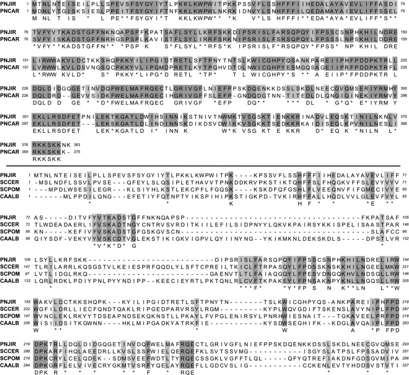
To address whether P. jirovecii contains a potential Rtt109 HAT, we performed an in silico search of the recently reported P. jirovecii genome (54, 55). A putative Pjrtt109 orthologue (GenBank accession number CCJ28444) was identified in a pairwise alignment with PcRtt109 (GenBank accession number ACR39370.1), showing 61% primary sequence conservation (Fig. 1, top). The moderate divergence between these two closely related species is not unexpected, as several features in the mitochondrial DNA of P. murina and P. carinii diverge from those of P. jirovecii (56), and this pattern of sequence similarity has also been observed between P. carinii and P. jirovecii dihydrofolate reductases (47, 57). In addition, there appears to be significant genetic diversity at several regions among P. jirovecii isolates, based on genetic analyses (58–61). The putative PjRtt109 sequence showed several conserved regions in comparison with other previously characterized fungal Rtt109 proteins, such as those of Saccharomyces cerevisiae (GenBank accession number Q07794), Schizosaccharomyces pombe (GenBank accession number Q9Y7Y5), and Candida albicans (GenBank accession number Q5AAJ8) (Fig. 1, bottom). These regions included the aspartic acid at amino acid position 84 in the conserved SKAD motif. Mutations of this specific residue result in nearly complete abolishment of in vitro H3 histone acetylation at the K56 position for both PcRtt109 and S. cerevisiae Rtt109 (ScRtt109) (30, 34). As with PcRtt109, the Pjrtt109 gene encodes a conserved lysine residue at amino acid position 221, which is analogous to the site for autoacetylation in yeast Rtt109 (62, 63). Furthermore, P. murina Rtt109 (GenBank accession number EMR10273) showed significant homology to both PjRtt109 and PcRtt109 (data not shown).
FIG 1.
PjRtt109 is an orthologue of PcRtt109 and is homologous to yeast and other fungal Rtt109 proteins involved in H3K56 acetylation. (Top) Pairwise sequence alignment of PjRtt109 and PcRtt109. (Bottom) Multiple-sequence alignment of fungal Rtt109 histone acetyltransferases. Alignments were performed using default CLUSTALW settings (MacVector 12.5.1). PNJIR, Pneumocystis jirovecii; PNCAR, Pneumocystis carinii; SCPOM, Schizosaccharomyces pombe; SACER, Saccharomyces cerevisiae; CAALB, Candida albicans. Dark shading indicates identical residues, and light shading indicates similar residues; asterisks denote similar residues in the consensus sequence.
Despite difficulties associated with studying Pneumocystis organisms in the laboratory, additional evidence supports the idea that Pjrtt109 is expressed by P. jirovecii. P. jirovecii organisms were obtained from a BAL fluid sample from a patient with PcP, and total RNA was extracted and amplified nonspecifically prior to sequencing (54). A total of 56 RNA-sequencing reads mapped unambiguously to the putative Pjrtt109 gene (http://www.ebi.ac.uk/ena/data/view/ERP001479). Additionally, the Pjrtt109 gene was present in the transcriptome assembly (HAAA01000299) (http://www.ebi.ac.uk/ena/data/view/HAAA01000299), which was assembled de novo using RNA-sequencing reads (O. Cissé, personal communication).
With this information, we next sought to confirm the presence of the putative Pjrtt109 gene in P. jirovecii organisms in vivo. To accomplish this, we extracted P. jirovecii genomic DNA from BAL fluid samples obtained from patients with confirmed cases of PcP at the Mayo Medical Center. These Pneumocystis infections were confirmed with our recently published single-copy nonnested PCR assay that distinguishes active PcP from simple Pneumocystis colonization (64). Primers based on the cDNA sequence of Pjrtt109 were mixed with either P. jirovecii genomic DNA or human genomic DNA isolated from healthy human lung cells. As expected, the Pjrtt109 primer set amplified a specific amplicon of the expected size with P. jirovecii genomic DNA as the template but not with human genomic DNA as the template (Fig. 2). As a positive control for human DNA quality, we also included a primer set based on the hGAPDH gene, which amplified an expected amplicon using human genomic DNA but not P. jirovecii genomic DNA (Fig. 2). These results strongly support the idea that P. jirovecii contains the Pjrtt109 gene and this gene is specifically represented in the P. jirovecii genome.
FIG 2.
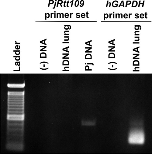
Pjrtt109 primer set amplifies a specific amplicon from P. jirovecii (Pj) genomic DNA but not human genomic DNA. hGAPDH, human glyceraldehyde-3-phosphate dehydrogenase.
Pjrtt109 restores H3K56ac levels and genotoxic resistance in rtt109-null yeast.
In S. cerevisiae, Rtt109 is required for H3K56ac and is associated with genotoxic resistance due to its role in replication-coupled nucleosome assembly (34). Pneumocystis species cannot be maintained using in vitro culture methods and cannot yet be manipulated genetically. To circumvent these issues, our group assesses Pneumocystis gene function using heterologous expression and complementation in other fungi such as S. cerevisiae (65, 66). In this manner, we evaluated the potential activities of Pjrtt109 in complementing the H3K56 acetylation defect in rtt109-null yeast by transforming this strain with either pYES2.1/V5-His/lacZ alone or the same vector containing full-length Pjrtt109. Western blots of yeast whole-cell extracts demonstrated that rtt109-null yeast cells were unable to acetylate H3K56. In contrast, Pjrtt109 cDNA complementation efficiently restored H3K56ac levels in rtt109-null yeast to levels seen in WT yeast (Fig. 3A).
FIG 3.

Heterologous expression of Pjrtt109 restores H3K56ac levels and genotoxic resistance in rtt109-null yeast. (A) Complementation of rtt109-null yeast with Pjrtt109 restores H3K56ac, as assessed by Western blots of yeast whole-cell extracts. WT, wild-type strain plus control vector; Δ, rtt109-null strain plus control vector; PJ, rtt109-null strain plus Pjrtt109 cDNA. (B) Complementation of rtt109-null yeast with Pjrtt109 restores genotoxic resistance, as assessed by the growth of yeast on solid medium. Tenfold serial dilutions of S. cerevisiae were spotted on minimal medium (with 2% galactose minus uracil [−URA]) alone or with the addition of MMS, CPT, or HU.
In addition, rtt109-null yeast demonstrated increased sensitivity to genotoxins such as MMS, CPT, and HU (34, 35). As anticipated for a putative Rtt109 HAT, Pjrtt109 complementation restored genotoxin resistance in rtt109-null yeast to the level in WT yeast (Fig. 3B). Taken together, these data strongly indicate that Pneumocystis-derived Pjrtt109 can function in promoting H3K56ac and genotoxin resistance in budding yeast, supporting the notion that PjRtt109 may perform similar functions in P. jirovecii in vivo.
PjRtt109 is an active HAT in vitro.
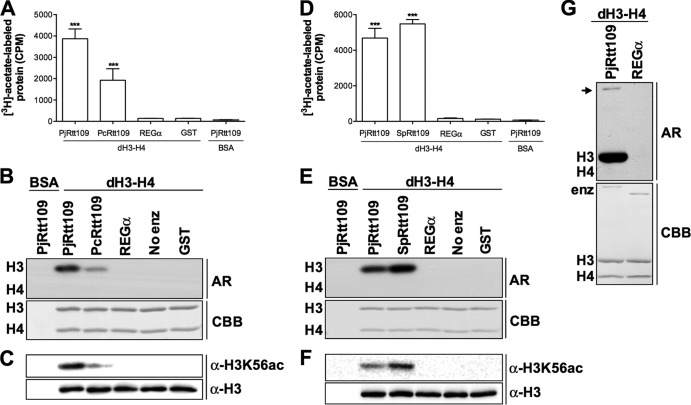
Based on the heterologous expression experiments that demonstrated Pjrtt109 functionality in vivo, we next sought to confirm the HAT activity of expressed PjRtt109 protein in vitro. Purified PjRtt109 protein demonstrated HAT activity comparable to that of its orthologue PcRtt109 in an in vitro [3H]acetyl-CoA HAT assay that quantified the amount of [3H]acetate incorporated into histone substrates (Fig. 4A). Autoradiographs of the reaction mixtures demonstrated that histone acetylation was confined to histone H3 and not histone H4, consistent with the known substrate profiles of previously characterized Rtt109 enzymes (Fig. 4B). Western blots verified that PjRtt109 catalyzed H3K56ac in vitro (Fig. 4C). To show that PjRtt109 was homologous to fungal Rtt109 HATs outside the Pneumocystis genus, the HAT activities of Pneumocystis jirovecii Rtt109 and Schizosaccharomyces pombe Rtt109 were also compared in vitro (30, 52, 67). The recombinant enzymes demonstrated comparable HAT activities (Fig. 4D). Autoradiographs (Fig. 4E) and Western blots (Fig. 4F) showed that both Rtt109 proteins catalyzed acetylation on histone H3 and both proteins were capable of acetylating H3K56 in vitro. Autoacetylation is an important regulatory element in ScRtt109 (62, 63), and low-level autoacetylation has been observed for PcRtt109 (30). As expected, we observed a band consistent with PjRtt109 autoacetylation using extended-exposure autoradiographs, demonstrating that autoacetylation is likely a conserved feature of Rtt109 HATs (Fig. 4G). These results demonstrate that PjRtt109 possesses functional Rtt109 HAT activity in vitro and that this activity is comparable to that of other fungal Rtt109 HATs. Such information is vital for the eventual development of Rtt109 inhibitors with the potential for therapeutic activity across a variety of clinically relevant fungal species that infect humans, such as P. jirovecii.
FIG 4.
PjRtt109 is an active HAT in vitro. (A) PjRtt109, expressed as a GST-fusion protein, shows in vitro HAT activity comparable to that of PcRtt109. ***, P < 0.001, compared with the REGα negative control. Shown are representative results from a single experiment, with similar results being obtained in at least two other independent experiments. (B) Reaction mixture aliquots, as shown in panel A, were resolved by SDS-PAGE and stained with CBB to demonstrate equal substrate and enzyme contents. Autoradiographs (AR) reveal that Rtt109-catalyzed histone acetylation is detected only on H3 and not on H4. (C) Western blot analysis of reaction mixture aliquots shows that PjRtt109, like PcRtt109, catalyzes H3K56ac in vitro. Equal substrate contents were verified with Ponceau S staining and Western blotting for H3. (D) PjRtt109 and SpRtt109 have similar HAT activities in vitro. (E) SDS-PAGE and CBB staining of reaction mixture aliquots, as shown in panel D, show equal protein contents. Autoradiographs show that PjRtt109 and PcRtt109 similarly catalyze the acetylation of H3, and H4 acetylation was not detected. (F) PjRtt109 and SpRtt109 both catalyze H3K56ac in vitro, as assessed by Western blotting. Equal substrate contents were verified with Ponceau S staining and anti-H3 Western blotting. (G) An extended-exposure autoradiograph using reaction mixture aliquots as shown in panel A demonstrates low levels of PjRtt109 autoacetylation (arrow).
PjRtt109 HAT activity is enhanced by the histone chaperone Asf1 in vitro.
Rtt109 is subject to complex regulation by histone chaperones both in vitro and in vivo. One such chaperone, Asf1, enhances the in vitro HAT activity of Rtt109 in both yeast and P. carinii (30, 31, 52, 68) and is required for H3K56ac in vivo (35). The addition of recombinant ScAsf1 to dH3–H4 tetramers significantly enhanced PjRtt109-catalyzed HAT activity in vitro (Fig. 5A). As expected, autoradiography confirmed that the enhanced acetylation was confined to histone H3 and not ScAsf1 or histone H4 (Fig. 5B). Western blots confirmed that this ScAsf1-mediated increase in PjRtt109-catalyzed histone acetylation in vitro led to an increase in H3K56ac (Fig. 5C). These results demonstrate that, like previously characterized Rtt109 HATs, PjRtt109 HAT activity in vitro is enhanced by the histone chaperone Asf1.
FIG 5.
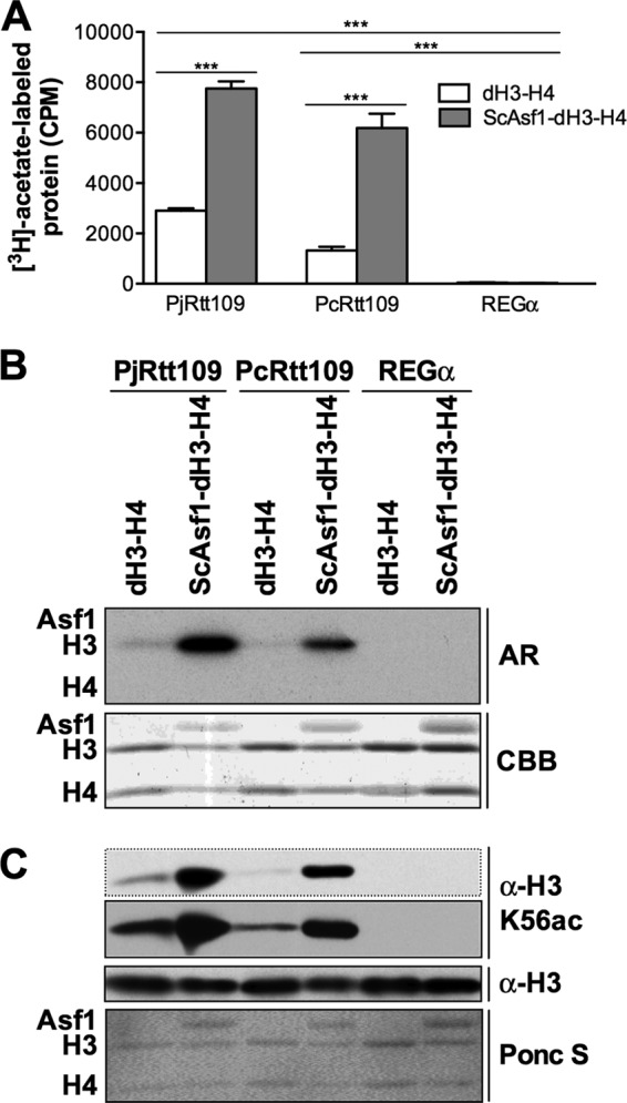
PjRtt109 HAT activity is enhanced by the histone chaperone Asf1 in vitro. (A) PjRtt109 HAT activity in vitro is enhanced by the addition of ScAsf1. ***, P < 0.001, pairwise comparisons between the dH3–H4 and ScAsf1-dH3–H4 substrates for each enzyme or pairwise comparisons with the REGα negative controls. Shown are representative results from a single experiment, with similar results being obtained in at least two other independent experiments. (B) Autoradiography of reaction mixture aliquots, as shown in panel A, demonstrates that acetylation is detected only on H3 and not on H4 or ScAsf1. Equal substrate histone contents were verified by CBB staining. (C) Western blotting of reaction mixture aliquots analogous to those shown in panel B versus H3K56ac, using nonradiolabeled acetyl-CoA as the substrate, was performed. Equal histone substrate contents were verified by Ponceau S staining and Western blotting.
PjRtt109 HAT activity is inhibited by small molecules in vitro.
Currently, there are no reports of small molecules that specifically inhibit PjRtt109 or PcRtt109 activity. In the absence of such compounds, we examined the effects of several previously reported HAT inhibitors on PjRtt109 enzymatic activity in vitro. Garcinol is a polyisoprenylated benzophenone natural product that is reported to inhibit several HATs, including p300/CBP-associated factor (PCAF) and p300, in the low micromolar range in vitro (69, 70). We recently showed that garcinol inhibits PcRtt109 HAT activity and yeast Rtt109-Vps75 in vitro at low micromolar concentrations (31, 42). Of note, we observed that garcinol inhibited PjRtt109 HAT activity in vitro in a dose-dependent manner (IC50, 2.7 ± 0.72 μM) (Fig. 6A and B). Compound 1 has been recently reported as a low-nanomolar inhibitor of yeast Rtt109 in vitro and does not inhibit the HATs p300 and GCN5 in vitro (43). Like garcinol, compound 1 showed dose-dependent inhibition of PjRtt109 HAT activity in vitro (IC50, 4.3 ± 3.9 μM) (Fig. 6A and B). Fluconazole, an inhibitor of fungal cytochrome 14α-demethylase with no reported activity against HATs, was used as a negative-control compound. Both garcinol and compound 1 inhibited PjRtt109 and PcRtt109 at low micromolar concentrations (Fig. 6B). Using the reaction mixtures for garcinol, we confirmed dose-dependent decreases in histone acetylation via autoradiography (Fig. 6C) and Western blotting for both H3K56ac and H3K27ac (Fig. 6D). These results demonstrate that PjRtt109 is capable of in vitro enzymatic inhibition by small molecules at low micromolar concentrations and that PjRtt109 and PcRtt109 are inhibited at similar compound concentrations under the conditions tested.
FIG 6.
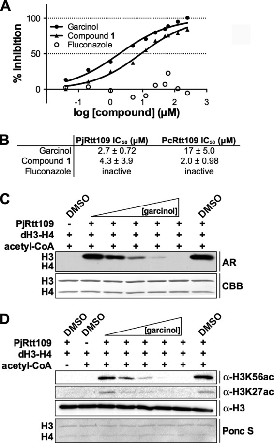
Reported small-molecule HAT inhibitors can inhibit PjRtt109 activity in vitro. (A) Inhibition of PjRtt109 activity by the reported HAT inhibitors garcinol (IC50, 5.9 μM) and compound 1 (IC50, 1.7 μM). Shown are representative results from a single experiment. (B) Comparison of IC50s for the compounds from panel A versus PjRtt109 and PcRtt109, tested under similar conditions. Values represent the average and standard deviation of three independent experiments. (C) Autoradiography of reaction mixture aliquots from panel A, confirming dose-dependent inhibition by garcinol of PjRtt109-catalyzed histone acetylation in vitro. Equal histone substrate contents were verified by CBB staining. (D) Western blot confirmation of dose-dependent inhibition by garcinol of PjRtt019-catalyzed H3K56ac and H3K27ac in vitro. Reaction mixture aliquots are analogous to those shown in panel C except that nonradiolabeled acetyl-CoA was used as the substrate. Equal histone substrate contents were verified by Ponceau S staining (representative membrane shown).
Effects of small-molecule inhibitors of HAT activity on Pneumocystis viability.
Finally, as support of the concept, we sought to investigate whether these reported small-molecule inhibitors of HAT activity might also alter Pneumocystis viability in vivo, using a previously described ATP-based viability assay (53). To investigate, we utilized freshly isolated P. carinii organisms, since these are readily available in the laboratory and because our prior experiments demonstrated comparable activities of the tested HAT inhibitors against PcRtt109 and PjRtt109 in vitro. Interestingly, the general HAT inhibitor garcinol demonstrated dose-dependent suppression of P. carinii viability, as measured by ATP contents (Fig. 7). In contrast, the recently reported yeast Rtt109 inhibitor compound 1 failed to induce any reductions of P. carinii viability under the conditions tested. Pentamidine and ampicillin served as positive- and negative-control compounds, respectively. The lack of observed activity for compound 1 is consistent with its lack of activity against C. albicans in vivo (43). However, the activity of the reported HAT inhibitor garcinol to suppress Pneumocystis viability does support the further investigation of this class of agents, as well as novel small molecules with more drug-like characteristics, potency, and target specificity, as potential antifungal antimicrobials with activity against Pneumocystis species.
FIG 7.
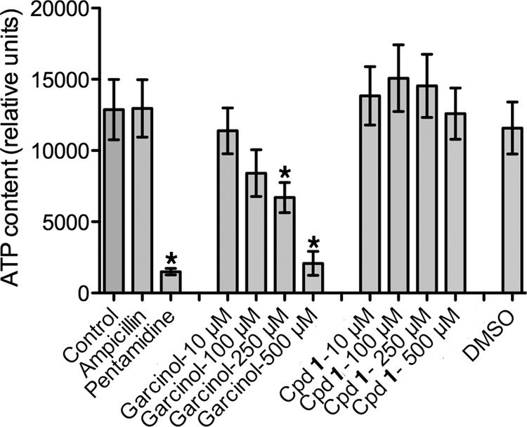
Effects of HAT inhibitors on Pneumocystis viability. Freshly isolated P. carinii organisms were maintained ex vivo for 72 h in test medium, and organism viability was determined by relative ATP contents. Ampicillin (10 μg/ml) and pentamidine (1 μl/ml) were included as relevant negative- and positive-control compounds. The reported pan-HAT inhibitor garcinol exhibited a significant dose-dependent reduction in P. carinii viability. In contrast, the recently described Rtt109 inhibitor compound 1 failed to alter P. carinii viability under the conditions tested. A DMSO diluent control was also used. Shown are the results of three independent experiments. *, P < 0.05, compared with control ATP levels without test agent.
DISCUSSION
In the current study, we demonstrate that P. jirovecii contains a functional Rtt109 orthologue with functions parallel to those exhibited by PcRtt109, which is an important early characterization step in the drug discovery process. We first identified the putative PjRtt109 orthologue by using a recently published P. jirovecii genome. We then showed that a Pjrtt109-specific primer set specifically amplified a product of the expected size when P. jirovecii genomic DNA was used as the template but not when human genomic DNA was used, which demonstrates that this gene is contained within the P. jirovecii genome. Because P. jirovecii cannot be cultured in vitro, we utilized heterologous expression to show that Pjrtt109 restores genotoxin resistance and H3K56ac in rtt109-null yeast, which demonstrates its functional importance as an Rtt109 HAT in this surrogate in vivo system. We were further able to synthesize full-length Pjrtt109 cDNA to study the function of recombinantly expressed PcRtt109 protein. We verified that PjRtt109 has HAT activity in vitro, comparable to that of its orthologue PcRtt109 and another fungal Rtt109, namely, SpRtt109. As in previous reports describing PcRtt109, the HAT activity of PjRtt109 was enhanced by the addition of the yeast histone chaperone Asf1, a salient feature of the well-characterized Rtt109-Vps75-Asf1 system in yeast. Finally, since small-molecule inhibition of Rtt109-catalyzed histone acetylation is hypothesized to be a potential novel epigenetically based antifungal therapy, we showed that the reported HAT inhibitors garcinol and compound 1 can inhibit PjRtt109 activity in vitro and these compounds inhibited PcRtt109 and PjRtt109 HAT activity comparably in vitro. Interestingly, garcinol, but not compound 1, also suppressed the viability of Pneumocystis organisms maintained ex vivo in viability medium.
Our observation that garcinol inhibits PjRtt109 and PcRtt109 as well as other HATs raises interesting follow-up questions about the target specificity of this natural product, its mechanism(s) of enzymatic inhibition, and its utility for additional in vivo studies. Garcinol has been reported to inhibit Pneumocystis and yeast Rtt109, as well as the HATs p300, PCAF, and GCN5, at low micromolar concentrations in vitro. It has also been reported to inhibit completely unrelated enzymes in vitro (71). This behavior may be explained by the chemical structure of garcinol, which contains an ortho-catechol group that has the potential to oxidize to form a thiol-reactive ortho-quinone (72). Being aware of this possibility, we have so far been unable to detect the presence of glutathione-garcinol adducts by UPLC-MS, although we admit that this does not definitively exclude the possible formation of these adducts (data not shown). Garcinol also can induce apoptosis, inhibit cancer cell growth, and modulate several important signaling pathways and has been shown to exert some protective effects (73–78). With regard to potential mammalian toxicity, garcinol has been studied in several animal experiments, in which it showed certain chemoprotective effects and was relatively well-tolerated by rodents (79–81). The overall in vitro data are consistent with garcinol being a relatively nonselective inhibitor, and this property may coincide with its ability to inhibit cancer cell growth in vitro. Given its in vitro activity profile and the relatively high concentrations used in our fungal viability assays, we cannot exclude the possibility of off-target effects contributing to the reduced viability of Pneumocystis upon exposure to garcinol. Therefore, we think that garcinol is not ideal for further in vivo experiments in its present form, because of its potential for off-target effects and the need for more-definitive mechanistic studies. More specific and potent chemical probes for HATs like Pneumocystis Rtt109 would be useful to better assess our therapeutic hypothesis in a cellular setting.
This demonstration that both PjRtt109 and PcRtt109 are functional HAT orthologues is significant for early Rtt109 drug discovery efforts for several reasons. First, it demonstrates that functional Rtt109 HATs are conserved across fungi. Second, it verifies that, in the absence of P. jirovecii culture systems, P. carinii may serve as an entirely suitable surrogate model for studying the Rtt109 system in the laboratory setting. In addition, the observation that reported small-molecule HAT inhibitors can disrupt PjRtt109 activity in vitro suggests that inhibitors developed for one species of Rtt109 may inhibit homologues across a variety of other fungal species, indicating that such a strategy may benefit a wide range of fungal infections. The discovery and development of more-potent small molecules targeting Rtt109 HATs will be useful to more rigorously address this speculation. Finally, our findings support further target characterization and drug development efforts with respect to PjRtt109, a potential epigenetic therapeutic target with clinical relevance as the specific cause of human PcP.
This study strongly validates the use of the P. carinii system for preclinical work of relevance for drug development eventually targeting human disease caused by P. jirovecii. We recently characterized the Rtt109-Vps75-Asf1 system in P. carinii, showing that PcRtt109 restores genotoxic resistance and H3K56ac levels in rtt109-null yeast. This system verifies many of the features found in the well-characterized S. cerevisiae and S. pombe Rtt109-Vps75-Asf1 systems in vitro, such as HAT activity being enhanced by the histone chaperone Asf1. As Rtt109 is conserved in fungi but not in mammals, specific inhibitors of Rtt109-catalyzed histone acetylation may eventually prove useful as novel antifungal targets across a number of relevant infections, including P. jirovecii-related pneumonia.
It is noteworthy that the general HAT inhibitor garcinol also reduced the in vitro viability of Pneumocystis organisms. Interestingly, the recently reported specific Rtt109 inhibitor compound 1 did not exhibit any suppression of Pneumocystis viability. Similar findings were noted when this compound was tested previously against C. albicans (43). There are several possible reasons for our observations, including possibly poor penetration of compound 1 into the fungal cytoplasm, the presence of active rapid export proteins that may limit internal accumulation of this agent, or compound instability under the testing conditions. Nonetheless, the wide presence of Rtt109 proteins across pathogenic fungi, the divergence from mammalian host HAT proteins, and our initial results on such agents reducing Pneumocystis viability continue to support additional searches for more-selective anti-Rtt109 agents that may be of therapeutic benefit for fungal infections in humans, including P. jirovecii pneumonia.
ACKNOWLEDGMENTS
We acknowledge Jessica Strasser and Subhashree Francis for chemical purification and characterization. We gratefully acknowledge Ousmane Cissé and Philippe Hauser, Institute of Microbiology, University Hospital Center (Lausanne, Switzerland), for assistance in verifying Pjrtt109 expression. The purified P. carinii organisms used in this study were generated with the assistance of Deanne Hebrink. Human-derived P. jirovecii organisms were obtained from the Mayo Clinical Microbiology Laboratory, with the assistance of Nancy Wengenack. The samples were stored as residual clinical waste specimens.
This work was supported by the Minnesota Partnership for Biotechnology and Medical Genomics (grant 73-01), the National Institutes of Health (grant R01HL-62150 to A.H.L.), and the Mayo Foundation for Medical Education and Research. J.L.D. was supported by the NIH Medical Scientist Training Program (grant T32 GM065841), a Pharmaceutical Research and Manufacturers of America Foundation predoctoral pharmacology/toxicology fellowship, and the Mayo Foundation.
The funders had no role in study design, data collection or analysis, the decision to publish, or preparation of the manuscript. The opinions and assertions contained herein belong to the authors and are not necessarily the official views of the funders.
We declare no conflicting interests.
J.L.D., T.K., A.H.L, and Z.Z. designed the experiments, J.L.D., T.K., J.H., and H.Z. performed the experiments, J.L.D., T.K., A.H.L., Z.Z., and M.A.W. analyzed the data, A.H.L., Z.Z., and M.A.W. contributed equipment and reagents, J.L.D. wrote the manuscript, and J.L.D., T.K., A.H.L., Z.Z., and M.A.W. contributed with revisions.
Footnotes
Published ahead of print 14 April 2014
REFERENCES
- 1.Huang L, Cattamanchi A, Davis JL, den Boon S, Kovacs J, Meshnick S, Miller RF, Walzer PD, Worodria W, Masur H, International HIV-Associated Opportunistic Pneumonias (IHOP) Study, Lung HIV Study 2011. HIV-associated Pneumocystis pneumonia. Proc. Am. Thorac. Soc. 8:294–300. 10.1513/pats.201009-062WR [DOI] [PMC free article] [PubMed] [Google Scholar]
- 2.Carmona EM, Limper AH. 2011. Update on the diagnosis and treatment of Pneumocystis pneumonia. Ther. Adv. Respir. Dis. 5:41–59. 10.1177/1753465810380102 [DOI] [PMC free article] [PubMed] [Google Scholar]
- 3.Krajicek BJ, Limper AH, Thomas CF. 2008. Advances in the biology, pathogenesis and identification of Pneumocystis pneumonia. Curr. Opin. Pulm. Med. 14:228–234. 10.1097/MCP.0b013e3282f94abc [DOI] [PubMed] [Google Scholar]
- 4.Cushion MT, Stringer JR. 2010. Stealth and opportunism: alternative lifestyles of species in the fungal genus Pneumocystis. Annu. Rev. Microbiol. 64:431–452. 10.1146/annurev.micro.112408.134335 [DOI] [PubMed] [Google Scholar]
- 5.Tasaka S, Tokuda H. 2012. Pneumocystis jirovecii pneumonia in non-HIV-infected patients in the era of novel immunosuppressive therapies. J. Infect. Chemother. 18:793–806. 10.1007/s10156-012-0453-0 [DOI] [PubMed] [Google Scholar]
- 6.Mekinian A, Durand-Joly I, Hatron P, Moranne O, Denis G, Queyrel V. 2011. Pneumocystis jirovecii colonization in patients with systemic autoimmune diseases: prevalence, risk factors of colonization and outcome. Rheumatology 50:569–577. 10.1093/rheumatology/keq314 [DOI] [PubMed] [Google Scholar]
- 7.Schmoldt S, Schuhegger R, Wendler T, Huber I, Söllner H, Hogardt M, Arbogast H, Heesemann J, Bader L, Sing A. 2008. Molecular evidence of nosocomial Pneumocystis jirovecii transmission among 16 patients after kidney transplantation. J. Clin. Microbiol. 46:966–971. 10.1128/JCM.02016-07 [DOI] [PMC free article] [PubMed] [Google Scholar]
- 8.Ewig S. 1996. The effect of prophylaxis on the outcome of HIV-associated Pneumocystis carinii pneumonia. Chest 109:586–587. 10.1378/chest.109.2.586 [DOI] [PubMed] [Google Scholar]
- 9.Monnet X, Vidal-Petiot E, Osman D, Hamzaoui O, Durrbach A, Goujard C, Miceli C, Bouree P, Richard C. 2008. Critical care management and outcome of severe Pneumocystis pneumonia in patients with and without HIV infection. Crit. Care 12:R28. 10.1186/cc6806 [DOI] [PMC free article] [PubMed] [Google Scholar]
- 10.Fei M, Kim E, Sant C, Jarlsberg L, Davis J, Swartzman A, Huang L. 2009. Predicting mortality from HIV-associated Pneumocystis pneumonia at illness presentation: an observational cohort study. Thorax 64:1070–1076. 10.1136/thx.2009.117846 [DOI] [PMC free article] [PubMed] [Google Scholar]
- 11.Fisk M, Sage E, Edwards S, Cartledge J, Miller R. 2009. Outcome from treatment of Pneumocystis jirovecii pneumonia with co-trimoxazole. Int. J. STD AIDS 20:652–653. 10.1258/ijsa.2009.008470 [DOI] [PubMed] [Google Scholar]
- 12.Mansharamani N, Garland R, Delaney D, Koziel H. 2000. Management and outcome patterns for adult Pneumocystis carinii pneumonia, 1985 to 1995: comparison of HIV-associated cases to other immunocompromised states. Chest 118:704–711. 10.1378/chest.118.3.704 [DOI] [PubMed] [Google Scholar]
- 13.Festic E, Gajic O, Limper A, Aksamit T. 2005. Acute respiratory failure due to Pneumocystis pneumonia in patients without human immunodeficiency virus infection: outcome and associated features. Chest 128:573–579. 10.1378/chest.128.2.573 [DOI] [PubMed] [Google Scholar]
- 14.Held J, Koch MS, Reischl U, Danner T, Serr A. 2011. Serum (1→3)-β-d-glucan measurement as an early indicator of Pneumocystis jirovecii pneumonia and evaluation of its prognostic value. Clin. Microbiol. Infect. 17:595–602. 10.1111/j.1469-0691.2010.03318.x [DOI] [PubMed] [Google Scholar]
- 15.Damiani C, Gal SL, Costa CD, Virmaux M, Nevez G, Totet A. 2013. Combined quantification of pulmonary Pneumocystis jirovecii DNA and serum (1→3)-β-d-glucan for differential diagnosis of Pneumocystis pneumonia and Pneumocystis colonization. J. Clin. Microbiol. 51:3380–3388. 10.1128/JCM.01554-13 [DOI] [PMC free article] [PubMed] [Google Scholar]
- 16.Steele C, Shellito JE, Kolls JK. 2005. Immunity against the opportunistic fungal pathogen Pneumocystis. Med. Mycol. 43:1–19. 10.1080/13693780400015360 [DOI] [PubMed] [Google Scholar]
- 17.Villegas LR, Kottom TJ, Limper AH. 2010. Characterization of PCEng2, a β-1,3-endoglucanase homolog in Pneumocystis carinii with activity in cell wall regulation. Am. J. Respir. Cell Mol. Biol. 43:192–200. 10.1165/rcmb.2009-0131OC [DOI] [PMC free article] [PubMed] [Google Scholar]
- 18.Gigliotti F, Wright T. 2005. Immunopathogenesis of Pneumocystis carinii pneumonia. Expert Rev. Mol. Med. 7:1–16. 10.1017/S1462399405010203 [DOI] [PubMed] [Google Scholar]
- 19.Kelly M, Shellito J. 2010. Current understanding of Pneumocystis immunology. Future Microbiol. 5:43–65. 10.2217/fmb.09.116 [DOI] [PMC free article] [PubMed] [Google Scholar]
- 20.Goodridge HS, Wolf AJ, Underhill DM. 2009. β-Glucan recognition by the innate immune system. Immunol. Rev. 230:38–50. 10.1111/j.1600-065X.2009.00793.x [DOI] [PMC free article] [PubMed] [Google Scholar]
- 21.Thomas CF, Jr, Limper AH. 2007. Current insights into the biology and pathogenesis of Pneumocystis pneumonia. Nat. Rev. Microbiol. 5:298–308. 10.1038/nrmicro1621 [DOI] [PubMed] [Google Scholar]
- 22.Huang L, Crothers K, Atzori C, Benfield T, Miller R, Rabodonirina M, Helweg-Larsen J. 2004. Dihydropteroate synthase gene mutations in Pneumocystis and sulfa resistance. Emerg. Infect. Dis. 10:1721–1728. 10.3201/eid1010.030994 [DOI] [PMC free article] [PubMed] [Google Scholar]
- 23.Kessl JJ, Hill P, Lange BB, Meshnick SR, Meunier B, Trumpower BL. 2004. Molecular basis for atovaquone resistance in Pneumocystis jirovecii modeled in the cytochrome bc1 complex of Saccharomyces cerevisiae. J. Biol. Chem. 279:2817–2824. 10.1074/jbc.M309984200 [DOI] [PubMed] [Google Scholar]
- 24.Walker DJ, Wakefield AE, Dohn MN, Miller RF, Baughman RP, Hossler PA, Bartlett MS, Smith JW, Kazanjian P, Meshnick SR. 1998. Sequence polymorphisms in the Pneumocystis carinii cytochrome b gene and their association with atovaquone prophylaxis failure. J. Infect. Dis. 178:1767–1775. 10.1086/314509 [DOI] [PubMed] [Google Scholar]
- 25.Sloand E, Laughon B, Armstrong M, Bartlett M, Blumenfeld W, Cushion M, Kalica A, Kovacs J, Martin W, Pitt E, Pesanti EL, Richards F, Rose R, Walzer P. 1993. The challenge of Pneumocystis carinii culture. J. Eukaryot. Microbiol. 40:188–195. 10.1111/j.1550-7408.1993.tb04902.x [DOI] [PubMed] [Google Scholar]
- 26.Beck JM, Newbury RL, Palmer BE. 1996. Pneumocystis carinii pneumonia in scid mice induced by viable organisms propagated in vitro. Infect. Immun. 64:4643–4647 [DOI] [PMC free article] [PubMed] [Google Scholar]
- 27.Bartlett MS, Fishman JA, Durkin MM, Queener SF, Smith JW. 1990. Pneumocystis carinii: improved models to study efficacy of drugs for treatment or prophylaxis of Pneumocystis pneumonia in the rat (Rattus spp.). Exp. Parasitol. 70:100–106. 10.1016/0014-4894(90)90089-U [DOI] [PubMed] [Google Scholar]
- 28.McFadden DC, Powles MA, Pittarelli LA, Schmatz DM. 1991. Establishment of Pneumocystis carinii in various mouse strains using natural transmission to initiate infection. J. Protozool. 38:126S–127S [PubMed] [Google Scholar]
- 29.Beck JM, Preston AM, Wilcoxen SE, Morris SB, Sturrock A, Paine R. 2009. Critical roles of inflammation and apoptosis in improved survival in a model of hyperoxia-induced acute lung injury in Pneumocystis murina-infected mice. Infect. Immun. 77:1053–1060. 10.1128/IAI.00967-08 [DOI] [PMC free article] [PubMed] [Google Scholar]
- 30.Kottom TJ, Han J, Zhang Z, Limper AH. 2011. Pneumocystis carinii expresses an active Rtt109 histone acetyltransferase. Am. J. Respir. Cell Mol. Biol. 44:768–776. 10.1165/rcmb.2009-0443OC [DOI] [PMC free article] [PubMed] [Google Scholar]
- 31.Pupaibool J, Kottom TJ, Bouchonville K, Limper AH. 2013. Characterization of the Pneumocystis carinii histone acetyltranferase chaperone proteins PcAsf1 and PcVps75. Infect. Immun. 81:2268–2275. 10.1128/IAI.01173-12 [DOI] [PMC free article] [PubMed] [Google Scholar]
- 32.Masumoto H, Hawke D, Kobayashi R, Verreault A. 2005. A role for cell-cycle-regulated histone H3 lysine 56 acetylation in the DNA damage response. Nature 436:294–298. 10.1038/nature03714 [DOI] [PubMed] [Google Scholar]
- 33.Tsubota T, Berndsen CE, Erkmann JA, Smith CL, Yang L, Freitas MA, Denu JM, Kaufman PD. 2007. Histone H3–K56 acetylation is catalyzed by histone chaperone-dependent complexes. Mol. Cell 25:703–712. 10.1016/j.molcel.2007.02.006 [DOI] [PMC free article] [PubMed] [Google Scholar]
- 34.Han J, Zhou H, Horazdovsky B, Zhang K, Xu R, Zhang Z. 2007. Rtt109 acetylates histone H3 lysine 56 and functions in DNA replication. Science 315:653–655. 10.1126/science.1133234 [DOI] [PubMed] [Google Scholar]
- 35.Driscoll R, Hudson A, Jackson SP. 2007. Yeast Rtt109 promotes genome stability by acetylating histone H3 on lysine 56. Science 315:649–652. 10.1126/science.1135862 [DOI] [PMC free article] [PubMed] [Google Scholar]
- 36.Chen CC, Carson JJ, Feser J, Tamburini B, Zabaronick S, Linger J, Tyler JK. 2008. Acetylated lysine 56 on histone H3 drives chromatin assembly after repair and signals for the completion of repair. Cell 134:231–243. 10.1016/j.cell.2008.06.035 [DOI] [PMC free article] [PubMed] [Google Scholar]
- 37.Li Q, Zhou H, Wurtele H, Davies B, Horazdovsky B, Verreault A, Zhang Z. 2008. Acetylation of histone H3 lysine 56 regulates replication-coupled nucleosome assembly. Cell 134:244–255. 10.1016/j.cell.2008.06.018 [DOI] [PMC free article] [PubMed] [Google Scholar]
- 38.Das C, Lucia M, Hansen K, Tyler J. 2009. CBP/p300-mediated acetylation of histone H3 on lysine 56. Nature 459:113–117. 10.1038/nature07861 [DOI] [PMC free article] [PubMed] [Google Scholar]
- 39.Tjeertes J, Miller K, Jackson S. 2009. Screen for DNA-damage-responsive histone modifications identifies H3K9Ac and H3K56Ac in human cells. EMBO J. 28:1878–1889. 10.1038/emboj.2009.119 [DOI] [PMC free article] [PubMed] [Google Scholar]
- 40.Wurtele H, Tsao S, Lépine G, Mullick A, Tremblay J, Drogaris P, Lee E-H, Thibault P, Verreault A, Raymond M. 2010. Modulation of histone H3 lysine 56 acetylation as an antifungal therapeutic strategy. Nat. Med. 16:774–780. 10.1038/nm.2175 [DOI] [PMC free article] [PubMed] [Google Scholar]
- 41.Lopes da Rosa J, Boyartchuk VL, Zhu LJ, Kaufman PD. 2010. Histone acetyltransferase Rtt109 is required for Candida albicans pathogenesis. Proc. Natl. Acad. Sci. U. S. A. 107:1594–1599. 10.1073/pnas.0912427107 [DOI] [PMC free article] [PubMed] [Google Scholar]
- 42.Dahlin JL, Sinville R, Solberg J, Zhou H, Han J, Francis S, Strasser JM, John K, Hook DJ, Walters MA, Zhang Z. 2013. A cell-free fluorometric high-throughput screen for inhibitors of Rtt109-catalyzed histone acetylation. PLoS One 8:e78877. 10.1371/journal.pone.0078877 [DOI] [PMC free article] [PubMed] [Google Scholar]
- 43.Lopes da Rosa J, Bajaj V, Spoonamore J, Kaufman PD. 2013. A small molecule inhibitor of fungal histone acetyltransferase Rtt109. Bioorg. Med. Chem. Lett. 23:2853–2859. 10.1016/j.bmcl.2013.03.112 [DOI] [PMC free article] [PubMed] [Google Scholar]
- 44.Gangjee A, Guo X, Queener SF, Cody V, Galitsky N, Luft JR, Pangborn W. 1998. Selective Pneumocystis carinii dihydrofolate reductase inhibitors: design, synthesis, and biological evaluation of new 2,4-diamino-5-substituted-furo[2,3-d]pyrimidines. J. Med. Chem. 41:1263–1271. 10.1021/jm970537w [DOI] [PubMed] [Google Scholar]
- 45.Anderson AC, Perry KM, Freymann DM, Stroud RM. 2000. The crystal structure of thymidylate synthase from Pneumocystis carinii reveals a fungal insert important for drug design. J. Mol. Biol. 297:645–657. 10.1006/jmbi.2000.3544 [DOI] [PubMed] [Google Scholar]
- 46.Cushion MT, Walzer PD, Ashbaugh A, Rebholz S, Brubaker R, Eynde JJV, Mayence A, Huang TL. 2006. In vitro selection and in vivo efficacy of piperazine- and alkanediamide-linked bisbenzamidines against Pneumocystis pneumonia in mice. Antimicrob. Agents Chemother. 50:2337–2343. 10.1128/AAC.00126-06 [DOI] [PMC free article] [PubMed] [Google Scholar]
- 47.Cody V, Pace J, Queener SF, Adair OO, Gangjee A. 2013. Kinetic and structural analysis for potent antifolate inhibition of Pneumocystis jirovecii, Pneumocystis carinii, and human dihydrofolate reductases and their active-site variants. Antimicrob. Agents Chemother. 57:2669–2677. 10.1128/AAC.00172-13 [DOI] [PMC free article] [PubMed] [Google Scholar]
- 48.Porollo A, Meller J, Joshi Y, Jaiswal V, Smulian AG, Cushion MT. 2012. Analysis of current antifungal agents and their targets within the Pneumocystis carinii genome. Curr. Drug Targets 13:1575–1585. 10.2174/138945012803530107 [DOI] [PMC free article] [PubMed] [Google Scholar]
- 49.Perez-Leal O, Moncada C, Clarkson AB, Merali S. 2011. Pneumocystis S-adenosylmethionine transport: a potential drug target. Am. J. Respir. Cell Mol. Biol. 45:1142–1146. 10.1165/rcmb.2011-0009OC [DOI] [PMC free article] [PubMed] [Google Scholar]
- 50.Levenstein M, Kadonaga J. 2002. Biochemical analysis of chromatin containing recombinant Drosophila core histones. J. Biol. Chem. 277:8749–8754. 10.1074/jbc.M111212200 [DOI] [PubMed] [Google Scholar]
- 51.Berndsen CE, Denu JM. 2005. Assays for mechanistic investigations of protein/histone acetyltransferases. Methods 36:321–331. 10.1016/j.ymeth.2005.03.002 [DOI] [PubMed] [Google Scholar]
- 52.Han J, Zhou H, Li Z, Xu R, Zhang Z. 2007. Acetylation of lysine 56 of histone H3 catalyzed by Rtt109 and regulated by Asf1 is required for replisome integrity. J. Biol. Chem. 282:28587–28596. 10.1074/jbc.M702496200 [DOI] [PubMed] [Google Scholar]
- 53.Cushion MT, Collins M, Hazra B, Kaneshiro ES. 2000. Effects of atovaquone and diospyrin-based drugs on the cellular ATP of Pneumocystis carinii f. sp. carinii. Antimicrob. Agents Chemother. 44:713–719. 10.1128/AAC.44.3.713-719.2000 [DOI] [PMC free article] [PubMed] [Google Scholar]
- 54.Cissé OH, Pagni M, Hauser PM. 2012. De novo assembly of the Pneumocystis jirovecii genome from a single bronchoalveolar lavage fluid specimen from a patient. mBio 4:e00428–12. 10.1128/mBio.00428-12 [DOI] [PMC free article] [PubMed] [Google Scholar]
- 55.Cushion MT, Keely SP. 2013. Assembly and annotation of Pneumocystis jirovecii from the human lung microbiome. mBio 4:e00224-13. 10.1128/mBio.00224-13 [DOI] [PMC free article] [PubMed] [Google Scholar]
- 56.Ma L, Huang D-W, Cuomo CA, Sykes S, Fantoni G, Das B, Sherman BT, Yang J, Huber C, Xia Y, Davey E, Kutty G, Bishop L, Sassi M, Lempicki RA, Kovacs JA. 2013. Sequencing and characterization of the complete mitochondrial genomes of three Pneumocystis species provide new insights into divergence between human and rodent Pneumocystis. FASEB J. 27:1962–1972. 10.1096/fj.12-224444 [DOI] [PMC free article] [PubMed] [Google Scholar]
- 57.Cody V, Pace J, Makin J, Piraino J, Queener SF, Rosowsky A. 2009. Correlations of inhibitor kinetics for Pneumocystis jirovecii and human dihydrofolate reductase with structural data for human active site mutant enzyme complexes. Biochemistry 48:1702–1711. 10.1021/bi801960h [DOI] [PMC free article] [PubMed] [Google Scholar]
- 58.Curran T, McCaughey C, Coyle PV. 2013. Pneumocystis jirovecii multilocus genotyping profiles in Northern Ireland. J. Med. Microbiol. 62:1170–1174. 10.1099/jmm.0.057794-0 [DOI] [PubMed] [Google Scholar]
- 59.Totet A, Duwat H, Magois E, Jounieaux V, Roux P, Raccurt C, Nevez G. 2004. Similar genotypes of Pneumocystis jirovecii in different forms of Pneumocystis infection. Microbiology 150:1173–1178. 10.1099/mic.0.26919-0 [DOI] [PubMed] [Google Scholar]
- 60.Matos O, Esteves F. 2010. Pneumocystis jirovecii multilocus gene sequencing: findings and implications. Future Microbiol. 5:1257–1267. 10.2217/fmb.10.75 [DOI] [PubMed] [Google Scholar]
- 61.Ripamonti C, Orenstein A, Kutty G, Huang L, Schuhegger R, Sing A, Fantoni G, Atzori C, Vinton C, Huber C, Conville PS, Kovacs JA. 2009. Restriction fragment length polymorphism typing demonstrates substantial diversity among Pneumocystis jirovecii isolates. J. Infect. Dis. 200:1616–1622. 10.1086/644643 [DOI] [PMC free article] [PubMed] [Google Scholar]
- 62.Albaugh BN, Arnold KM, Lee S, Denu JM. 2011. Autoacetylation of the histone acetyltransferase Rtt109. J. Biol. Chem. 286:24694–24701. 10.1074/jbc.M111.251579 [DOI] [PMC free article] [PubMed] [Google Scholar]
- 63.Stavropoulos P, Nagy V, Blobel G, Hoelz A. 2008. Molecular basis for the autoregulation of the protein acetyl transferase Rtt109. Proc. Natl. Acad. Sci. U. S. A. 105:12236–12241. 10.1073/pnas.0805813105 [DOI] [PMC free article] [PubMed] [Google Scholar]
- 64.Wilson JW, Limper AH, Grys TE, Karre T, Wengenack NL, Binnicker MJ. 2011. Pneumocystis jirovecii testing by real-time polymerase chain reaction and direct examination among immunocompetent and immunosuppressed patient groups and correlation to disease specificity. Diagn. Microbiol. Infect. Dis. 69:145–152. 10.1016/j.diagmicrobio.2010.10.021 [DOI] [PMC free article] [PubMed] [Google Scholar]
- 65.Hauser P, Lo Presti L, Cockell M, Cerutti L, Simanis V. 2006. Analysis of Pneumocystis carinii gene function by complementation in yeast mutants. J. Eukaryot. Microbiol. 53(Suppl 1):S149–S150. 10.1111/j.1550-7408.2006.00210.x [DOI] [PubMed] [Google Scholar]
- 66.Grigore D, Meade JC. 2006. Functional complementation of the yeast P-type H-ATPase, PMA1, by the Pneumocystis carinii P-type H-ATPase, PCA1. J. Eukaryot. Microbiol. 53:157–164. 10.1111/j.1550-7408.2006.00089.x [DOI] [PubMed] [Google Scholar]
- 67.Xhemalce B, Miller KM, Driscoll R, Masumoto H, Jackson SP, Kouzarides T, Verreault A, Arcangioli B. 2007. Regulation of histone H3 lysine 56 acetylation in Schizosaccharomyces pombe. J. Biol. Chem. 282:15040–15047. 10.1074/jbc.M701197200 [DOI] [PubMed] [Google Scholar]
- 68.Adkins MW, Carson JJ, English CM, Ramey CJ, Tyler JK. 2007. The histone chaperone anti-silencing function 1 stimulates the acetylation of newly synthesized histone H3 in S-phase. J. Biol. Chem. 282:1334–1340. 10.1074/jbc.M608025200 [DOI] [PubMed] [Google Scholar]
- 69.Balasubramanyam K, Altaf M, Varier R, Swaminathan V, Ravindran A, Sadhale P, Kundu T. 2004. Polyisoprenylated benzophenone, garcinol, a natural histone acetyltransferase inhibitor, represses chromatin transcription and alters global gene expression. J. Biol. Chem. 279:33716–33726. 10.1074/jbc.M402839200 [DOI] [PubMed] [Google Scholar]
- 70.Arif M, Pradhan SK, Thanuja GR, Vedamurthy BM, Agrawal S, Dasgupta D, Kundu TK. 2009. Mechanism of p300 specific histone acetyltransferase inhibition by small molecules. J. Med. Chem. 52:267–277. 10.1021/jm800657z [DOI] [PubMed] [Google Scholar]
- 71.Koeberle A, Northoff H, Werz O. 2009. Identification of 5-lipoxygenase and microsomal prostaglandin E2 synthase-1 as functional targets of the anti-inflammatory and anti-carcinogenic garcinol. Biochem. Pharmacol. 77:1513–1521. 10.1016/j.bcp.2009.02.005 [DOI] [PubMed] [Google Scholar]
- 72.Baell JB, Holloway GA. 2010. New substructure filters for removal of pan assay interference compounds (PAINS) from screening libraries and for their exclusion in bioassays. J. Med. Chem. 53:2719–2740. 10.1021/jm901137j [DOI] [PubMed] [Google Scholar]
- 73.Ahmad A, Sarkar SH, Bitar B, Ali S, Aboukameel A, Sethi S, Li Y, Bao B, Kong D, Banerjee S, Padhye SB, Sarkar FH. 2012. Garcinol regulates EMT and Wnt signaling pathways in vitro and in vivo, leading to anticancer activity against breast cancer cells. Mol. Cancer Ther. 11:2193–2201. 10.1158/1535-7163.MCT-12-0232-T [DOI] [PMC free article] [PubMed] [Google Scholar]
- 74.Weng M-S, Liao C-H, Yu S-Y, Lin J-K. 2011. Garcinol promotes neurogenesis in rat cortical progenitor cells through the duration of extracellular signal-regulated kinase signaling. J. Agric. Food Chem. 59:1031–1040. 10.1021/jf104263s [DOI] [PubMed] [Google Scholar]
- 75.Cheng A-C, Tsai M-L, Liu C-M, Lee M-F, Nagabhushanam K, Ho C-T, Pan M-H. 2010. Garcinol inhibits cell growth in hepatocellular carcinoma Hep3B cells through induction of ROS-dependent apoptosis. Food Funct. 1:301–307. 10.1039/c0fo00134a [DOI] [PubMed] [Google Scholar]
- 76.Hong J, Kwon SJ, Sang S, Ju J, Zhou JN, Ho CT, Huang MT, Yang CS. 2007. Effects of garcinol and its derivatives on intestinal cell growth: inhibitory effects and autoxidation-dependent growth-stimulatory effects. Free Radic. Biol. Med. 42:1211–1221. 10.1016/j.freeradbiomed.2007.01.016 [DOI] [PubMed] [Google Scholar]
- 77.Liao CH, Sang S, Ho CT, Lin JK. 2005. Garcinol modulates tyrosine phosphorylation of FAK and subsequently induces apoptosis through down-regulation of Src, ERK, and Akt survival signaling in human colon cancer cells. J. Cell. Biochem. 96:155–169. 10.1002/jcb.20540 [DOI] [PubMed] [Google Scholar]
- 78.Oike T, Ogiwara H, Torikai K, Nakano T, Yokota J, Kohno T. 2012. Garcinol, a histone acetyltransferase inhibitor, radiosensitizes cancer cells by inhibiting non-homologous end joining. Int. J. Radiat. Oncol. Biol. Phys. 84:815–821. 10.1016/j.ijrobp.2012.01.017 [DOI] [PubMed] [Google Scholar]
- 79.Chen X, Zhang X, Lu Y, Shim J-Y, Sang S, Sun Z, Chen X. 2012. Chemoprevention of 7,12-dimethylbenz[a]anthracene (DMBA)-induced hamster cheek pouch carcinogenesis by a 5-lipoxygenase inhibitor, garcinol. Nutr. Cancer 64:1211–1218. 10.1080/01635581.2012.718032 [DOI] [PMC free article] [PubMed] [Google Scholar]
- 80.Yoshida K, Tanaka T, Hirose Y, Yamaguchi F, Kohno H, Toida M, Hara A, Sugie S, Shibata T, Mori H. 2005. Dietary garcinol inhibits 4-nitroquinoline 1-oxide-induced tongue carcinogenesis in rats. Cancer Lett. 221:29–39. 10.1016/j.canlet.2004.08.016 [DOI] [PubMed] [Google Scholar]
- 81.Tanaka T, Kohno H, Shimada R, Kagami S, Yamaguchi F, Kataoka S, Ariga T, Murakami A, Koshimizu K, Ohigashi H. 2000. Prevention of colonic aberrant crypt foci by dietary feeding of garcinol in male F344 rats. Carcinogenesis 21:1183–1189. 10.1093/carcin/21.6.1183 [DOI] [PubMed] [Google Scholar]


