Abstract
Mesophilic Crenarchaeota (also known as Thaumarchaeota) are ubiquitous and abundant in marine habitats. However, very little is known about their metabolic function in situ. In this study, salt marsh sediments from New Jersey were screened via stable isotope probing (SIP) for heterotrophy by amending with a single 13C-labeled compound (acetate, glycine or urea) or a complex 13C-biopolymer (lipids, proteins or growth medium (ISOGRO)). SIP incubations were done at two substrate concentrations (30–150 μM; 2–10 mg ml−1), and 13C-labeled DNA was analyzed by terminal restriction fragment length polymorphism (TRFLP) analysis of 16S rRNA genes. To test for autotrophy, an amendment with 13C-bicarbonate was also performed. Our SIP analyses indicate salt marsh crenarchaea are heterotrophic, double within 2–3 days and often compete with heterotrophic bacteria for the same organic substrates. A clone library of 13C-amplicons was screened to find matches to the 13C-TRFLP peaks, with seven members of the Miscellaneous Crenarchaeal Group and seven members from the Marine Group 1.a Crenarchaeota being discerned. Some of these crenarchaea displayed a preference for particular carbon sources, whereas others incorporated nearly every 13C-substrate provided. The data suggest salt marshes may be an excellent model system for studying crenarchaeal metabolic capabilities and can provide information on the competition between crenarchaea and other microbial groups to improve our understanding of microbial ecology.
Keywords: Crenarchaea, stable isotope probing, Thaumarchaea, TRFLP, 16S rRNA gene profiling
Introduction
For many years it was assumed that archaea were either methanogens or extremophiles. However, in the last few decades, small ribosomal RNA (rRNA) subunit and 16S rRNA gene analysis of environmental samples revealed the presence of archaea in aerobic, marine environments at moderate temperatures (DeLong, 1992; Fuhrman et al., 1992). These archaea (mostly belonging to the subdomain Crenarchaeota) have been found in soils (Birtrim et al., 1997), polar seas (Murray et al., 1998), estuaries (Abreu et al., 2001), caves (Gonzalez et al., 2006), a wide variety of oceanic settings (DeLong et al., 1994; Stein and Simon, 1996; Massana et al., 1997; Massana et al., 2000; Karner et al., 2001), deep-sea sediments (Vetriani et al., 1999; Teske et al., 2002; Søresnson and Teske, 2006; Biddle et al., 2008) and salt marshes (Nelson et al., 2009). Although the crenarchaeota are globally ubiquitous and frequently abundant in various environments, relatively little is known about their metabolic capabilities.
Recent evidence suggests some crenarchaea possess the ammonium monooxygenase gene and may have a role in ammonia oxidation (Venter et al., 2004; Francis et al., 2005; Schleper et al., 2005; Treusch et al., 2005). Therefore, many studies on their metabolism have focused on uptake of inorganic carbon. For example, 13C-bicarbonate incorporation into crenarchaeal lipids was observed in waters of the North Sea, the deep waters of the North Atlantic and the Pacific Gyre (Wuchter et al., 2003; Herndl et al., 2005; Ingalls et al., 2006). In addition, genomic sequencing results indicate an uncultivated marine crenarchaeote contained genes associated with a modified 3-hydroxypropionate cycle for autotrophic carbon assimilation (Hallam et al., 2006). In contrast, there is also evidence of archaeal heterotrophy as described by Ouverney and Fuhrman (2000), where up to 60% of the crenarchaeota in the deep Mediterranean and Pacific accumulate amino acids as measured by STARFISH (MICRO-FISH) methods. Likewise, a study by Biddle et al. (2006) determined that deep-sea sedimentary crenarchaeota are mostly heterotrophic based on stable isotope signatures of carbon in archaeal membrane lipids. Finally, metagenomic research on open ocean Group IA crenarchaeaota found genes for 3-hydroxypropionate carbon fixation and oligopeptide transport, suggesting amino acid uptake in addition to fixing inorganic carbon for cellular needs (Martin-Cuadrado et al., 2008). Unfortunately, these bulk approaches or metagenomic studies cannot elucidate the particular substrates used by specific crenarchaea under a given condition, nor can they provide information on the doubling time of crenarchaea in situ. A method is needed, such as stable isotope probing (SIP; Radajewski et al., 2000), which directly links carbon utilization with specific members of the mesothermal crenarchaea community.
In this report, SIP experiments are presented showing 13C-incorporation of various organic substrates by salt marsh sediment-associated members of the Miscellaneous Crenarchaeota Group (Inagaki et al., 2003) and Marine Group I (Vetriani et al., 2003). Our results demonstrate salt marsh crenarchaeota are capable of assimilating a wide array of organic carbon substrates, which are also utilized by bacterial populations in situ, suggesting competition between the two domains. In addition, there is evidence of resource partitioning for urea and whole proteins. Interestingly, a minimum incubation of 5 days was required to obtain unambiguous SIP signal for the crenarchaea, suggesting a relatively long (2–2.5 day) doubling time compared with the bacterial community. These findings suggest both top-down and bottom-up mechanisms are allowing for the stable coexistence of crenarchaea and bacteria in salt marsh settings.
Materials and methods
Surficial, salt marsh sediment was collected using a sterile syringe from the tidal levee at Hooks Creek in Cheesequake State Park, NJ, USA. Approximately 10 g of sediment was placed in 170 ml glass serum vials, filled to the top with sterile-filtered (0.2 μm Supor, Pall, Port Washington, NY, USA) site water as in Kerkhof et al. (2011). To determine heterotrophic activity, the SIP microcosms were amended with a single 13C-labeled compound (acetate, glycine or urea) or a single 13C-labeled biopolymer (methanol extract of algal lipids/pigments, extract of algal proteins) or complex growth medium (ISOGRO, Sigma-Aldrich, St Louis, MO, USA). Each organic substrate was added at two concentrations: 30 or 150 μM for the simple organics and 2 or 10 mg ml−1 for the biopolymers/growth medium. The vials were sealed, mixed, incubated for 3–14 days in the dark and turned once per day to ensure exposure of 13C-substrate to the sediment. SIP controls included no amendment and 12C-urea amendments. For assessing autotrophic activity, the experiment was repeated using 5 mM 13C-bicarbonate and a 5-day incubation, with 12C-labeled substrates or no-amendment microcosms as controls. This concentration of bicarbonate (2.5 times that of seawater) raised the pH of the site water from 7.6 to 8.3, which is close to the range normally observed in seawater (7.6–8.2; Emerson and Hedges, 2008).
After each SIP incubation, the sediment was collected by centrifugation at 16 000 g for 1 min to remove any liquid, then immediately frozen in liquid nitrogen. DNA was extracted by phenol–chloroform methods and fractionated by isopycnic cesium chloride gradient ultracentrifugation at 200 000 g for 36 h, with 13C-labeled Halobacterium salinarum as a carrier when detecting bacterial DNA or 13C-Escherichia coli DNA when targeting archaeal DNA (Gallagher et al., 2005). After ultracentrifugation, the 12C- and 13C-bands were collected by pipette and amplified by PCR using 5′-fluorescently labeled, archaea-specific (21F, 5′-TTCCGGTTGATCCYGCCGGA-3′/958R, 5′-YCCGGCGTTGAMTCCAATT-3′) and crenarchaeota-specific (7F, 5′-TTCCGGTTGATCCYGCCGGACC-3′/518R, 5′-GCTGGTWTTACCGCGGCGGCTGA-3′) or bacteria-specific forward/reverse primers (27F, 5′-AGAGTTTGATCCTGGCTCAG-3′/1100R, 5′-AGGGTTGCGCTCGTTG-3′) targeting the 16S rRNA gene. Seven nanograms of amplicon were digested with Mnl I in 20 μl volumes for 6 h at 37 °C, ethanol precipitated and resuspended in deionized formamide with ROX 500 size standard (Applied Biosystems, Foster City, CA, USA) as in McGuinness et al. (2006). Terminal restriction fragment length polymorphism (TRFLP) fingerprinting (Avaniss-Aghajani et al., 1994) was carried out on an ABI 310 genetic analyzer (Applied Biosystems) using Genescan software (Applied Biosystems) with a peak detection of 25 arbitrary fluorescent units.
To determine the phylogenetic affiliation of the various 13C-crenarchaeal peaks, a crenarchaeal 13C-amplicon clonal library was constructed using the Topo TA cloning kit, as per the manufacturer's instruction (Invitrogen, Carlsbad, CA, USA). One hundred recombinant clones were screened in a multiplex format as in Babcock et al. (2007), to find clones (500 bp) matching the 13C-SIP peaks. Twenty four clones were identified in this way and sequenced via Sanger methods using M13 primers (Genewiz Inc., South Plainfield, NJ, USA), producing 14 unique sequences (<99% similarity). These crenarchaeal sequences were initially screened by BLAST to find nearest matches, and a maximum likelihood phylogenetic tree was reconstructed using 433 unambiguously aligned bases among 44 taxa with Geneious analysis software (Guindon and Gascuel, 2003; Drummond et al., 2009).
Results
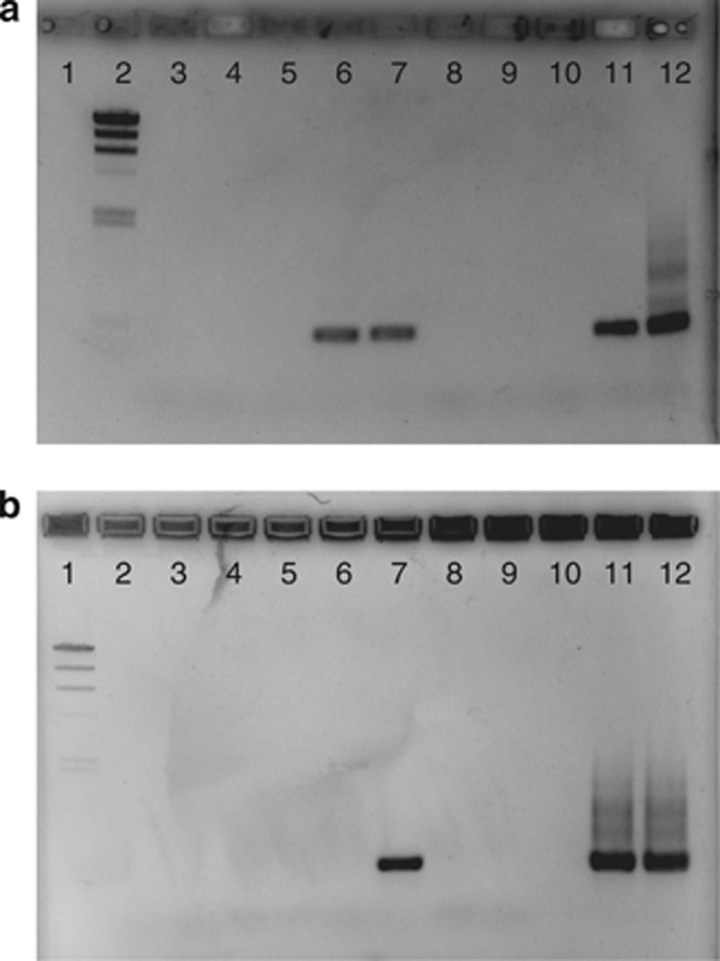
SIP is predicated on detecting a PCR signal in the 13C-carrier band when 13C-substrates are added, and not detecting a PCR signal in the 13C-carrier when no substrate or 12C-substrates are added, as shown in Figure 1a. In this incubation, crenarchaeal amplicons are only detected when 13C-lipids/pigments (lanes 1a-6 and 7) or 13C-ISOGRO at the higher concentration (lane 1a-11) had been added to the incubations. With carrier DNA alone (lane 1a-4), when no substrate has been added (lane 1a-5), or with 13C-whole proteins in the incubations (lanes 1a-8 and 9), no crenarchaeal PCR signal was observed. For all SIP incubations, a minimum incubation time of 5 days was required to observe this unambiguous 13C-uptake by crenarchaea. The 13C-genome replication was detected at both high and low concentrations of 13C-acetate, glycine, urea and the algal lipid/pigment extract. 13C-DNA synthesis was only detected at higher concentrations for 13C-ISOGRO. A summary of crenarchaeal 13C-incorporation is presented in Table 1.
Figure 1.
Agarose gel of crenarchaeal 16S rRNA gene amplicons from 13C-bands: a1) empty; a2) lambda DNA; a3) negative; a4) carrier; a5) no substrate; a6) 13C-lipid (2 mg ml−1); a7) 13C-lipid (10 mg ml−1); a8) 13C-protein (2 mg ml−1); a9) 13C-protein (10 mg ml−1); a10) 13C-ISOGRO (2 mg ml−1); a11) 13C-ISOGRO (10 mg ml−1); a12) positive control; b1) lambda DNA; b2) negative; b3) 13C-carrier; b4) T0 (time=0; sample taken before amendment); b5) no substrate; b6) 12C-urea; b7) 13C-urea; b8) 12C-bicarbonate; b9) 13C-bicarbonate; b10) empty; b11) inhibitor test (positive plus sample); b12) positive control.
Table 1. Summary of crenarchaeal TRFs representing at least 2% of the total community profile on the various 13C-carbon amendments.
| TRF |
Acetate |
Glycine |
Urea |
Lipids |
Proteins |
ISOGRO |
Bicarbonate | ||||||
|---|---|---|---|---|---|---|---|---|---|---|---|---|---|
| Length (bp) | Low | High | Low | High | Low | High | Low | High | Low | High | Low | High | |
| 58 | + | + | |||||||||||
| 66 | + | + | |||||||||||
| 76 | + | +++ | |||||||||||
| 78 | ++ | + | |||||||||||
| 84 | + | + | |||||||||||
| 89 | + | ++ | +++ | + | +++ | + | +++ | ||||||
| 91 | + | ++ | +++ | +++ | |||||||||
| 93 | + | + | + | ++ | ++ | ||||||||
| 101* | + | +++ | + | + | +++ | + | + | + | |||||
| 112 | + | + | |||||||||||
| 115* | +++ | + | +++ | +++ | +++ | +++ | +++ | ||||||
| 123* | ++ | + | |||||||||||
| 126 | + | + | |||||||||||
| 136 | + | ||||||||||||
| 152 | + | ||||||||||||
| 158* | +++ | + | + | + | + | + | |||||||
| 164 | + | ||||||||||||
| 166 | + | + | |||||||||||
| 173 | + | ||||||||||||
| 178 | + | + | |||||||||||
| 182 | + | + | |||||||||||
| 188 | |||||||||||||
| 192 | + | + | |||||||||||
| 207 | |||||||||||||
| 213 | + | ||||||||||||
| 217 | + | + | |||||||||||
| 223* | ++ | ||||||||||||
| 244 | + | + | |||||||||||
| 250* | +++ | + | + | ++ | |||||||||
| 252* | +++ | + | |||||||||||
Abbreviation: TRF, terminal restriction fragment.
The plus signs indicate: (+) 250–5000 arbitrary fluorescence units (a.f.u.); (++) 5000–10000 a.f.u.; (+++) >10 000 a.f.u. The asterisks (*) are TRFs represented in the clonal library.
Bacterial 13C-incorporation could also be detected in the SIP experiments. In 83% of the microcosms, bacterial 13C-DNA synthesis was observed, except for the low concentrations of urea and ISOGRO (Table 2). The relatively long SIP incubation time (5 days) most likely allowed for extensive cross-feeding between the bacteria (Gallagher et al., 2005) and the generation of 13C-CO2 (data not shown). To test whether the crenarchaea were actually taking up 13CO2 rather than the 13C-labeled substrates during our SIP experiment, a 13C-bicarbonate incubation was also conducted. The results are presented in Figure 1b. The 13C-bicarbonate treatment did not yield any crenarchaeal amplicon in the 13C-band (lane 1b-9). A test for PCR inhibitors by spiking a positive DNA sample with 13C-DNA from the bicarbonate incubation indicated no PCR suppression from this extract (lane 1b-11). These results demonstrate the SIP crenarchaeal signal resulted from the incorporation of 13C-organics, not 13C-labeled bicarbonate produced by bacterial respiration.
Table 2. Summary of bacterial TRFs representing at least 2% of the total community profile on the various 13C-carbon amendments.
| TRF | Acetate | Glycine | Urea | Lipids | Proteins | ISOGRO | ||||||
|---|---|---|---|---|---|---|---|---|---|---|---|---|
|
Length (bp) |
Low |
High |
Low |
High |
Low |
High |
Low |
High |
Low |
High |
Low |
High |
| 63 | + | + | ||||||||||
| 66 | + | |||||||||||
| 89 | ++ | |||||||||||
| 94 | + | |||||||||||
| 101 | + | |||||||||||
| 103 | + | + | ||||||||||
| 106 | ++ | |||||||||||
| 108 | + | |||||||||||
| 113 | + | |||||||||||
| 117 | + | |||||||||||
| 119 | + | + | ++ | + | ||||||||
| 126 | + | ++ | + | + | ||||||||
| 128 | + | |||||||||||
| 130 | + | + | + | |||||||||
| 134 | + | + | + | + | + | |||||||
| 138 | + | |||||||||||
| 142 | + | |||||||||||
| 145 | + | |||||||||||
| 148 | + | + | + | + | ||||||||
| 150 | + | + | + | |||||||||
| 153 | + | + | ||||||||||
| 162 | + | + | ||||||||||
| 164 | + | +++ | ++ | + | +++ | |||||||
| 167 | + | ++ | + | + | ||||||||
| 169 | + | + | ||||||||||
| 172 | + | |||||||||||
| 177 | + | |||||||||||
| 180 | + | + | + | + | ||||||||
| 182 | + | + | + | + | ||||||||
| 186 | + | + | + | ++ | + | |||||||
| 189 | + | ++ | ||||||||||
| 192 | +++ | + | +++ | |||||||||
| 198 | +++ | + | ||||||||||
| 200 | +++ | + | + | |||||||||
| 204 | +++ | + | + | + | ||||||||
| 206 | + | + | ||||||||||
| 208 | ++ | + | + | |||||||||
| 210 | + | + | + | + | + | |||||||
| 213 | + | + | + | + | + | |||||||
| 215 | + | |||||||||||
| 216 | + | + | + | + | + | + | ||||||
| 219 | + | + | ||||||||||
| 225 | + | + | + | |||||||||
| 227 | + | |||||||||||
| 235 | ++ | |||||||||||
| 237 | + | |||||||||||
| 240 | + | + | + | + | + | + | ||||||
| 246 | +++ | |||||||||||
| 248 | + | + | ||||||||||
| 250 | + | ++ | + | +++ | ++ | +++ | ||||||
Abbreviation: TRF, terminal restriction fragment.
The plus signs indicate: (+) 250–5000 arbitrary fluorescence units (a.f.u.); (++) 5000–10 000 a.f.u.; (+++) >10 000 a.f.u.
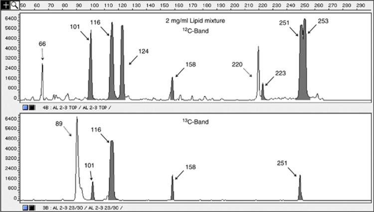
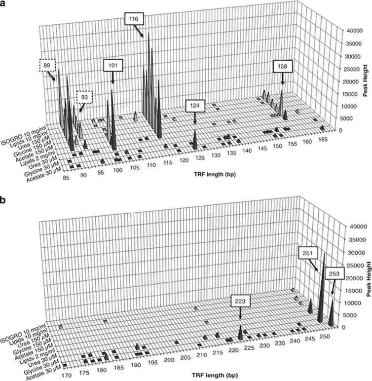
To ascertain which specific members of the crenarchaeal community were actively synthesizing DNA from the 13C-carbon sources, TRFLP analysis of the 16S rRNA gene amplicons was performed (see example in Figure 2). A small number of crenarchaeal peaks (5–10) was found to dominate the overall community profiles for both the 12C- and 13C-bands (Supplementary Figures 1–4). For lipids, TRFs 66, 124, 220, 223 and 253 bp were present in the 12C-fraction, but were not detected in the 13C-fraction. Other TRFs (101, 116, 158, 251 bp) were detected in both 12C- and 13C-fractions, whereas the TRF 89 bp was mainly observed only in the 13C-fraction. A compilation of all crenarchaeal fingerprints from our SIP experiments is provided in Figure 3. It can be seen that many of the 13C-TRFs (89, 93, 116 and 158 bp) are detected in nearly all SIP experiments, with the exception of the 30 μM glycine, urea and acetate treatments (Figure 3a). There was a general pattern of lower peak area for the lower concentrations of 13C-substrates and higher peak area for the higher concentration amendments for these particular TRFs. In contrast, other crenarchaeal TRFs were only detected in the 13C-labeled community under specific treatments. For example, TRFs (124, 223, 253 bp) were found in the 30 μM acetate-amended microcosms, but did not appear to be active in any other treatment. Overall, the crenarchaeota present in the salt marsh sediment appear to be much less diverse (in terms of operational taxonomic units) than the bacteria. The average number of peaks at the lower TRFLP peak detection settings in the archaeal 13C-community was 23±5, whereas the bacterial 13C-community profiles averaged 42±14 (data not shown).
Figure 2.
Example of TRFLP profiles of crenarchaeal 16S rRNA genes in 12C- and 13C-bands from lipid amendment. Highlighted peaks (gray) are represented in the clonal library.
Figure 3.
A compilation of crenarchaeal fingerprints with TRFs from 85 to 169 bp (a) and 170–254 bp (b) in length. Major peak sizes are indicated by boxes (solid, detected in the clone library; dashed, not detected). Light shades indicate higher concentration amendments; dark shades indicate lower concentration amendments.
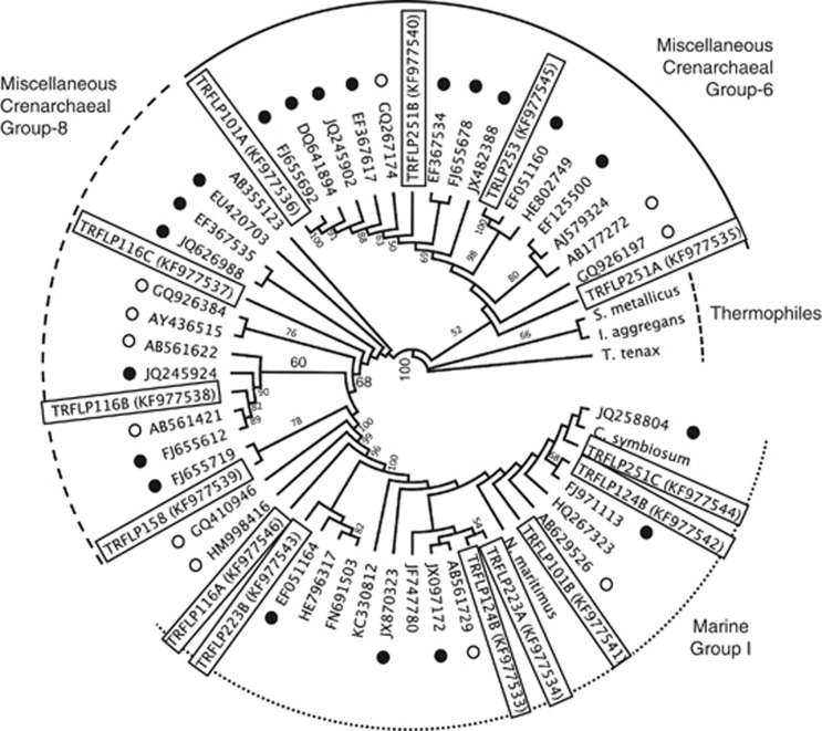
Screening of a clone library from the 13C-bands yielded seven of the observed TRFLP peaks within our SIP profiles and have been highlighted in gray (Figure 2). Specifically, three clones matched the 101 bp peak (two unique sequences), five clones matched the 116 bp peak (three unique sequences), four clones matched the 124 bp peak (two unique sequences), one clone matched the 159 bp peak, four clones matched the 223 bp peak (two unique sequences), six clones matched the 251 bp peak (three unique sequences) and one clone matched the 253 bp peak. Three major peaks (66, 89 and 93 bp) were not detected in our clonal library. The cloned crenarchaeal 16S rRNA genes were sequenced, aligned and a phylogenetic tree was re-constructed using maximum likelihood methods (Figure 4). Seven of these sequenced clones grouped with uncultured crenarchaeota from marine environments, belonging to the Miscellaneous Crenarcheotal Group (MCG-6 and MCG-8; Kubo et al., 2012). The other seven clones detected in our clone library grouped with members of the Marine Group 1.a Crenarchaeota. Several of the peaks in our TRFLP profiles yielded more than one clone, suggesting that archaeal diversity may be more extensive than our profiles indicate, and additional enzyme digests are required for higher TRFLP resolution.
Figure 4.
Phylogenetic estimation using Maximum Likelihood (PhyML) tree with support from 100 bootstrap runs indicated. The clones in this study are indicated with TRF size and GenBank accession numbers. Black dots represent sequences found in coastal or estuarine sediments. Open dots signify sequences from the marine deep biosphere.
Discussion
SIP methodology has only recently been used to elucidate the metabolic potential of archaea. Most of these studies have focused on autotrophy and ammonia oxidation in various environments such as rice paddies (Lu and Conrad, 2005), soils (Adair and Schwartz, 2011; Pratscher et al., 2011; Lu and Jia, 2013) and in freshwater sediment (Wu et al., 2013). However, there are also reports of heterotrophic activity from an acidic fen (Hamberger et al., 2008) and an estuarine setting in the UK (Webster et al., 2010). The fen study utilized soil samples suspended in a minimal salt media, pre-incubated for 8 days and supplemented with 13C-xylose or 13C-glucose. After 13 days, most archaeal clones associated with the 13C-fraction were methanogens, whereas 25% of the colonies screened (n=16) were identified as being crenarchaea. Likewise, in the Severn Estuary SIP study, various sediments were added to a minimal salt media and amended with 13C-glucose, 13C-acetate and 13CO2 for up to 14 days (Webster et al., 2010). There was no archaeal CO2 uptake observed in sediment taken from the methanogenic zone, and after 13C-glucose amendment the aerobic zones were highly similar in the 12C- and the 13C-fractions. However, in sediments from the sulfate-reducing zone amended with 13C-acetate, active members of the Miscellaneous Crenarchaeal Group were detected, which were clearly enriched when compared with the 12C-fraction.
In this report, SIP microcosms were made using filter-sterilized site water and the incubations were concluded at 5 days. The crenarchaeota of the Cheesequake salt marsh were found to be heterotrophic, with no 13C-uptake from bicarbonate over the time frame of the experiment. Certain crenarchaeal TRFs were detected in the 13C-fraction from nearly every microcosm amended with organic 13C-carbon, regardless of the type of substrate provided. This suggests these particular crenarchaea are able to assimilate a wide variety of organic substrates: simple metabolic intermediates (acetate), amino acids (glycine), organic nitrogen compounds (urea), lipids and pigments, and complex mixtures of biopolymers (ISOGRO). Other crenarchaeal TRFs, however, were only detected in the 13C-bands from microcosms amended with a specific substrate (acetate or urea) and may have more restricted metabolic capabilities.
Interestingly, bacterial and crenarchaeal populations seem to be competing for the same resources in our SIP incubations. Members from both domains were active on acetate, glycine, lipids, and high levels of urea and ISOGRO (Tables 1, 2). The only resource partitioning for active microbes was observed in microcosms amended with whole proteins (used exclusively by bacteria) and at low concentrations of urea (used exclusively by crenarchaea). The former result was surprising, considering recent single-cell genome sequencing of a member of the Miscellaneous Crenarchaeal Group (MCG) from marine sediment discovered several genes for extracellular cysteine peptidases (Lloyd et al., 2013). This finding underscores the need for direct incubations (such as SIP) to determine metabolic capabilities, in addition to assessing genetic potential by sequence analysis at the genome level. Likewise, it was surprising that bacteria did not take up any carbon from urea at low concentrations, whereas the crenarchaea did. There is ample evidence of bacteria possessing urease genes (Collier et al., 2009; Solomon et al., 2010) and reports of Campylobacter nitrofigilis isolated from Spartina with urease activity along the United States eastern seaboard (McClung et al., 1983). While no bacterial activity was detected at 30 μM on urea, there was bacterial activity at 150 μM urea. This result implies the crenarchaea are taking up virtually all of the urea at the lower concentration, preventing the bacteria from accessing this substrate for heterotrophic (or autotrophic) growth.
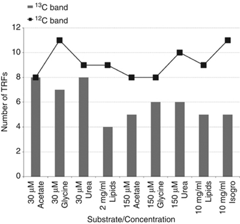
While the type of 13C-substrate influenced the crenarchae replicating their DNA, the concentration of 13C-substrate also had a marked effect on the profiles of the active crenarchaeal community. Specifically, low concentrations of substrate produced more active TRFs compared with microcosms amended with high concentrations (Figure 5). Higher concentrations of acetate (150 μM) produced communities that were dominated by three TRFs (101, 116 and 158 bp) comprising 68% of the community profile. High concentrations of glycine (150 μM) yielded an active community in which two of the same TRFs (116 and 158 bp) accounted for 80% of the community profile, whereas 150 μM urea yielded 89 and 116 bp peaks, which also dominated the community profile (80%). There are several possible explanations for this result. One is that these particular crenarchaea are very successful at competing for substrates at high concentration and their transport systems internally mobilize virtually all of the substrate. This prevents other microorganisms from accessing the 13C-organic carbon and synthesizing DNA. If this is the case, there must be a tipping point between low concentrations (tens of micromolar) and higher concentrations (hundreds of micromolar), which triggers a hoarding response by certain crenarchaea. Another possibility is that minor TRFs in the 13C-carrier band are having their signal suppressed by the dominant TRF in the high concentration SIP experiment. Although our fingerprinting methods are highly reproducible (McGuinness et al., 2006; Tuorto et al., 2013), there is a suppression of smaller peaks in the TRFLP profile when screening clone libraries (Babcock et al., 2007) or samples where one microorganism is >10% of the community. This same mechanism may be inhibiting the detection of other active crenarchaea that are not synthesizing large amounts of DNA in our SIP incubations. Another explanation is that low concentrations of some carbon compounds, such as urea, may not be bioavailable to the bacteria. Our low concentration ISOGRO additions did not produce any 13C-archaeal or 13C-bacterial signal. However, we know that 2 mg ml−1 of ISOGRO in liquid culture will allow for bacterial growth. It is possible that the salt marsh sediments are preventing the bioremineralization of proteins and other biomolecules at low ISOGRO concentrations, as has been reported by Keil et al. (1994). Alternatively, uptake of 13C-substrates at low concentrations may be observed given a longer incubation.
Figure 5.
Total number of TRFs in the 12C-band (line) and in the 13C-band (gray bars) for peaks representing more than 2% of the community profile.
From the incubation time of our SIP experiments, it is also possible to estimate crenarchaeal growth parameters in situ. Assuming two doubling events are required for complete 13C-labeling of DNA and detection in our 100%-labeled 13C-carrier band, this suggests salt marsh crenarchaea have a doubling time on the order of 2–2.5 days. Prior studies have measured the growth rate of cultured ammonia-oxidizing crenarchaea (Könneke et al., 2005) and/or inferred doubling times by determining copy numbers of 16S rRNA (Park et al., 2010). These reports indicate a maximum growth rate of ammonia oxidizing archaea (AOA) in liquid medium at 25–28 °C between 0.57 and 0.78 per day (or a doubling rate between 1.28 and 1.75 days). In contrast, our findings are consistent with prior observations of slightly slower growth rates in sediment (0.2 per day, Mosier et al. (2012)). However, these results may be an overestimate of the potential growth rate, as SIP requires that all precursor pools for DNA synthesis become uniformly labeled with 13C for detection.
In conclusion, many mesothermal Crenarchaea (Thaumarchaea) in salt marsh sediments were found to assimilate a wide variety of organic 13C-substrates to replicate their genomes. Other crenarchaea were much more selective in the 13C-carbon sources used for growth. The SIP approach demonstrates salt marshes can be used for studying crenarchaeal metabolic capabilities and may help in determining laboratory conditions for isolating these heterotrophic microorganisms. Once in pure culture, traditional microbiological techniques can be utilized to improve our understanding of crenarchaeal physiological capabilities. Finally, experiments that directly link carbon-utilization patterns with specific members of the microbial community can provide insight into the competitive relationships between crenarchaea and bacteria and improve our understanding of microbial ecology.
Acknowledgments
The authors wish to thank Dr S.K. Rhee for his preliminary work on culturing Crenarchaeota in our lab and the Cheesequake State Park rangers for their cooperation during this project. This research was partially supported by funds from Rutgers University and from the National Science Foundation (1131022) in Ocean Technology and Interdisciplinary Coordination to LJK and Dr Jingang Yi.
The authors declare no conflict of interest.
Footnotes
Supplementary Information accompanies this paper on The ISME Journal website (http://www.nature.com/ismej)
Supplementary Material
References
- Abreu C, Jurgens G, De Marco P, Saana A, Bordalo AA. Crenarchaeota and Euryarchaeota in temperate estuarine sediments. J Appl Microbiol. 2001;90:713–718. doi: 10.1046/j.1365-2672.2001.01297.x. [DOI] [PubMed] [Google Scholar]
- Adair KL, Schwartz E.2011Stable isotope probing with 18O-water to investigate growth and mortality of ammonia oxidizing bacteria and archaea in soilIn: Klotz MG, (ed).Methods in Enzymology: Research on Nitrification and Related Processes Academic Press: San Diego, CA, USA; 155–169. [DOI] [PubMed] [Google Scholar]
- Avaniss-Aghajani E, Jones K, Chapman D, Brunk C. A molecular technique for identification of bacteria using small subunit ribosomal RNA sequences. Biotechniques. 1994;17:144–146. [PubMed] [Google Scholar]
- Babcock DA, Wawrik B, Paul JH, McGuinness LR, Kerkhof LJ. Rapid screening of a large insert BAC library for specific 16S rRNA genes using TRFLP. J Microbiol Methods. 2007;71:156–161. doi: 10.1016/j.mimet.2007.07.015. [DOI] [PubMed] [Google Scholar]
- Biddle JF, Lipp JS, Lever MA, Lloyd KG, Sørenson KB, Anderson R, et al. Heterotrophic Archaea dominate sedimentary subsurface ecosystems off Peru. Proc Natl Acad Sci USA. 2006;103:3846–3851. doi: 10.1073/pnas.0600035103. [DOI] [PMC free article] [PubMed] [Google Scholar]
- Biddle JF, Fitz-Gibbon S, Schuster SC, Brenchley JE, House CH. Metagenomic signatures of the Peru Margin subseafloor biosphere show a genetically distinct environment. Proc Natl Acad Sci USA. 2008;105:10583–10588. doi: 10.1073/pnas.0709942105. [DOI] [PMC free article] [PubMed] [Google Scholar]
- Birtrim SB, Donohue TJ, Handelsman J, Roberts GP, Goodman RM. Molecular phylogeny of Archaea from soil. Proc Natl Acad Sci USA. 1997;94:277–282. doi: 10.1073/pnas.94.1.277. [DOI] [PMC free article] [PubMed] [Google Scholar]
- Collier JL, Baker KM, Bell SL. Diversity of urea-degrading microorganisms in open-ocean and estuarine planktonic communities. Environ Microbiol. 2009;12:3118–3131. doi: 10.1111/j.1462-2920.2009.02016.x. [DOI] [PubMed] [Google Scholar]
- DeLong EF. Archaea in coastal marine environments. Proc Natl Acad Sci USA. 1992;89:5685–5689. doi: 10.1073/pnas.89.12.5685. [DOI] [PMC free article] [PubMed] [Google Scholar]
- DeLong EF, Wu KY, Prézelin BB, Jovine RVM. High abundance of Archaea in Antarctic marine picoplankton. Nature. 1994;371:695–697. doi: 10.1038/371695a0. [DOI] [PubMed] [Google Scholar]
- Drummond AJ, Ashton B, Cheung M, Heled J, Kearse M, Moir R, et al. 2009. Geneious v3.8 and v4.6, available from: http://www.geneious.com .
- Emerson S, Hedges J.2008Chemical Oceanography and the Marine Carbon Cycle Cambridge University Press; p453 [Google Scholar]
- Francis CA, Roberts KJ, Beman JM, Santoro AE, Oakley BB. Ubiquity and diversity of ammonia- oxidizing archaea in water columns and sediments of the ocean. Proc Natl Acad Sci USA. 2005;102:14683–14688. doi: 10.1073/pnas.0506625102. [DOI] [PMC free article] [PubMed] [Google Scholar]
- Fuhrman JA, McCallum K, Davis AA. Novel major archaebacterial group from marine plankton. Nature. 1992;356:148–149. doi: 10.1038/356148a0. [DOI] [PubMed] [Google Scholar]
- Gallagher E, McGuinness L, Phelps C, Young LY, Kerkhof LJ. 13C carrier DNA shortens the incubation time needed to detect benzoate-utilizing denitrifying bacteria by stable-isotope probing. Appl Environ Microbiol. 2005;71:5192–5196. doi: 10.1128/AEM.71.9.5192-5196.2005. [DOI] [PMC free article] [PubMed] [Google Scholar]
- Gonzalez JM, Portilo MC, Saiz-Jimenez C. Metabolically active Crenarchaeota in Altamira Cave. Naturwissenschaften. 2006;93:42–45. doi: 10.1007/s00114-005-0060-3. [DOI] [PubMed] [Google Scholar]
- Guindon S, Gascuel O. A simple, fast, and accurate algorithm to estimate large phylogenies by maximum likelihood. Syst Biol. 2003;52:696–704. doi: 10.1080/10635150390235520. [DOI] [PubMed] [Google Scholar]
- Hallam SJ, Mincer TJ, Schleper C, Preston CM, Roberts K, Richardson PM, et al. Pathways of carbon assimilation and ammonia oxidation suggested by environmental genomic analyses of marine Crenarchaeota. PLoS Biol. 2006;4:520–536. doi: 10.1371/journal.pbio.0040095. [DOI] [PMC free article] [PubMed] [Google Scholar]
- Hamberger A, Horn MA, Dumont MG, Murrell JC, Drake HL. Anaerobic consumers of monosaccharides in a moderately acidic fen. Appl Environ Microbiol. 2008;74:3112–3120. doi: 10.1128/AEM.00193-08. [DOI] [PMC free article] [PubMed] [Google Scholar]
- Herndl GJ, Reinthaler T, Teira E, van Aken H, Veth C, Pernthaler A, et al. Contribution of Archaea to total prokaryotic production in the deep Atlantic Ocean. Appl Environ Microbiol. 2005;71:2303–2309. doi: 10.1128/AEM.71.5.2303-2309.2005. [DOI] [PMC free article] [PubMed] [Google Scholar]
- Inagaki F, Suzuki M, Takai K, Oida H, Sakamoto T, Aoki K, et al. Microbial communities associated with geological horizons in coastal subseafloor sediments from the Sea of Okhotsk. Appl Environ Microbiol. 2003;69:7224–7235. doi: 10.1128/AEM.69.12.7224-7235.2003. [DOI] [PMC free article] [PubMed] [Google Scholar]
- Ingalls AE, Shah SR, Hansman RL, Aluwihare LI, Santos GM, Druffel ERM, et al. Quantifying archaeal community autotrophy in the mesopelagic ocean using natural radiocarbon. Proc Natl Acad Sci USA. 2006;103:6442–6447. doi: 10.1073/pnas.0510157103. [DOI] [PMC free article] [PubMed] [Google Scholar]
- Karner MB, DeLong EF, Karl DM. Archaeal dominance in the mesopelagic zone of the Pacific Ocean. Nature. 2001;409:507–510. doi: 10.1038/35054051. [DOI] [PubMed] [Google Scholar]
- Keil RG, Montluçon DB, Prahl FG, Hedges JI. Sorptive preservation of labile organic matter in marine sediments. Nature. 1994;370:549–552. [Google Scholar]
- Kerkhof LJ, Williams KH, Long PE, McGuinness LR. Phase preference by active, acetate-utilizing bacteria at the Rifle CO integrated field research challenge site. Environ Sci Techol. 2011;45:1250–1256. doi: 10.1021/es102893r. [DOI] [PubMed] [Google Scholar]
- Könneke M, Bernhard AE, de la Torre JR, Walker CB, Waterbury JB, Stahl DA. Isolation of an autotrophic ammonia-oxidizing marine archaeon. Nature. 2005;437:543–546. doi: 10.1038/nature03911. [DOI] [PubMed] [Google Scholar]
- Kubo K, Lloyd KG, Biddle JF, Amann R, Teske A, Knittel K. Archaea of the Miscellaneous Crenarchaeotal Group are abundant, diverse and widespread in marine sediments. ISME J. 2012;6:1949–1965. doi: 10.1038/ismej.2012.37. [DOI] [PMC free article] [PubMed] [Google Scholar]
- Lloyd KG, Schreiber L, Petersen DG, Kjeldsen KU, Lever MA, Steen AD, et al. Predominant archaea in marine sediments degrade detrital proteins. Nature. 2013;496:215–218. doi: 10.1038/nature12033. [DOI] [PubMed] [Google Scholar]
- Lu Y, Conrad R. In situ stable isotope probing of methanogenic archaea in the rice rhizosphere. Science. 2005;309:1088–1090. doi: 10.1126/science.1113435. [DOI] [PubMed] [Google Scholar]
- Lu L, Jia Z. Urease gene-containing Archaea dominate autotrophic ammonia oxidation in two acid soils. Environ Microbiol. 2013;15:1795–1809. doi: 10.1111/1462-2920.12071. [DOI] [PubMed] [Google Scholar]
- Martin-Cuadrado AB, Rodriguez-Valera F, Moreira D, Alba JC, Ivars-Martínez E, Henn MR, et al. Hindsight in the relative abundance, metabolic potential and genome dynamics of uncultivated marine archaea from comparative metagenomic analyses of bathypelagic plankton of different oceanic regions. ISME J. 2008;2:865–886. doi: 10.1038/ismej.2008.40. [DOI] [PubMed] [Google Scholar]
- Massana R, Murray AE, Preston CM, DeLong EF. Vertical distribution and phylogenetic characterization of marine planktonic Archaea in the Santa Barbara Channel. Appl Environ Microbiol. 1997;63:50–56. doi: 10.1128/aem.63.1.50-56.1997. [DOI] [PMC free article] [PubMed] [Google Scholar]
- Massana R, DeLong EF, Pedrós-Alió C. A few cosmopolitan phylotypes dominate planktonic archaeal assemblages in widely different oceanic provinces. Environ Microbiol. 2000;66:1777–1787. doi: 10.1128/aem.66.5.1777-1787.2000. [DOI] [PMC free article] [PubMed] [Google Scholar]
- McClung CR, Patriquin DG, Davis RE. Campylobacter nitrofigilis sp. nov., a nitrogen-fixing bacterium associated with roots of Spartina alternaflora loisel. IJSEM. 1983;33:605–612. [Google Scholar]
- McGuinness LM, Salganik M, Vega L, Pickering KD, Kerkhof LJ. Replicability of bacterial communities in denitrifying bioreactors as measured by PCR/T-RFLP analysis. Environ Sci Technol. 2006;40:509–515. doi: 10.1021/es050900l. [DOI] [PubMed] [Google Scholar]
- Mosier AC, Lund MB, Francis CA. Ecophysiology of an ammonia-oxidizing archaeon adapted to low-salinity habitats. Microb Ecol. 2012;64:955–963. doi: 10.1007/s00248-012-0075-1. [DOI] [PubMed] [Google Scholar]
- Murray AE, Preston CM, Massana R, Taylor LT, Blakis A, Wu K, et al. Seasonal and spatial variability of bacterial and archaeal assemblages in the coastal waters near Anvers Island, Antarctica. Appl Environ Microbiol. 1998;64:2585–2595. doi: 10.1128/aem.64.7.2585-2595.1998. [DOI] [PMC free article] [PubMed] [Google Scholar]
- Nelson KA, Moin NS, Bernhard AE. Archaeal diversity and the prevalence of Crenarchaeota in salt marsh sediments. Appl Environ Mircobiol. 2009;75:4211–4215. doi: 10.1128/AEM.00201-09. [DOI] [PMC free article] [PubMed] [Google Scholar]
- Ouverney CC, Fuhrman JA. Marine planktonic archaea take up amino acids. Appl Environ Microbiol. 2000;66:4829–4833. doi: 10.1128/aem.66.11.4829-4833.2000. [DOI] [PMC free article] [PubMed] [Google Scholar]
- Park BJ, Park SJ, Yoon DN, Schouten S, Sinninghe Damsté JS, Rhee SK. Cultivation of autotrophic ammonia-oxidzing archaea from marine sediments in co-culture with sulfur-oxidizing bacteria. Appl Environ Microbiol. 2010;76:7575–7587. doi: 10.1128/AEM.01478-10. [DOI] [PMC free article] [PubMed] [Google Scholar]
- Pratscher J, Dumont MG, Conrad R. Ammonia oxidation coupled to CO2 fixation by archaea and bacteria in an agricultural soil. Proc Natl Acad Sci USA. 2011;108:4170–4175. doi: 10.1073/pnas.1010981108. [DOI] [PMC free article] [PubMed] [Google Scholar]
- Radajewski S, Ineson P, Parekh NR, Murrell JC. Stable-isotope probing as a tool in microbial ecology. Nature. 2000;403:646–649. doi: 10.1038/35001054. [DOI] [PubMed] [Google Scholar]
- Schleper C, Jurgens G, Jonuscheit M. Genomic studies of uncultivated archaea. Nat Rev Microbiol. 2005;3:479–488. doi: 10.1038/nrmicro1159. [DOI] [PubMed] [Google Scholar]
- Solomon CM, Collier JL, Berg GM, Glibert PM. Role of urea in microbial metabolism in aquatic system: a biochemical and molecular review. Aquat Microb Ecol. 2010;59:67–88. [Google Scholar]
- Søresnson KB, Teske A. Stratified communities of active archaea in deep marine subsurface sediments. Appl Environ Microbiol. 2006;72:4596–4603. doi: 10.1128/AEM.00562-06. [DOI] [PMC free article] [PubMed] [Google Scholar]
- Stein JL, Simon MI. Archaeal ubiquity. Proc Natl Acad Sci USA. 1996;93:6228–6230. doi: 10.1073/pnas.93.13.6228. [DOI] [PMC free article] [PubMed] [Google Scholar]
- Teske A, Hinrichs KU, Edgcomb V, Gomez ADV, Kysela D, Sylva SP, et al. Microbial diversity of hydrothermal sediments in the Guaymas Basin: evidence for anaerobic methanotrophic communities. Appl Environ Microbiol. 2002;68:1994–2007. doi: 10.1128/AEM.68.4.1994-2007.2002. [DOI] [PMC free article] [PubMed] [Google Scholar]
- Treusch AH, Leininger S, Kletzin A, Schuster SC, Klenk HP, Schleper C. Novel genes for nitrite reductase and Amo-related proteins indicate a role of uncultivated mesophilic crenarchaeota in nitrogen cycling. Environ Microbiol. 2005;7:1985–1995. doi: 10.1111/j.1462-2920.2005.00906.x. [DOI] [PubMed] [Google Scholar]
- Tuorto SJ, Darias P, McGuinness LR, Panikov N, Zhang T, Häggblom MM, et al. Bacterial Genome Replication at Subzero Temperatures in Permafrost. ISME J. 2013;8:139–149. doi: 10.1038/ismej.2013.140. [DOI] [PMC free article] [PubMed] [Google Scholar]
- Venter JC, Remington K, Heidelberg JF, Halpern AL, Rusch D, Eisen JA, et al. Environmental genome shotgun sequencing of the Sargasso Sea. Science. 2004;304:66–74. doi: 10.1126/science.1093857. [DOI] [PubMed] [Google Scholar]
- Vetriani C, Jannasch H, MacGregor B, Stahl D, Reysenbach A. Population structure and phylogenetic characterization of marine benthic Archaea in deep-sea sediments. Appl Environ Microbiol. 1999;65:4375–4384. doi: 10.1128/aem.65.10.4375-4384.1999. [DOI] [PMC free article] [PubMed] [Google Scholar]
- Vetriani C, Tran H, Kerkhof L. Fingerprinting microbial assemblages from the oxic/anoxic chemocline of the Black Sea. Appl Environ Microbiol. 2003;69:6481–6488. doi: 10.1128/AEM.69.11.6481-6488.2003. [DOI] [PMC free article] [PubMed] [Google Scholar]
- Webster G, Rinna J, Roussel EG, Fry JC, Weightman AJ, Parkes RJ. Prokaryotic functional diversity in different biochemical depth zones in tidal sediments of the Severn Estuary, UK, revealed by stable-isotope probing. FEMS Microbiol Ecol. 2010;72:179–197. doi: 10.1111/j.1574-6941.2010.00848.x. [DOI] [PubMed] [Google Scholar]
- Wuchter C, Schouten S, Boschker HTS, Damsté JSS. Bicarbonate uptake by marine Crenarchaeota. FEMS Microbiol Lett. 2003;219:203–207. doi: 10.1016/S0378-1097(03)00060-0. [DOI] [PubMed] [Google Scholar]
- Wu Y, Ke X, Hernandez M, Wang B, Dumont MG, Jia Z, et al. Autotrophic growth of bacterial and archaeal ammonia oxidizers in freshwater sediment microcosm incubated at different temperatures. Appl Environ Microbiol. 2013;79:3076–3084. doi: 10.1128/AEM.00061-13. [DOI] [PMC free article] [PubMed] [Google Scholar]
Associated Data
This section collects any data citations, data availability statements, or supplementary materials included in this article.


