

Abstract
Gastroparesis is a condition which results in delayed gastric emptying without gastric outflow tract obstruction. Gastrointestinal involvement in diabetes can present in various forms such as oesophageal dysmotility, gastro-oesophageal reflux disease, gastroparesis, enteropathy, non-alcoholic fatty liver disease and glycogenic hepatopathy. Gastroparesis is the most common gastric motility disorder complicating long-standing diabetes. It can sometimes lead to mechanical obstruction as a result of formation of bezoars. Phytobezoars are the most common type of bezoar and are composed of indigestible food, vegetable fibre or seeds. Poor pyloric function and decreased acid formation predisposes phytobezoars formation in patients with diabetic gastroparesis. An 80-year-old patient with diabetes in our presentation developed gastric outlet obstruction due to impaction of phytobezoar over the pylorus.
Background
Gastroparesis results in delayed gastric emptying without true gastric outflow tract obstruction. Important causes leading to gastroparesis are diabetes, idiopathic, postsurgical1 scleroderma, Ehlers-Danlos syndrome and Parkinson's disease.2 Gastrointestinal (GI) involvement in diabetes can present in various ways such as oesophageal dysmotility, gastro-oesophageal reflux disease, gastroparesis, enteropathy, non-alcoholic fatty liver disease and glycogenic hepatopathy. Gastroparesis presents with features of early satiety, nausea, vomiting and epigastric fullness.3 The pathophysiology of gastroparesis in diabetes mellitus is not well understood.4 Long-standing diabetes mellitus for more than 10 years predisposes a patient to gastroparesis and usually is associated with involvement of the retina, kidney and nerves. Gastroparesis is reported to occur in 5–12% of patients with long-standing diabetes.5 In diabetic gastroparesis, basic mechanisms of gastric emptying are affected, due to the involvement of the vagus nerve (autonomic neuropathy).6 Gastric outlet obstruction can occur as a result of mechanical gastroduodenal obstruction or motility disorders. Mechanical obstruction can be benign or malignant. Benign obstruction is usually secondary to peptic ulcer disease with or without secondary stricture.7 Malignancy is commonly caused due to cancer affecting the distal stomach or the duodenum.8 Gastroparesis is the most common gastric motility disorder complicating long-standing diabetes. Other causes of gastroparesis could be idiopathic, viral or medications.9 It can sometimes lead to mechanical obstruction as a result of the formation of bezoars. These bezoars are collections of indigestible foreign material which are accumulated anywhere in the gastrointestinal tract.10 Phytobezoars are the most common type of bezoar and is composed of indigestible fruit, vegetable fibres or seeds.11 These are formed in diabetic gastroparesis due to poor pyloric function and decrease in acid formation.12 A patient in our presentation developed mechanical gastric outlet obstruction due to impaction of phytobezoars over the pylorus. Association of diabetic gastroparesis with bezoar formation is well known. This case has also revealed the same, but this type of cases are not been reported from northeastern part of India, so it was worth reporting this case as a reminder of common clinical association.
Case presentation
An 80-year-old man, non-alcoholic and non-smoker, came to medicine outpatient department with symptoms of continuous vomiting containing undigested food material for the past 4 days with pain over the epigastrium. He reported nausea, early satiety and vague epigastric discomfort and fullness for the past 6 months with intermittent self-induced vomiting. He also gave a history of weight loss. On taking detailed medical history he was found to be a known case of diabetes mellitus and hypertension for the past 30 years and was on medical management in the form of glimepiride 2 mg, metformin 1000 mg, telmisartan 40 mg, aspirin 75 mg and atorvastatin 10 mg. On examination, his pulse was 102 bpm, blood pressure 100/70 mm Hg, and was found dehydrated and ill-looking. There was mild pallor with no icterus, cyanosis, clubbing, pedal oedema or lymphadenopathy. The patient's per abdomen examination revealed distended epigastrium with tenderness and mild guarding and increased bowel sounds. Cardiovascular, respiratory, central nervous system and genitourinary systems were normal. He was admitted with provisional diagnosis of gastric outlet obstruction/intestinal obstruction and was started on conservative management and worked up further. The patient's laboratory reports showed a haemoglobin concentration of 10 g%, total leucocyte count 5200 differential leucocyte count P72% L23% M2% E3% with normal platelet counts. Blood urea was 68 mg/dL, creatinine 1.5 mg/dL with normal electrolytes. Random blood sugar was 248 mg/dL, glycosylated haemoglobin of 7.3% and urine report revealed, sugar 1+, protein 1+ with no ketone bodies. His fasting blood sugar was 131 mg% and the postprandial sugar level was 218 mg%. His funduscopy showed diabetic retinopathy. Other tests such as liver function test, lipid profile and thyroid profile were normal. An X-ray of the abdomen did not show any features of intestinal obstruction. An X-ray of the chest was normal. An ultrasound of the abdomen revealed only distended stomach with fluid and food matters. Upper gastrointestinal endoscopy showed large food bolus (bezoar) impacted over the pylorus (figure 1) causing complete gastric outlet obstruction with stagnant fluid and food material over the fundus and body of the stomach. Removal of the bezoar was tried but could not be removed as it was tightly impacted. He was referred to another well-equipped centre, where the impacted bezoar was removed endoscopically with difficulty using polypectomy snare (figure 2). Duodenum was found to be normal and patent (figure 3). Rapid urease test for Helicobacter pylori was negative. After endoscopic intervention and removal of impacted bezoar, the patient improved dramatically, vomiting stopped and tolerance to food increased but sense of epigastric fullness and early satiety persisted. He underwent barium meal study which showed delayed gastric emptying. He was started on domperidone 10 mg thrice daily half an hour before meals. Dietary modification was advised in the form of low-fibre, low-residue and low-fat diet with small frequent meals with proper mastication and strict glycaemic control. Significant improvement was seen with this treatment and the patient was advised to follow-up regularly.
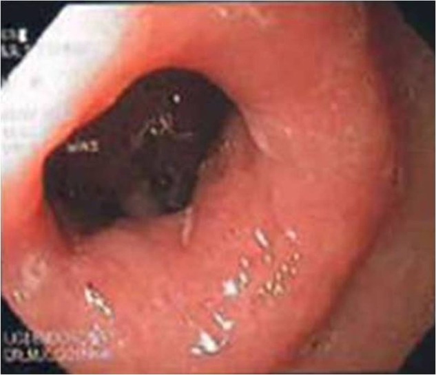
Figure 1.

Endoscopy showing large phytobezoar impacted over the pylorus.
Figure 2.

Process of removal of impacted phytobezoar.
Figure 3.
Normal pylorus and duodenum after removal of phytobezoar.
Treatment
Endoscopic removal of impacted bezoar was performed. The patient was advised to take prokinetic drug domperidone 10 mg thrice daily before food and dietary modification was advised in the form of low-fibre, low-residue and low-fat diet with small frequent meals with proper mastication and strict glycaemic control. Significant improvement was seen with this treatment and the patient was advised to follow-up regularly. The patient is in regular follow-up since the past 6 months and till now there is no recurrence of symptoms.
Outcome and follow-up
Significant improvement was seen with the treatment and the patient is in regular follow-up for the past 6 months and till now there is no recurrence.
Discussion
Gastroparesis is the most common gastric motility disorder and is often seen in long-standing diabetes mellitus.9 Specific-genetic mutations resulting in failure of proper migration and differentiation of enteric neurones are thought to be the cause of gastroparesis. Decreased nitric oxide production in patients with diabetes may also lead to intestinal dysfunction manifesting as gastroparesis, diarrhoea or constipation.13 Autonomic dysfunction in these patients also predisposes to gastroparesis.6 This can be complicated by gastric outlet obstruction as a result of formation of bezoars. Bezoars are concretions of indigestible foreign material trapped in the GI tract. These are most commonly formed in the stomach followed by small bowel. Colon, rectum or oesophagus are some unusual locations.10 On searching publications, we found various case reports of bezoars causing gastric outlet obstruction.11 14 15 Bezoars are known to form in the stomach in patients with altered gastric physiology, impaired gastric emptying, conditions causing decreased acid production, previous gastric surgery and gastric outlet obstruction. Ingestion of large quantities of indigestible food and poor mastication also contributes to its formation. Our patient developed gastric outlet obstruction due to impaction of phytobezoar over the pylorus. Diabetes-induced gastroparesis predisposed bezoar formation in our case which was removed endoscopically.
The incidence of bezoars is reported to be 0.4%. They are classified in four types according to the composition of materials. These are phytobezoars, trichobezoars, medication bezoars and lactobezoars. Phytobezoars are the most common type and are composed of indigestible vegetable fibres, skin or seeds of fruits.11 Usually they present with nausea, vomiting, halitosis, gastric outlet obstruction, perforation, abdominal pain and bleeding. Rarely, they present with features of small bowel obstruction.16 Some phytobezoars may remain silent and can be discovered incidentally. Barium X-ray, ultrasonography, CT scan of the abdomen or endoscopy are the useful modalities to diagnose this condition. Upper GI endoscopy is considered to be the gold standard for the diagnosis of gastric bezoars.17 Removal of the mass and prevention of recurrence are the two objectives of treatment. Conservative management includes endoscopic removal16 or fragmentation of the mass with lithotripsy, snares or laser therapy. Use of papain, cellulase, injection and irrigation with coca-cola are also found to be very helpful to reduce the size or even lead to dissolution of gastric phytobezoars. Surgical management in the form of gastrotomy or laparoscopic manipulation is indicated for huge bezoars.11 14 18 Phytobezoar resolution with coca-cola administration alone or with combined endoscopic techniques is found to be a quite successful treatment. It obviates the need of surgery in most of the patients.19 To prevent recurrence, dietary modification in the form of low-fibre, low-residue and low-fat diet with small frequent meals along with proper mastication of food and prokinetic drugs is advised. Strict glycaemic control is essential as poor glycaemic control is independently associated with delayed gastric emptying and reduced antral contractility.20
Learning points.
Gastroparesis should be kept in mind while handing cases of patients with long-standing diabetes reporting dyspeptic symptoms.
Gastric motility disorder can sometimes present with acute gastric outlet obstruction as a result of formation of bezoars.
Dietary modification can help to prevent recurrence of bezoar formation in all patients with long-standing diabetes with features of gastroparesis.
Footnotes
Competing interests: None.
Patient consent: Obtained.
Provenance and peer review: Not commissioned; externally peer reviewed.
References
- 1.Steven JE, Jones KL, Rayner CK, et al. Pathophysiology and pharmacotherapy of gastroparesis: current and future perspectives. Expert Opin Pharmacother 2013;14:1171–86 [DOI] [PubMed] [Google Scholar]
- 2. Gastroparesis—your guide to gastroparesis—causes of gastroparesis. Heartburn.about.com.
- 3.Krishna B, Babu S, Waker J, et al. Gastrointestinal complication of diabetes mellitus. World J Diabetes 2013;4:51–63 [DOI] [PMC free article] [PubMed] [Google Scholar]
- 4.Vanormelingen C, Tack J, Andrews CN. Diabetic gastroparesis. Br Med Bull 2013;105:213–30 [DOI] [PubMed] [Google Scholar]
- 5.Camilleri M. Diabetic gastroparesis. N Engl J Med 2007;356:820–9 [DOI] [PubMed] [Google Scholar]
- 6.Watkins CC, Sawa A, Jaffrey S, et al. Insulin restores neuronal nitric oxide synthase expression and function that is lost in diabetic gastropathy. J Clin Invest 2000;106:373–84 [DOI] [PMC free article] [PubMed] [Google Scholar]
- 7.Wang YR, Ricther JE, Dempsey DT. Trends and outcomes of hospitalizations for peptic ulcer disease in the United States, 1993 to 2006. Ann Surg 2010;251:51–8 [DOI] [PubMed] [Google Scholar]
- 8.Dormann A, Meisner S, Verin N, et al. Self expanding metals stents for gastroduodenal malignancies; systematic review of their clinical effectiveness. Endoscopy 2004;36:543–50 [DOI] [PubMed] [Google Scholar]
- 9.Abell TL, Bernstein RK, Cutts T, et al. Treatment of gastroparesis: a multidisciplinary clinical review. Neurogastroenterol Motil 2006;18:263–83 [DOI] [PubMed] [Google Scholar]
- 10.Byrne WJ. Foreign bodies, bezoars, and caustic ingestion. Gastrointest Endosc Clin North Am 1994;4:99–104 [PubMed] [Google Scholar]
- 11.Chen SL, Chun FT, Yen CP, et al. Successful treatment with a combination of endoscopic injection and irrigation with coca cola for gastric bezoar-induced gastric outlet obstruction. J Chin Med Assoc 2008;71:49–52 [DOI] [PubMed] [Google Scholar]
- 12.Emerson AP. Foods high in fiber and phytobezoar formation. J Am Diet Assoc 1987;87:1675–7 [PubMed] [Google Scholar]
- 13.Smith DS, Williams CS, Ferris CD. Diagnosis and treatment of chronic gastroparesis and chronic intestinal pseudo-obstruction. Gastroenterol Clin North Am 2003;32:619–58 [DOI] [PubMed] [Google Scholar]
- 14.Guner A, Izzettin K, Adem A, et al. Gastric outlet obstruction due to duodenal bezoar: a case report. Int J Surg Case Rep 2012;3:523–5 [DOI] [PMC free article] [PubMed] [Google Scholar]
- 15.Singh SK, Marupaka SK. Duodenal date seeds bezoar: a very unusual case of partial gastric outlet obstruction. Australas Radiol 2007;51 (Spec No.):B126–9 [DOI] [PubMed] [Google Scholar]
- 16.Chisholm EM, Leong HT, Chung SC, et al. Phytobezoar: an uncommon cause of small bowel obstruction. Ann R Coll Surg Engl 1992;74:342–4 [PMC free article] [PubMed] [Google Scholar]
- 17.Gelrud D, Gelrud M. Gastric bezoars. 2008 Up to date. www.uptodate.com. [Google Scholar]
- 18.Zhang RL, Yang ZL, Fan BG. Huge gastric disopyrobezoar: a case report and review of literatures. World J Gastroenterol 2008;14:152–4 [DOI] [PMC free article] [PubMed] [Google Scholar]
- 19.Ladas SD, Kamberoglou D, Karamanolis G, et al. Systematic review: coca-cola can effectively dissolve gastric phytobezoars as a first-line treatment. Aliment Phasmacol Ther 2013;37:169–73 [DOI] [PubMed] [Google Scholar]
- 20.Samsom M, AkkermansL M, Jebbink RJ, et al. Gastrointestinal motor mechanisms in hyperglycaemia induced delayed gastric emptying in type 1 diabetes mellitus. Gut 1997;40:641–6 [DOI] [PMC free article] [PubMed] [Google Scholar]