Abstract
It is known that fluoride produces oxidative stress. Inflammation in bone tissue and an impairment of the respiratory chain of liver have been described in treatments with fluoride. Whether the impairment of the respiratory chain and oxidative stress are related is not known. The aim of this work was to study the effects of fluoride on the production of superoxide radical, the function of the respiratory chain and the increase in oxidative stress in ROS 17/2.8 osteoblastic cells. We measured the effect of fluoride (100 µM) on superoxide production, oxygen consumption, lipid peroxidation and antioxidant enzymes activities of cultured cells following the treatment with fluoride. Fluoride decreased oxygen consumption and increased superoxide production immediately after its addition. Furthermore, chronic treatment with fluoride increased oxidative stress status in osteoblastic cells. These results indicate that fluoride could damage bone tissue by inhibiting the respiratory chain, increasing the production of superoxide radicals and thus of the others reactive oxygen species.
Introduction
Fluoride (F-) is a typical double-edged weapon for human beings. On one hand, its daily administration prevents from tooth cavities and has a mitogenic action on osteoblasts [1]. On the other hand, F- chronic exposure has been demonstrated to be toxic and to cause fluorosis. Several studies have described an increase in bone mineral density in treatments with F- [2]. However, the bone is poorly mineralized and exhibits inflammatory foci [3], which could explain the lack of beneficial effects in treatments with sodium fluoride (NaF). It has been demonstrated that 100 µM of F- decreased the proliferation of osteoblasts and induced apoptosis through the production of reactive oxygen species (ROS) [4].
The generation of ROS, lipid peroxidation and altered antioxidant defence systems are considered to play an important role in the toxic effects of F-. Although damaging effects of F- and ROS production are well documented, the cellular mechanisms by which F- induces ROS formation in bone tissue is still unknown [5].
Mitochondria are considered to be the major source of intracellular reactive oxygen species [6]. The mitochondrial electron transport chain is a major site of superoxide radicals’ production, followed by formation of hydrogen peroxide (H2O2), which can be converted into the reactive hydroxyl free radical causing oxidative damage [7]. Most oxygen consumed (98%) by cells is used in mitochondria [8] so a key parameter of mitochondrial function is the value of oxygen uptake rate (VO2). Changes in the oxygen availability or alterations in the electron transport can increase superoxide production.
Previous studies performed in bacteria have demonstrated that F- can be extracelularly protonated to form hydrofluoric acid that freely diffuses through the membrane [9]. Therefore, F- could enter mitochondria following a similar mechanism. We have previously demonstrated that the treatment with F- produced oxidative stress and decreased VO2 in liver [10]. However the link between the two processes was not found and there is scarce evidence about the effects of therapeutically used concentrations of F- on bone tissue or cells. The aim of this study was to assess the effects of F- on the production of superoxide radical, oxygen consumption and oxidative stress in ROS 17/2.8 osteoblastic cells. Fluoride concentrations used in the experiments described in this paper were within the range of plasma concentrations (10–100 µM) found after the intake of a therapeutic dose of F- (3–20 mgF-/Kg bw. day) [11], [12] or water with high fluoride concentration [13].
Materials and Methods
Cell Culture
ROS 17/2.8 osteoblastic cell line was developed by Dr. Gideon Rodan and kindly donated by Dr. Ricardo Boland (Universidad Nacional Del Sur, Argentina) [14], [15]. Cells were grown in DMEM/Ham’s F-12 medium (1∶1) (Invitrogen, Carslbad, CA, USA) containing 10% inactivated fetal bovine serum (PAA, Pasching, Austria), 2 mM Glutamine (Invitrogen), 100 Units Penicillin/mL and 100 µg streptomycin/mL (Invitrogen), at 37°C in a humidified atmosphere of 5% CO2.
Fluoride Treatments
Acute experiments
These experiments were carried out to study the effects of F- when it immediately contacts with cells and mitochondria. For this purpose, F- was added to cells or isolated mitochondria while were respiring. Briefly, subconfluent ROS 17/2.8 cells were trypsinized and immediately transferred to an oxygen chamber at a density of 1.6×10⁶ cells/mL. Basal VO2 was measured for 1 min. Afterwards, the effect of F- (10, 50 or 100 µM) on VO2 was measured. The experiment was repeated seven times for each F- concentration. Then, isolated mitochondria from subconfluent ROS 17/2.8 cells were obtained and resting mitochondrial VO2 (state 4) and active VO2 (state 3) were measured before and after the addition of 100 µM of F-. Finally, superoxide production was studied in isolated mitochondria before and after the addition of 100 µM of F-.
Chronic experiment
This treatment mimics a situation where daily doses of F- are chronically consumed. It has been reported that after an oral F- dose there is a peak plasma concentration of approximately 100 µM followed by a fast return to basal concentration levels [11], [16]. In order to reproduce more precisely the in vivo situation, ROS 17/2.8 cells were exposed to a daily 15-min pulse of NaF (100 µM, F-treated cells) or water (control cells) for three consecutive days. After each pulse, culture medium was removed and cells were further incubated for 24 h in fresh medium. After three days, cells were released by trypsinization and used for VO2 measurements in intact cells, activity of respiratory chain complexes and oxidative stress indexes. The volume of F-solution (treated cells) or distilled water (control cells) was 0.1% of the total culture medium. The chronic experiment was repeated three times.
Oxidative stress indexes were measured in Control and F- groups. Isolated mitochondria were obtained from control and F-treated groups (see below) to measure respiratory complexes activities.
The results of the chronic experiment are expressed as percentage of the respective control group (water-exposed), considered as 100%.
Measurement of Oxygen Uptake Rate (VO2)
VO2 was measured in cell suspensions and in isolated mitochondria using a hermetically sealed oxygen measure chamber equipped with a Clark-type electrode maintained at 37°C [17]. Data were recorded with software designed in the laboratory (Biomedical data acquisition suite 1.0). VO2 from cell suspensions was measured in growth medium. VO2 from isolated mitochondria was measured in a reaction medium consisting of 0.23 M mannitol, 0.07 M sucrose, 20 mM Tris-HCl, 1 mM EDTA, 3 mM MgCl2 and 5 mM KH2PO4/K2HPO4 (pH 7.40). Succinate 6 mM was used as substrate to measure resting respiration (state 4) and 1 mM ADP was added to measure active respiration (state 3). Respiratory control was calculated as the relationship between state 3 respiration and state 4 respiration. To be sure mitochondrial respiration was being measured; 25 mM of KCN (complex IV inhibitor) was added at the end of the measurement.
Isolation of Mitochondria
Mitochondria were isolated from ROS 17/2.8 cells as previously described by Boveris A. [18]. Briefly, cells were lysed by sonication in a MSTE medium consisting of 0.23 M mannitol, 0.07 M sucrose, 10 mM Tris-HCl, 1 mM EDTA, pH 7.40 (3 pulses, 5 seconds, 30% amplitude) using a Vibra-Cell VCX130 device (Sonics & Materials, Newton, CT, USA). Resulting lysates were centrifuged at 500 g for 10 min to discard nuclei and cell debris. The sediment was discarded and the supernatant was centrifuged at 11000 g for 10 min to obtain the enriched mitochondrial fraction. Purity of isolated mitochondria was assessed by determining lactate dehydrogenase activity; only mitochondria with less than 10% impurity were used [19], [20]. The total protein concentration of the obtained fractions was measured using a commercial kit based on the red pyrogallol-molibdate method [21] (ProtiU/LCR, Wiener Lab, Rosario, Argentina).
Preparation of Submitochondrial Particles
Submitochondrial particles were obtained for measurement of the activity of respiratory complexes and superoxide production. For this purpose, previously obtained mitochondria were resuspended in MSTE buffer, frozen and thawed three times, and homogenized through the passage of the suspension through a 27G needle 15 mm in length and 0.1 mm in outer diameter [19], [20].
Activity of Respiratory Complexes
The determination of the activities of NADH-cytochrome c reductase (Complex I–III) and succinate-cytochrome c reductase (Complex II–III) was based on the reduction of cytochrome c3+ to cytochrome c2+ and was followed spectrophotometrically at 550 nm for 2 min at 30°C. The reaction mixture was composed of 100 mM buffer H2KPO4/HK2PO4 pH 7.40, mitochondrial membranes (0.02 mg protein/ml), 0.5 mM KCN (to inhibit the activity of complex IV), 25 µM cytochrome c3+, and 5 mM NADH or 0.2 mM succinate. The activities were calculated as µmol.min−1.mg protein−1 [22].
The determination of the activity of cytochrome oxidase (Complex IV) was based on the oxidation of cytochrome c2+ to cytochrome c3+ and followed spectrophotometrically at 550 nm for 1 min at 30°C. The reaction mixture was composed of 100 mM buffer KH2PO4/K2HPO4 pH 7.40, mitochondrial membranes (0.1 mg protein/ml) and 40 µM cytochrome c2+. As this reaction follows a pseudo-first order kinetic mechanism, the constant (k = min−1) of the one-phase exponential decay was used as a measure of the activity of the complex, expressed as min−1.mg protein−1 [23].
Measurement of Superoxide Production by Submitochondrial Particles
Superoxide production was measured by the SOD-inhibitable oxidation of adrenaline to adrenochrome [24]. The assay medium (pH 7.40) contained 0.23 M mannitol, 0.07 M sucrose, 20 mM Tris-HCl, 1 mM adrenaline and 0.5 mg/mL submitochondrial particles. Superoxide production was iniciated with 7 mM succinate. Formation of adrenochrome was followed spectrophotometrically at 480 nm (ε = 4.0 mM/cm) for 2 min.
For the acute experiment, superoxide production was measured before and after the addition of 100 µM of F-.
Measurement of Glutathione Peroxidase (GPx) and Catalase (CAT) Activities
Glutathione peroxidase (GPx) activity was determined in cell lysates following the method of Lawrence and Burk [25] employing 0.25 mM hydrogen peroxide as substrate. The assay medium contained 50 mM buffer KH2PO4/K2HPO4 pH 7.40, 1 mM EDTA, 1 mM NaN3, 0.2 mM NADPH, 1 mM GSH, and 1 U/ml glutathione reductase. The consumption of NADPH was followed spectrophotometrically at 340 nm. The resulting activity was expressed in µmol.min-1.mg of protein-1.
Catalase (CAT) activity was evaluated following Aebi’s method [26]. The principle of the assay is based on the determination of the rate constant of hydrogen peroxide decomposition by catalase enzyme. The decomposition of the substrate hydrogen peroxide was monitored spectrophotometrically at 240 nm for 3 min in a reaction medium consisting of 100 mM phosphate buffer (pH 7.40) and 20 mM hydrogen peroxide.
Measurement of Malondialdehyde (MDA)
MDA, the marker of extent lipid peroxidation, was estimated as thiobarbituric acid reactive substances (TBARS) level by the method of Ohkawa [27]. The principle of the method is based on the spectrophotometric measurement of the complex that appeared during thiobarbituric acid’s reaction with MDA. Briefly, cells were lysed by sonication (3 pulses, 5 seconds, 30% amplitude). Resulting lysates were incubated for 1 h at 95°C in a reaction medium consisting of 14 mM sodium dodecyl sulfate, 1.25 M acetic acid and 18 mM thiobarbituric acid. Then samples were centrifuged at 1000 g for 5 min and supernatants were subjected to deproteinization with 0.31 M of trichloroacetic acid. Finally, samples were centrifuged at 1000 g for 10 min and the absorbances of supernatants were measured at 532 nm. The amount of TBARS was expressed in nmol.mg of protein-1.
Measurement of Oxidized/Total Glutathione Ratio
Oxidized/total glutathione ratio (GSSG/TGSH) was calculated as the quotient between the intracellular oxidized (GSSG) and total glutathione (TGSH) contents determined spectrophotometrically by the enzymatic recycling procedure of Tietze [28], as modified by Griffith [29]. Briefly, cells were scraped in phosphate saline buffer (125 mM, pH 7.50) supplemented with EDTA (6.3 mM) and lysed by sonication (3 pulses, 5 seconds, 30% amplitude). Resulting lysates were subjected to deproteinization with sulfosalicylic acid (10%) and used for the determination of TGSH or derivatized by incubation with 2-vinylpyridine and triethanolamine (3 µl and 5 µl for 100 µl of deproteinized supernatant respectively) for 1 h at 30°C. Derivatized samples were used for GSSG. Reactions were carried out in phosphate saline buffer-EDTA as above described. NADPH (0.21 M) and dithionitrobenzoic acid (0.6 mM) were used as substrates. 2 and 20 µl of sample were used for TGSH and GSSG respectively and reactions were started by addition of glutathione reductase (0.5 units). TGSH and GSSG contents were quantified through the rate of formation of a coloured derivative of dithionitrobenzoic acid measured spectrophotometrically at 412 nm.
Statistical Analysis
Differences among groups were analysed with One-way analysis of variance (ANOVA) followed by Bonferroni’s post test. Students t-test was used to compare two independent samples. Differences were considered significant if p<0.05. All data in text and figures are provided as mean ± SD. Shapiro and Wilk test demonstrated normal distribution of the samples and Fligner test demonstrated equality of variances of each group. Therefore, parametric tests were used. Data analyses were performed with the package agricolae [30] for R.2.14.1 [31].
Results
Acute Experiments
After the addition of 10, 50 or 100 µM of F- a significant decrease in cells VO2 was observed for all F- concentrations (Figure 1). The most important inhibition (40.4%) was assessed when cells were exposed to 100 µM of F-. On the basis of the results obtained, we decided to use the highest F- concentration in subsequent experiments.
Figure 1. Oxygen uptake rate (VO2) in ROS 17/2.8 cells.
VO2 in the absence (Basal) or presence (NaF) of different concentrations of F- (10, 50, 100 µM). Points and segments represent mean and SD respectively. *Significant differences compared to Basal VO2 (before the exposure to F), Paired Student’s t-test, n = 7, p<0.05.
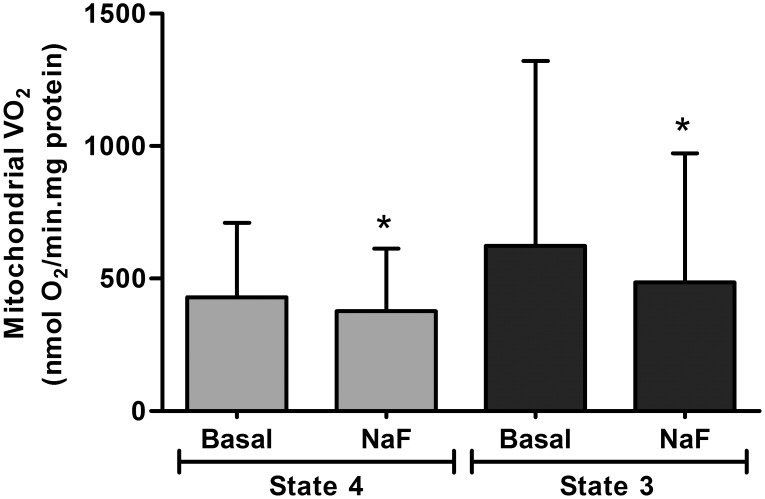
The addition of 100 µM of F- to active mitochondria produced a significant decrease in mitochondrial respiration, both in state 4 and state 3 (Figure 2). These results coincided with previous VO2 inhibition observed in the osteoblasts and demonstrated that F- inhibits the respiratory chain immediately after its addition.
Figure 2. Oxygen uptake rate (VO2) in isolated mitochondria.
Fluoride effects on VO2 of isolated mitochondria in states 4 and 3 in the absence (Basal) or presence of 100 µM of F- (NaF). Bars and segments represent mean and SD respectively. *Significantly different from Basal of the corresponding state. Paired Student’s t test, n = 12, p<0.05.
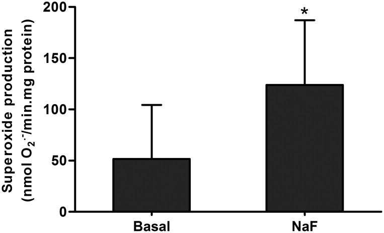
Finally, superoxide production in the absence or presence of F- was assessed to verify whether fluoride-inhibition of the respiratory chain increases the production of superoxide radicals. The results shown in Figure 3 demonstrate a significant increase in superoxide production after the addition of 100 µM of F- to submitochondrial particles.
Figure 3. Superoxide production by isolated mitochondria.
Fluoride effects on superoxide production of submitochondrial particles before (Basal) and after adding 100 µM of F- (NaF). Bars and segments represent mean and SD respectively. *Significantly different of NaF compared to Basal. Paired Student’s t test, n = 8, p<0.05.
Chronic Experiment
F- decreased VO2 of osteoblastic cells after 3 pulses of NaF 100 µM of 15 min duration (Figure 4).
Figure 4. Oxygen uptake rate (VO2) after chronic exposure to fluoride.
VO2 in ROS 17/2.8 cells after chronic treatment with15-min daily pulses of NaF 100 µM (F-) or distilled water (Control) for three consecutive days. Measurements (n = 3) were performed in duplicate and data are presented as percentage (%) of the Control group. Bars and segments represent mean and SD respectively. *Significantly different to Control group, Student’s t test, p<0.05.
The analysis of mitochondrial complexes revealed that there was a decrease of all respiratory complexes activities after the third pulse of F- in F- group. A significant inhibition along time was observed for complex IV reaching an 80% inhibition in F- group (Table 1).
Table 1. Respiratory complexes activities of isolated mitochondria.
| Control | F- | |
| C I–III | 100±28.6 | 72±28.0 |
| C II–III | 100±11.7 | 66±31.6 |
| C IV | 100±13.3 | 20±26.2* |
Activities of complex I–III (C I–III), complex II-III (C II–III) and complex IV (C IV) of isolated mitochondria from experimental groups expressed as percentage of the Control group. Data are shown as mean ± SD (n = 3). *Significantly different to Control, Student’s t test, p<0.05.
Finally, oxidative stress measurements revealed a significant increase in the levels of TBARS of F-exposed cells compared to Control group (Table 2). The GSSG/TGSH ratio was also increased in cells exposed to F-. Finally, no changes in antioxidants enzymes activities were observed after three pulses with F-.
Table 2. Oxidative stress indexes of ROS 17/2.8 cells.
| Control | F- | |
| CAT | 100±35.1 | 97±30.2 |
| GPx | 100±62.7 | 118±63.0 |
| TBARS | 100±13.4 | 181±61.8* |
| GSSG/TGSH | 100±3.1 | 140±7.7* |
Catalase (CAT) and glutathione peroxidase (GPx) activities, TBARS levels and GSSG/TGSH ratio in Control and F-treated ROS 17/2.8 cells. Data are expressed as percentage (%) of the Control group and represent three different biological experiments (n = 3). Values are mean ± SD. *Significantly different to Control group, Student’s t test, p<0.05.
Discussion
The increase in oxidative stress damage caused by F- is well documented [32], but the mechanisms involved in ROS generation are still unknown. One possible explanation is that F- could trigger oxidative stress via inhibition of the pentose phosphate oxidative pathway [33]. In addition, F- induced apoptosis by oxidative stress-induced lipid peroxidation, causing the release of cytochrome c through HL-60 cells mitochondria [34]. Presently, no mechanism for mitochondrial ROS generation by F- in osteoblasts has been proposed. The present contribution has been aimed at investigating whether F- could modify the activity of the respiratory chain in osteoblasts-like cells, changing the rate of production of oxygen reactive species.
We observed that as soon as F- reaches the cells (acute experiment), it significantly inhibits their respiration measured as VO2. Also, F- inhibits mitochondrial VO2 in both state 4 and state 3 and increases superoxide production. According with these results, the oxygen uptake rate also decreased in chronic treatments with F-. The increase in superoxide radicals as soon as F- reaches the mitochondrion could explain the increase in GSSG/TGSH ratio due to an augmented amount of peroxides reacting with reduced glutathione and producing higher amounts of oxidized glutathione [35]. The antioxidant enzymes activities were not modified after three pulses of F- and the decreased in GSSG/TGSH could explain the increase in lipid peroxidation after three pulses of F-.
Recently it has been reported that the constant exposure to F- for 72 h is able to induce apoptosis in osteoblasts through increasing oxidative stress. In that work F- was administered to osteoblasts in constant concentration for 72 h [36]. In the experiments described in our paper, osteoblasts are exposed for a few minutes to a high concentration of F-, as it occurs in vivo. Although F- levels returned to basal levels 24 h after each pulse of F-, an inhibition of oxygen consumption and respiratory complexes activities and an increase in oxidative stress status were observed.
The results shown in this paper also demonstrate that chronic administration of F- produces a significant decrease in mitochondrial respiratory chain activity. F- treatment significantly inhibited complex IV and partially decreased (30% inhibition) complex I-II and complex I-III activities. As complex I and III are the main sites of superoxide radical synthesis [37], F- could enhance its production by inhibiting the mitochondrial activity at respiratory chain level. F- inhibition at complex IV level could enhance the formation of ubiquinone radical which in turns can react with molecular oxygen increasing superoxide radical production [38]. When the respiratory chain is inhibited (in complex IV in this case), the electron supply reduces the ubiquinone (Q) pool and in the presence of large proton motive force, the electrons are forced back from reduced ubiquinone (QH2) into complex I (by electron reverse transport), which has two possible sites of superoxide production: the flavin in the NADH-oxidizing site and the ubiquinone-reducing site [39]. Recently it has been demonstrated that when succinate is used as electron donor, most superoxide is produced at the ubiquinone reduction site [40].
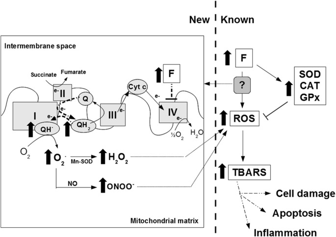
Taking all these results into account, we are able to describe the effect of F- on mitochondrial ROS production and its relationship with oxidative stress and inflammation (Figure 5). After an oral dose, F- may inhibit the respiratory chain, increasing the production of superoxide radical (by the possible mechanism explained before) and thereby of hydroxide peroxide and peroxynitrite [7]. Antioxidant enzymes activities cannot prevent increased free radical formation. Therefore, there is an increase in ROS that finally produce oxidations in membranes and damage the cell macromolecules (as seen by the increase in lipid peroxidation) and may be the cause of the inflammatory foci observed in the bone. It has already been demonstrated that ROS production induced inflammatory gene expression in alveolar macrophages [41], fibroblasts [42] and kidney [43]. Therefore, bone inflammatory foci could be enhanced via ROS-dependent activation of pro-inflammatory genes.
Figure 5. A possible mechanism of fluoride-induced oxygen radicals production.
New (left): knowledge generated by this paper. Thick black arrows and thick dashed lines indicate what happens when fluoride arrives at mitochondrial membrane. Known (right): known effects of fluoride on oxidative stress and antioxidant enzymes. F: fluoride; ROS: reactive oxygen species; SOD: superoxide dismutase: CAT: catalase; GPx: glutathione peroxidase; TBARS: thiobarbituric acid reactive substances, I: Complex I-NADH dehydrogenase, II: Complex II-Succinate dehydrogenase, III: Complex III-Cytochrome bc1 complex, IV: Complex IV-Cytochrome c oxidase, cyt c: Cytochrome c, Q: Ubiquinone, QH2: Reduced ubiquinone, O2 .: superoxide radical, H2O2: hydroxide peroxide, NO: Nitric oxide, ONOO−: peroxynitrite.
Supporting Information
Raw data from each figure and table. In each case, a data table and a legend which explains in detail what the data are, where they come from, the number of repetitions done and the test used for analysis are shown. Regarding the figures, beside each table the corresponding graph is shown.
(XLS)
Acknowledgments
We thank Lucas RM Brun for technical assistance.
Data Availability
The authors confirm that all data underlying the findings are fully available without restriction. Relevant data are included within the Supporting Information files.
Funding Statement
This paper was funded by Consejo Nacional de Investigaciones Científicas y Técnicas (CONICET) Grant 112-200801-00462 (http://www.conicet.gov.ar). The funders had no role in study design, data collection and analysis, decision to publish, or preparation of the manuscript.
References
- 1. Caverzasio J, Palmer G, Bonjour JP (1998) Fluoride: mode of action. Bone 22: 585–589. [DOI] [PubMed] [Google Scholar]
- 2. Briancon D, Meunier PJ (1981) Treatment of osteoporosis with fluoride, calcium and vitamin D. Orthop Clin North Am. 12: 629–648. [PubMed] [Google Scholar]
- 3. Brun LR, Roma SM, Pérez F, Rigalli A (2012) Presencia de inflamación en el tejido óseo de ratas inducida por fluoruro de sodio. Actual Osteol 8: 19–28. [Google Scholar]
- 4. Wang Z, Yang X, Yang S, Ren G, Ferreri M, et al. (2011) Sodium fluoride suppress proliferation and induce apoptosis through decreased insulin-like growth factor-I expression and oxidative stress in primary cultured mouse osteoblasts. Arch Toxicol 85: 1407–1417. [DOI] [PubMed] [Google Scholar]
- 5. Gazzano E, Bergandi L, Riganti C, Aldieri E, Doublier S, et al. (2010) Fluoride effects: the two faces of janus. Curr Med Chem 17: 2431–2441. [DOI] [PubMed] [Google Scholar]
- 6. Forman HJ, Azzi A (1997) On the virtual existence of superoxide anions in mitochondria: thoughts regarding its role in pathophysiology. FASEB J 11: 374–375. [DOI] [PubMed] [Google Scholar]
- 7. Sohal RS, Weindruch R (1996) Oxidative stress, caloric restriction, and aging. Science 273: 59–63. [DOI] [PMC free article] [PubMed] [Google Scholar]
- 8. Nathan AT, Singer M (1999) The oxygen trail: tissue oxygenation. Brit Med Bull 55: 95–108. [DOI] [PubMed] [Google Scholar]
- 9. Eisenberg AD, Marquis RE (1981) Enhanced transmembrane proton conductance in Streptococcus mutans GS-5 due to ionophores and fluoride. Antimicrob Agents Chemother 19: 807–812. [DOI] [PMC free article] [PubMed] [Google Scholar]
- 10. Fina BL, Brance ML, Brun LR, Rigalli A (2012) Fluoride inhibits in vitro and in vivo oxygen consumption and induces oxidative stress. Fluoride 45: 325–330. [Google Scholar]
- 11. Trautner K, Einwag J (1988) Human plasma fluoride levels following intake of dentifrices containing aminefluoride or monofluorophosphate. Archs oral Biol 33: 543–546. [DOI] [PubMed] [Google Scholar]
- 12. Rigalli A, Ballina JC, Beinlich A, Alloatti R, Puche RC (1994) Pharmacokinetic differences between sodium fluoride and sodium monofluorophosphate and comparative bone mass increasing activity of both compounds in the rat. Arzneim Forsch 44: 762–766. [PubMed] [Google Scholar]
- 13. Ekstrand J (1978) Relationship between fluoride in the drinking water and the plasma fluoride concentration in man. Caries Res 12: 123–127. [DOI] [PubMed] [Google Scholar]
- 14. Majeska RJ, Rodan SB, Rodan GA (1980) Parathyroid hormoneresponsive clonal cell lines from rat osteosarcoma. Endocrinology 107: 1494–1503. [DOI] [PubMed] [Google Scholar]
- 15. Towler DA, Bennett CD, Rodan GA (1994) Activity of the rat osteocalcin basal promoter in osteoblastic cells is dependent upon homeodomain and CP1 binding motifs. Mol Endocrinol 8(5): 614–624. [DOI] [PubMed] [Google Scholar]
- 16. Rigalli A, Alloatti R, Menoyo I, Puche RC (1995) Comparative study of the effect of sodium fluoride and sodium monofluorophosphate on glucose homeostasis in the rat. Arzneim Forsch 45: 289–292. [PubMed] [Google Scholar]
- 17. Fatt I (1968) The oxygen electrode: some special applications. Ann N Y Acad Sci 148: 81. [DOI] [PubMed] [Google Scholar]
- 18. Boveris A, Costa LE, Cadenas E, Poderoso JJ (1999) Regulation of mitochondrial respiration by adenosine diphosphate, oxygen and nitric oxide synthase. Methods Enzymol 301: 188–198. [DOI] [PubMed] [Google Scholar]
- 19. Boveris A, Chance B (1973) The mitochondrial generation of hydrogen peroxide. Biochem J 134: 707–716. [DOI] [PMC free article] [PubMed] [Google Scholar]
- 20. Chance B, Sies H, Boveris A (1979) Hydroperoxide metabolism in mammalian organs. Physiol Rev 59: 527–605. [DOI] [PubMed] [Google Scholar]
- 21. Watanabe N, Kamei S, Ohkubo A, Yamanaka M, Ohsawa S, et al. (1986) Urinary protein as measured with a pyrogallol red-molybdate complex, manually and in a Hitachi 726 automated analyzer. Clin Chem 32: 1551–1554. [PubMed] [Google Scholar]
- 23. Antunes F, Boveris A, Cadenas E (2004) On the mechanism and biology of cytochrome oxidase inhibition by nitric oxide. Proc Natl Acad Sci U S A 101: 16774–16779. [DOI] [PMC free article] [PubMed] [Google Scholar]
- 24. Boveris A (1984) Determination of the production of superoxide radicals and hydrogen peroxide in mitochondria. Methods Enzymol 105: 429–435. [DOI] [PubMed] [Google Scholar]
- 25. Lawrence RA, Burk RF (1976) Glutathione peroxidase activity in selenium-deficient rat liver. Biochem Biophys Res Commun 71: 952–958. [DOI] [PubMed] [Google Scholar]
- 26. Aebi H (1984) Catalase in vitro. Methods Enzymol 105: 121–126. [DOI] [PubMed] [Google Scholar]
- 27. Ohkawa H, Ohishi N, Yagi K (1979) Assay for lipid peroxides in animal tissues by thiobarbituric acid reaction. Anal Biochem 95: 351–358. [DOI] [PubMed] [Google Scholar]
- 28. Tietze F (1969) Enzymic method for quantitative determination of nanogram amounts of total and oxidized glutathione: applications to mammalian blood and other tissues. Anal Biochem 27: 502–522. [DOI] [PubMed] [Google Scholar]
- 29. Griffith OW (1980) Determination of glutathione and glutathione disulfide using glutathione reductase and 2-vinylpyridine. Anal Biochem 106: 207–212. [DOI] [PubMed] [Google Scholar]
- 30.Felipe de Mendiburu (2012) Agricolae: Statistical Procedures for Agricultural Research. R package version 1.1–3. Available: http://CRAN.R-project.org/package=agricolae.
- 31.R Development Core Team (2011) R: A language and environment for statistical computing. R Foundation for Statistical Computing, Vienna, Austria. ISBN 3-900051-07-0, URL http://www.R-project.org.
- 32. Xu H, Wang CH, Zhao ZT, Zhang WB, Li GS (2008) Role of oxidative stress in osteoblasts exposed to sodium fluoride. Biol Trace Elem Res 123: 109–115. [DOI] [PubMed] [Google Scholar]
- 33. Bergandi L, Aina V, Garetto S, Malavasi G, Aldieri E, et al. (2010) Fluoride-containing bioactive glasses inhibit pentose phosphate oxidative pathway and glucose 6-phosphate dehydrogenase activity in human osteoblasts. Chem Biol Interact 183: 405–415. [DOI] [PubMed] [Google Scholar]
- 34. Anuradha CD, Kanno S, Hirano S (2001) Oxidative damage to mitochondria is a preliminary step to caspase-3 activation in fluoride-induced apoptosis in HL-60 cells. Free Radic Biol Med 31: 367–373. [DOI] [PubMed] [Google Scholar]
- 35. Sies H (1999) Glutathione and its role in cellular functions. Free Radic Biol Med 27: 916–921. [DOI] [PubMed] [Google Scholar]
- 36. Wang Z, Yang X, Yang S, Ren G, Ferreri M, et al. (2011) Sodium fluoride suppress proliferation and induce apoptosis through decreased insulin-like growth factor-I expression and oxidative stress in primary cultured mouse osteoblasts. Arch Toxicol 85: 1407–1417. [DOI] [PubMed] [Google Scholar]
- 37. Cadenas E, Boveris A, Ragan CI, Stoppani AO (1977) Production of superoxide radicals and hydrogen peroxide by NADH-ubiquinone reductase and ubiquinol-cytochrome c reductase from beef-heart mitochondria. Arch Biochem Biophys 180: 248–257. [DOI] [PubMed] [Google Scholar]
- 38. Boveris A, Cadenas E, Stoppani AO (1976) Role of ubiquinone in the mitochondrial generation of hydrogen peroxide. Biochem J 156: 435–444. [DOI] [PMC free article] [PubMed] [Google Scholar]
- 39. Murphy MP (2009) How mitochondria produce reactive oxygen species. Biochem J 417(1): 1–13. [DOI] [PMC free article] [PubMed] [Google Scholar]
- 40. Quinlan CL, Perevoshchikova IV, Hey-Mogensen M, Orr AL, Brand MD (2013) Sites of reactive oxygen species generation by mitochondria oxidizing different substrates. Redox Biol 1(1): 304–312. [DOI] [PMC free article] [PubMed] [Google Scholar]
- 41. Eberlein M, Scheibner KA, Black KE, Collins SL, Chan-Li Y, et al. (2008) Anti-oxidant inhibition of hyaluronan fragment-induced inflammatory gene expression. J Inflamm 5: 20. [DOI] [PMC free article] [PubMed] [Google Scholar]
- 42. Chen AC, Arany PR, Huang YY, Tomkinson EM, Sharma SK, et al. (2011) Low-level laser therapy activates NF-kB via generation of reactive oxygen species in mouse embryonic fibroblasts. PLoS One 6: e22453. [DOI] [PMC free article] [PubMed] [Google Scholar]
- 43. Wang Y, John R, Chen J, Richardson JA, Shelton JM, et al. (2009) IRF-1 promotes inflammation early after ischemic acute kidney injury. J Am Soc Nephrol 20: 1544–1555. [DOI] [PMC free article] [PubMed] [Google Scholar]
Associated Data
This section collects any data citations, data availability statements, or supplementary materials included in this article.
Supplementary Materials
Raw data from each figure and table. In each case, a data table and a legend which explains in detail what the data are, where they come from, the number of repetitions done and the test used for analysis are shown. Regarding the figures, beside each table the corresponding graph is shown.
(XLS)
Data Availability Statement
The authors confirm that all data underlying the findings are fully available without restriction. Relevant data are included within the Supporting Information files.


