Abstract
To correlate symptoms of deviated nasal septum (DNS) and chronic rhinosinusitis with the findings of nasal endoscopy and computed tomographic (CT) imaging. To evaluate the influence of degree of septal angle deviation on the severity of lateral nasal wall abnormalities. A prospective study was conducted on 67 patients with clinical evidence of DNS and chronic sinusitis attending ENT OPD between January 2012 and September 2013. All these patients underwent nasal endoscopy and CT scan PNS coronal sections. Direction and degree of DNS was recorded. Range of sinus mucosal thickening on CT scan films was also recorded. Chronic sinusitis is common in the age group between 21 and 40 years (50.74 %) with male preponderance (55.22 %), chief symptoms being nasal obstruction (86.56 %), headache (73.13 %) and nasal discharge (52.23 %). Left sided DNS is more common (64.17 %). Most of the patients have moderate DNS, i.e. 6°–10° (56.7 %), followed by severe (22.4 %) and then mild (20.9 %). DNS results in compensatory structural changes in the turbinates and/or lateral nasal wall which causes ostiomeatal complex (OMC) obstruction resulting in sinusitis. Contralateral concha bullosa and ethmoid bulla prominence was noted. Maxillary sinus is most commonly affected sinus (73.13 %). Patients with increasing septal angles were associated with a higher incidence of maxillary sinus mucosal changes (p < 0.05). Present study reemphasized the concept that septal deviation causes obstruction at OMC which results in an increased incidence and severity of bilateral chronic sinus disease.
Keywords: Deviated nasal septum, Chronic rhinosinusitis, CT scan PNS, Angle of septal deviation, Ostiomeatal complex
Introduction
Septal deviation refers to convexities of the septum to one side with accompanying deformities of the midline structures. A marked deviation of portion or of entire nasal septum may cause not only obstructed nasal breathing but also disease within the lateral nasal wall and consecutively in paranasal sinuses [1]. Patients with increasing nasal septal deviation are associated with a higher incidence of ostiomeatal complex (OMC) obstruction [7]. OMC obstruction in the direction of septal angulation is attributable to the nasal septal deformity, however, contralateral OMC obstruction is related to middle turbinates and lateral nasal wall abnormalities [7].
Nasal septal deviation disturbs nasal physiology, not always, but together with conchal hypertrophy or other anatomical variations [1]. It could narrow the middle meatus by pushing the concha laterally [5]. Besides nasal obstruction, it exerts pressure to neighbouring structures. This in turn disturbs the drainage pathways, affects the mucosal ciliary function by contact and leads to obstruction and secondary nasal infections in all sinuses by disturbing normal mucus drainage [1, 4, 6]. An increased incidence and severity of bilateral chronic sinus disease was present with increasing deviations of the septum (p < 0.05) [3, 7]
Various studies showed that it is a deviated septum, which results in the OMC disease and compensatory conchal hypertrophy and enlarged ethmoid bulla. But in very few studies angle of septal deviation has been measured and the effect of increasing angle of septal deviation on lateral wall of nose has been evaluated.
Nasal endoscopy and computed tomography (CT) scan has made an enormous impact in regional imaging and it has increased a surgeon’s ability to depict accurately the status of structures within the paranasal sinus region and in delineating the location and extent of pathology [6]. The CT scan is the gold standard investigation in all sinus diseases and it gives detailed bony anatomy of the ostiomeatal area and serves as an anatomic roadmap for the operating surgeon [11, 14].
The intention of this study is to evaluate the nasal septal deviation by measuring angle of septal deviation and to study what is the role and relation of degree of septal deviation on lateral wall of nose and sinus diseases by both endoscopic and computed tomographic evaluation in patients of chronic rhinosinusitis.
Methods
This study is a prospective cross-sectional observation, carried out on 67 patients attending the Department of Otorhinolaryngology, Gandhi Medical College and Hamidia group of Hospitals, Bhopal (M.P.), India, from January 2012 to September 2013. All these patients with clinical evidence of chronic rhinosinusitis were evaluated with nasal endoscopy and CT scan PNS (coronal sections).
Exclusion Criteria
Patients with acute sinusitis, allergic sinusitis, asthma, cystic fibrosis, immune deficiency, metabolic diseases or malignant diseases or those who had previously undergone nasal or sinus surgery, or maxillofacial trauma cases, and children <8 years of age were excluded from the study [8].
Inclusion Criteria
All patients of both sexes with symptoms of DNS and chronic rhinosinusitis e.g. headache, nasal obstruction, nasal discharge, hyposmia or facial pain.
Out of 67 patients 37 were male and 30 were female. The youngest patient was 9 year old and the oldest was 60 year old.
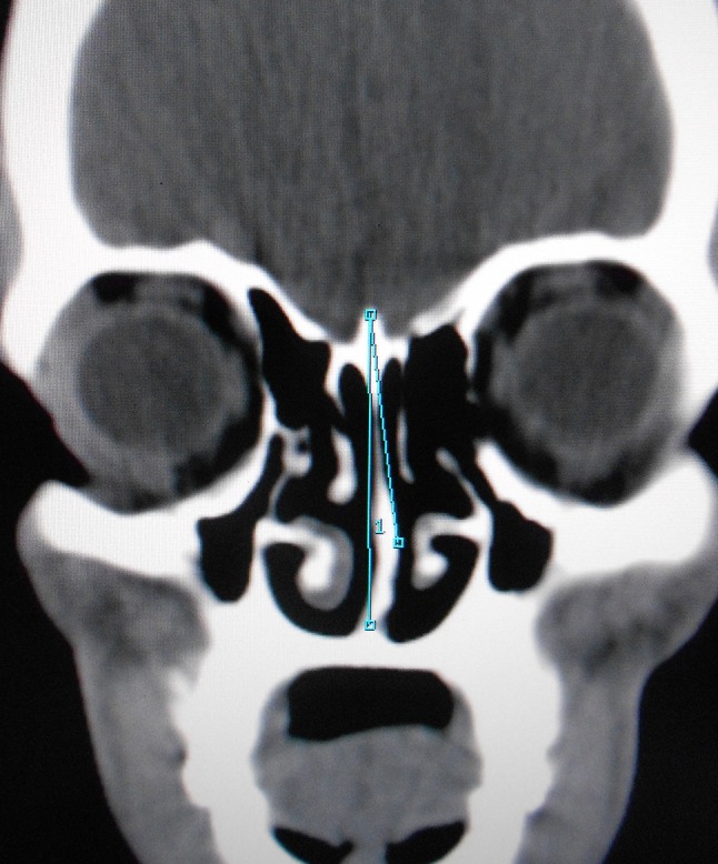
All patients symptomatically identified for chronic sinusitis were subjected to detailed clinical and radiological examination, diagnostic nasal endoscopy and CT scan PNS coronal sections. Angle of nasal septal deviation was measured in each case and its effect was noted on lateral nasal wall both ipsilaterally and contralaterally. The coronal CT image that best defined the OMC was utilized for the calculation of the direction and degree of septal deviation. The superior insertion of the nasal septum at the crista galli, its inferior insertion at the level of the anterior nasal spine, and most prominent point of nasal septal deviation were all identified and clearly marked on the respective film (Fig. 1). The resultant angle was noted. The relationship between nasal septal deviation, unilateral or bilateral occurrence of concha bullosa and bulla ethmoidalis and sinusitis were evaluated using Analysis of Variance (ANOVA). Correlation between angle of deviation and range of mucosal hypertrophy of the paranasal sinuses were evaluated with Fischer’s Exact Test.
Fig. 1.

Measurement of septal angle
Results
In present study, out of 67 cases, 37 cases (55.22 %) were male while 30 cases (44.78 %) were female. Male:female ratio 1.23:1. Nearly half of the cases (50.74 %) were in the age group of 21–40 years, 28.35 % in age group 10–20 years, 19.4 % cases in the age group of 41–60 and 1.5 % below 10 years of age (Table 1).
Table 1.
Age distribution
| Age groups | No. of cases | Percentage |
|---|---|---|
| <10 | 1 | 1.5 |
| 10–20 | 19 | 28.35 |
| 21–40 | 34 | 50.74 |
| 41–60 | 13 | 19.4 |
| >60 | – | – |
| Total | 67 | 100 |
Among the clinical symptoms of the patients, the most common presenting symptoms were nasal obstruction (86.56 % cases) and headache (73.13 % cases) followed by nasal discharge (52.23 %) as suggested by Table 2. On nasal endoscopy, left sided DNS was found to be more common (64.17 %).
Table 2.
Clinical symptoms
| Symptoms | No. of patients | Percentage |
|---|---|---|
| Nasal obstruction | 58 | 86.56 |
| Headache | 49 | 73.13 |
| Nasal discharge | 35 | 52.23 |
| Sneezing | 11 | 16.42 |
| Epistaxis | 07 | 10.45 |
| Throat discomfort | 20 | 29.85 |
| Ear manifestations | 20 | 29.85 |
In all 67 patients in the study group, CT scan PNS coronal section was done by taking 5 mm slice thickness. The patients were divided into three categories according to their degree of septal deviation to facilitate the presentation and comparison of data. Group I compared the patients with septal angles between 0° and 5°, where as group II and group III accommodated patients with angles ranging between 6° and 10° and >11°, respectively. 14 patients were included in group I, 38 in group II, and 15 patients in group III according to this categorization. This implies that most patients had septal angulation in moderate range (56.7 %), followed by severe (22.4 %), then mild (20.9 %) (Table 3).
Table 3.
Nasal septal deviations
| Group I (0°–5°) | Group II (6°–10°) | Group III (11°–15°) | Total | |
|---|---|---|---|---|
| No. | 14 | 38 | 15 | 67 |
| Percentage | 20.9 | 56.7 | 22.4 | 100 % |
Table 4 shows the result of OMC obstruction in relation to the three groups of angle of septal deviation. There was no apparent statistically significant difference in OMC obstruction between ipsilateral and contralateral sides in relation to direction of septal deviation in group I, though the obstruction was a bit higher on contralateral side in groups II and III. In evaluating middle turbinate abnormalities as a function of these three groups of septal deviations, significant difference in contralateral turbinates was noted in group II patients. There was a significantly increased incidence of concha bullosa on the contralateral side. Similar findings were realized for middle turbinates opposite to the direction of septal deviation for concha bullosa in group I and group III patients. Lateral nasal wall abnormalities, specifically prominent bulla ethmoidalis with OMC impingement, revealed a similar pattern of contralateral predominance. A statistically significant increase in predominance of ethmoid bulla was observed on the side opposite to the direction of septal deviation. There was no corresponding difference in comparing ipsilateral and contralateral discrepancies in uncinate process deviation (Table 4).
Table 4.
Lateral nasal wall abnormalities verses degree of septal deviation
| Group I (0°–5°) | Group II (6°–10°) | Group III (11°–15°) | ||||
|---|---|---|---|---|---|---|
| Ipsi lateral | Contra lateral | Ipsi lateral | Contra lateral | Ipsi lateral | Contra lateral | |
| OMC obstruction | 3 | 2 | 8 | 12 | 7 | 8 |
| Concha bullosa | 0 | 5 | 3 | 12 | 0 | 2 |
| Bulla ethmodalis | 0 | 2 | 3 | 9 | 1 | 4 |
| Uncinate deviation | 0 | 1 | 1 | 2 | 0 | 2 |
Mucosal abnormality detected on CT scan PNS coronal view, ranged from minimal mucosal thickening to total sinus opacification [13]. The most frequently involved sinus area was the maxillary sinus (73.13 %), followed by anterior ethmoid sinus region (53.73 %). Sphenoid sinus was least commonly involved (16.42 %) (Table 5). Highly significant statistical correlation was found between angle of deviation of nasal septum and sinus mucosal hypertrophy, as detected on CT scan PNS (coronal sections). Patients with increasing angles were associated with a higher incidence of maxillary sinus mucosal changes (p < 0.05). But this was not the same for ethmoid, frontal and sphenoid sinuses (Table 6).
Table 5.
CT detection of mucosal abnormalities
| Site of involvement | No. of patients | Percentage |
|---|---|---|
| Frontal sinus | 29 | 43.28 |
| Anterior ethmoid sinus | 36 | 53.73 |
| Posterior ethmoid sinus | 19 | 28.36 |
| Maxillary sinus | 49 | 73.13 |
| Sphenoid sinus | 11 | 16.42 |
Table 6.
Correlation between angle of deviation and maxillary sinus mucosal changes
| Angle of deviation | Range of mucosal hypertrophy | Total | |||
|---|---|---|---|---|---|
| 0 | +1 | +2 | +3 | ||
| 0°–5° | 6 | 8 | 6 | 0 | 20 |
| 6°–10° | 9 | 0 | 14 | 3 | 26 |
| 11°–15° | 3 | 0 | 9 | 9 | 21 |
| Total | 18 | 8 | 29 | 12 | 67 |
0 No mucosal hypertrophy; +1 minimal mucosal thickening; +2 partial opacification; +3 complete sinus opacification
Discussion
Ostiomeatal complex is a gateway to sinus disease. As a consequence of various anatomic and functional relationships immediately adjacent to the OMC, proliferation of disease into the anterior sinuses is natural [4, 12]. Nasal endoscopic examination in conjunction with CT scan has proven to be ideal combination in recent years. CT imaging of sinonasal region has become the gold standard in the evaluation of patients with chronic sinusitis [11, 14]. Its ability to accurately map out the bony and soft tissue anatomy of the paranasal sinuses has proven invaluable to the endoscopic surgeon in the diagnostic workup of a surgical candidate.
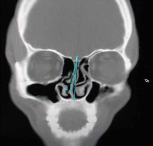
In present study authors found that the effect of nasal septal deviation on OMC obstruction is higher on contralateral side (17.91 %) as compared to ipsilateral side (11.94 %). This is in the group II, i.e., moderate angle of deviation (Fig. 2). While in group I and III, i.e., mild and severe degrees of septal deviation, there was no apparent statistically significant difference between ipsilateral and contraleteral sides in relation to direction of septal deviation.
Fig. 2.

Lt. Bulla ethmoidalis & Lt. Concha bullosa with Lt. OMC obstruction and Rt. Septal spur
Yousem et al. [16] evaluated the morphologic features that predispose to sinusitis and concluded that patients with evidence of sinusitis on CT scanning had a higher degree of septal deviation than did those without. They further showed that rates of sinusitis were not significantly different ipsilateral and contralateral to the side of the septal deviation.
Similarly, Calhoun et al. [4] examined the paranasal sinus CT images of both asymptomatic and symptomatic patients. These authors clearly showed a strong correlation between septal deviation and sinus disease, although the degree of septal deviation was never qualified. They further documented a significant association with OMC obstruction and ethmoid sinus disease only on the side to which the septum was deviated, in contradiction to Yousem et al.
Elahi et al. [7] evaluated the nasal septum along with paraseptal structural and pathological entities in an adult population with a clinical diagnosis of chronic rhinosinusitis. They showed ipsilaterally, that is, in the direction of septal deviation, OMC and sinus disease is directly attributable to the septal deviation in the absence of any other discernable factor. While on the side opposite the direction of septal deviation, a prominent bulla ethmoidalis and various middle turbinate abnormalities have been shown to be the cause of OMC obstruction. They also showed increasing OMC disease bilaterally with increasing septal deviation.
The results of present study were parallel to those of the study of Elahi et al. [7]. It has been shown that bilateral OMC obstruction and disease in anterior group of sinuses occurs without any discrepancy on the either sides of septal deviation. Authors assumed that ipsilateral OMC and sinus diseases are directly attributable to the septal deviation. While on the side opposite to the direction of septum, bulla ethmoidalis and various middle turbinate abnormalities have shown to be the cause of OMC obstruction.
Authors have observed in the present study that with increasing angle of deviation of septum there is statistically significant increase in both middle turbinate and lateral nasal wall abnormalities on the contralateral side. Incidence of concha bullosa and paradoxical middle turbinate being 28.35 and 1.5 % respectively on contralateral side, as compared to 4.5 and 0 % on ipsilateral sides. While incidence of bulla ethmoidalis being 22.37 % on contralateral side as compared to 5.97 % on ipsilateral side. But there was no corresponding difference in comparing ipsilateral and contralateral discrepancies in uncinate process deviation.
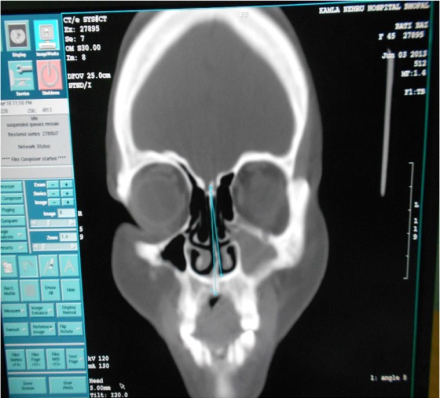
True concha bullosa (pneumatization of both the vertical lamella and the inferior bulbous portion) in the normal population has a reported prevalence of 4–15.7 % [2]. In patients with chronic sinusitis, the prevalence of concha bullosa increases to almost 33 % [9, 17]. In present study, concha bullosa was seen in 37.3 % of the cases (25 patients), which is consistent with above mentioned studies (Fig. 3).
Fig. 3.

B/L Bulla ethmoidalis & B/L Concha bullosa with Lt. Maxillary sinusitis
A markedly medially bent or folded uncinate process with a corresponding area of extensive contact with the middle turbinate is one of the most frequent pathological findings in patients with chronic sinusitis [15]. In a previous study of 114 cases Milczuk et al. [12] found the uncinate process to be laterally bent in 11 cases (9.64 %). The exact prevalence of these variations and their relation to sinus disease is not determined.
In present study authors observed bent uncinate process in 8.9 % of the cases. Medially bent uncinate process was seen in 7.5 % cases while laterally bent uncinate process was seen in 1.5 % cases.
A large ethmoidal bulla may contribute to sinus disease by obstructing the infundibulum or middle meatus or by being primarily diseased and filled with pus, cysts or polyps [15]. The exact prevalence is not known [6, 10]. In present study authors observed enlarged ethmoidal bulla in 28.35 % of the cases, on CT Scanning.
Distribution of mucosal abnormalities in each paranasal sinus was determined by coronal sections of CT scan PNS. As recommended by Som [13], mucosa is normally not visible on CT film, so any imaging of the paranasal sinus mucosa was considered abnormal. Accordingly, mucosal abnormalities ranged from minimal mucosal thickening to total sinus opacification.
Mucosal abnormalities were most frequently noted in the maxillary sinus region (73.13 %) in present study, while Bolger et al. [2] in their study of 202 cases reported the maxillary sinus to be involved in 77.7 % of the cases. This was followed by anterior ethmoid sinuses which were involved in 53.73 % of the cases in present series, in comparison to the 84.3 % in the study by Bolger et al. Frontal sinus was involved in 14.75 % of the cases in the study by Bolger et al. while in present series it was involved in 43.28 % of the cases. In the Bolger series posterior ethmoid sinuses and sphenoid sinus were involved in 38.6 and 25.4 % cases respectively, in comparison to 28.36 and 16.42 % cases respectively in present study. Kennedy and Zinreich [9], also noted the anterior ethmoid region to be the most frequently involved area of the paranasal sinuses (78 %). Maxillary sinus was involved in 66 % of cases in their study, while posterior ethmoids in 31 % of cases. Frontal and sphenoid sinuses were involved in 34 and 16 % of cases respectively.
In present study, authors find maxillary sinus to be more commonly involved than anterior ethmoid sinuses, which is similar to study done by Mamatha et al. [11], they found 67.5 % maxillary sinus involvement, 32.5 % ethmoid sinus involvement and 25 % frontal sinus involvement. Unethical antibiotic usage is a possible source of bias in these investigations. The full effect of such therapy on CT scan outcome is unknown. It is logical to assume, however, that antibiotics would reduce the prevalence of mucosal abnormalities. Hence the actual prevalence might be even higher than reported.
Conclusion
Deviated septum most probably results in compensatory structural changes in the middle turbinates and/or lateral nasal wall. Increasing angles of septal deviation are associated with bilateral sinus disease and contralateral middle turbinate abnormalities and ethmoid bulla prominence. Maxillary sinus is the most commonly involved sinus region, followed by anterior ethmoid sinuses and frontal sinuses. Patients with increasing angles are associated with a higher incidence of maxillary sinus mucosal changes.
Hence, present study reemphasized the concept that obstruction at OMC is the key factor for causation of chronic sinusitis, which is secondary to septal deviation.
Acknowledgments
Conflict of interest
The authors declare that they have no conflict of interest.
Contributor Information
V. K. Poorey, Email: drvkpoorey@gmail.com
Neha Gupta, Email: nehagupta.vision@gmail.com.
References
- 1.Atkas D, Kalcioglu MT, Kutlu R, Ozturan O, Oncel S. The relationship between the concha bullosa, nasal septal deviation and sinusitis. Rhinology. 2003;41:103–106. [PubMed] [Google Scholar]
- 2.Bolger WE, Butzin CA, Parsons DS. Paranasal sinus bony anatomic variations and mucosal abnormalities: CT analysis for endoscopic sinus surgery. Laryngoscope. 1991;101:56–64. doi: 10.1288/00005537-199101000-00010. [DOI] [PubMed] [Google Scholar]
- 3.Buyukertan M, Keklikoglu N, Kokten G. A morphometric consideration of nasal septal deviations by people with paranasal complaints; a computed tomography study. Rhinology. 2002;41:21–24. [PubMed] [Google Scholar]
- 4.Calhoun KH, Waggenspack GA, Simpson CB, Hokanson JA, Bailey BJ. CT evaluation of the paranasal sinuses in symptomatic and asymptomatic populations. Otolaryngol Head Neck Surg. 1991;104(4):480–483. doi: 10.1177/019459989110400409. [DOI] [PubMed] [Google Scholar]
- 5.Davis WE, Templer J, Parsons DS. Anatomy of the paranasal sinuses. Otolaryngol Clin North Am. 1996;29(1):57–91. [PubMed] [Google Scholar]
- 6.de Araújo Neto SA, Martins PDSL, Souza AS, Baracat ECE, Nanni L 2006 The role of ostiomeatal complex anatomical variants in chronic rhinosinusitis. Radiologia Brasileira 39:3. São Paulo May/June 2006. ISSN 0100-3984
- 7.Elahi MM, Frenkiel S, Fageeh N. Paraseptal structural changes and chronic sinus disease in relation to the deviated septum. J Otolaryngol. 1997;26(4):236–240. [PubMed] [Google Scholar]
- 8.Keles B, Ozturk K, Unaldi D, Arbag H, Ozer B. Is there any relationship between nasal septal deviation and concha bullosa. Eur J Gen Med. 2010;7(4):359–364. [Google Scholar]
- 9.Kennedy DW, Zinreich SJ, Rosenbaum AE, Johns ME. Functional endoscopic sinus surgery—theory & diagnostic evaluation. Arch Otolaryngol. 1985;111:576–582. doi: 10.1001/archotol.1985.00800110054002. [DOI] [PubMed] [Google Scholar]
- 10.Laine FJ, Smoker WR. The ostiomeatal unit and endoscopic surgery: anatomy, variations, and imaging findings in inflammatory diseases. AJR Am J Roentgenol. 1992;159:849–857. doi: 10.2214/ajr.159.4.1529853. [DOI] [PubMed] [Google Scholar]
- 11.Mamatha H, Shamasunder NM, Bharathi MB, Prasanna LC. Variations of ostiomeatal complex and its applied anatomy: a CT scan study. Indian J Sci Technol. 2010;3:8. [Google Scholar]
- 12.Milczuk HA, Dalley RW, Wessbacher FW, Richardson MA. Nasal and PNS anomalies in children with chronic sinusitis. Laryngoscope. 1993;103(3):247–252. doi: 10.1288/00005537-199303000-00002. [DOI] [PubMed] [Google Scholar]
- 13.Som PM. CT of paranasal sinuses. Neuro Radiol. 1985;27:189–201. doi: 10.1007/BF00344487. [DOI] [PubMed] [Google Scholar]
- 14.Stammberger H, Hawke M. Essentials of endoscopic sinus surgery. 1. St. Louis, MO: Mosby; 1993. p. 43. [Google Scholar]
- 15.Stammberger H, Wolf G. Headaches and sinus diseases : the endoscopic approach. Ann Otol Rhinol Laryngol. 1988;97(Suppl 134):3–23. doi: 10.1177/00034894880970s501. [DOI] [PubMed] [Google Scholar]
- 16.Yousem DM, Kennedy DW, Rosenberg S. Ostiomeatal complex risk factors for sinusitis: CT evaluation. J Otolaryngol. 1991;20(6):419–424. [PubMed] [Google Scholar]
- 17.Zinreich SJ. Paranasal sinus imaging. Otolaryngol Head Neck Surg. 1990;103(5):863–868. doi: 10.1177/01945998901030S505. [DOI] [PubMed] [Google Scholar]


