Abstract
Purpose
Osteoarthritis in combination with rotator cuff deficiency following previous shoulder stabilisation surgery and after failed surgical treatment for chronic anterior shoulder dislocation is a challenging condition. The aim of this study was to analyse the results of reverse shoulder arthroplasty in such patients.
Methods
Thirteen patients with a median follow-up of 3.5 (range two to eight) years and a median age of 70 (range 48–82) years were included. In all shoulders a tear of at least one rotator cuff tendon in combination with osteoarthritis was present at the time of arthroplasty. The Constant score, shoulder flexion and external and internal rotation with the elbow at the side were documented pre-operatively and at the final follow-up. Pre-operative, immediate post-operative and final follow-up radiographs were analysed. All complications and revisions were documented.
Results
Twelve patients were either satisfied or very satisfied with the procedure. The median Constant score increased from 26 points pre-operatively to 67 points at the final follow-up (p = 0.001). The median shoulder flexion increased significantly from 70° to 130° and internal rotation from two to four points (p = 0.002). External rotation did not change significantly (p = 0.55). Glenoid notching was present in five cases and was graded as mild in three cases and moderate in two. One complication occurred leading to revision surgery.
Conclusions
Reverse arthroplasty leads to high satisfaction rates for patients with osteoarthritis and rotator cuff deficiency who had undergone previous shoulder stabilisation procedures. The improvements in clinical outcome as well as the radiographic results seem to be comparable with those of other studies reporting on the outcome of reverse shoulder arthroplasty for other conditions.
Keywords: Reverse shoulder arthroplasty, Shoulder instability, Rotator cuff, Anterior instability, Shoulder, Arthroplasty
Introduction
Recurrent anterior shoulder dislocation is a challenging pathology, which can be treated conservatively but often requires surgical stabilisation. Multiple stabilisation procedures have been developed in recent years [1–7], including open or arthroscopic Bankart repairs (with or without capsular shifts) and bone block procedures.
Osteoarthritis as well as rotator cuff deficiency may be observed after these procedures although the shoulder remains stable after treatment [8, 9]. It has been described that the degenerative changes of the glenohumeral joint are associated with the pathology itself and do not seem to be related to the treatment option which was chosen [9, 8, 10]. Non-constrained anatomic shoulder arthroplasty (total shoulder replacement or hemiarthroplasty) can obtain a good clinical result in patients with arthritis after instability, although revision rates between 18 and 35 % have been reported [11–15].
Various redislocation rates have been described after anatomic shoulder replacement in the above-mentioned studies [11–14]. The use of semi-constrained reverse shoulder arthroplasty could theoretically reduce the rate of recurrent dislocation due to the inherent stability of this implant design [16]. Moreover, in cases of insufficient rotator cuff, favourable results have been described compared to non-constrained anatomic shoulder arthroplasty [17, 18].
The purpose of this study was therefore to analyse the clinical and radiographic results and complications of reverse shoulder arthroplasty in patients with osteoarthritis of the shoulder and rotator cuff deficiency who had undergone previous surgical stabilisation procedures for recurrent anterior shoulder instability.
Methods
Demographics
Between July 2000 and December 2009, 15 patients (15 shoulders) underwent reverse arthroplasty after previous surgical shoulder stabilisation procedures. All of the patients had end-stage post-instability osteoarthritis of the affected shoulders in combination with rotator cuff deficiency. Surgeries were performed in two specialised shoulder centres. Inclusion criteria were (1) osteoarthritis and rotator cuff deficiency after previous surgery for recurrent anterior shoulder instability, (2) treatment with the same reverse shoulder arthroplasty (Tornier, St. Ismier, France) after the initial instability procedure and (3) a minimum follow-up of two years. Patients with locked dislocations were not included.
Of the patients, two died before having a follow-up examination and therefore 13 patients (13 shoulders) were included in the study. There were eight men and five women. The median age at the time of surgery was 70 (range 48–82) years. The right shoulder was operated ten times and the dominant shoulder 11 times. The mean follow-up was 3.5 (range two to eight) years. The mean time delay between the first surgery and arthroplasty was 15 (range one to 49) years and the mean number of dislocations before the stabilisation procedures was three (range one to ten). Eleven patients had had one previous surgical procedure and two patients had had two. The patients with one previous surgery had undergone a Trillat procedure [2] in five cases, a Latarjet procedure [3] in two cases, a Bankart repair in combination with a capsular shift in three cases and an Eden-Hybinette procedure [5] in one case. A Trillat procedure was performed when a non-repairable rotator cuff tear with upward migration of the humeral head was present. In the two patients who underwent two operations, Latarjet procedures were performed after a failed Bankart repair in one and a failed Trillat procedure in the other. Three patients had recurrent anterior subluxations after a Trillat procedure.
Clinical protocol
All patients underwent a clinical examination before surgery and at the most recent follow-up review. Active shoulder flexion and external rotation with the elbow at the side were measured in degrees. Internal rotation was graded according to the posterior region the thumb could reach. The Constant score [19] with its subgroups (pain, activity, mobility and strength) was recorded. The maximum score consists of 100 points. The maximum points for the subgroups are 15 for pain, 20 for activity, 40 for mobility and 25 for strength. Patient’s satisfaction at the final follow-up was rated as very satisfied, satisfied, somewhat disappointed and disappointed.
Radiographic protocol
Anteroposterior (AP) and axillary radiographs were performed before surgery and at the most recent follow-up examination. The presence of osteoarthritis in the affected shoulder joints was rated according to the method of Samilson and Prieto [20] on X-rays performed immediately before arthroplasty. This classification system describes three degrees of severity: (1) evidence of an inferior humeral and/or glenoid osteophyte less than 3 mm in height was rated as mild, (2) evidence of an osteophyte between 3 and 7 mm in height and slight glenohumeral joint irregularity as moderate and (3) evidence of an osteophyte >7 mm in height and narrowing of the glenohumeral joint space and subchondral sclerosis as severe. In all cases a pre-operative computed tomography (CT) scan was performed, and the fatty infiltration of the subscapularis, supraspinatus and infraspinatus muscles was analysed according to the method of Goutallier et al. [21].
The humeral components were analysed for radiolucent lines as described by Melis et al. [22]. The shaft was divided into seven zones and radiolucent lines were graded as <2 mm or >2 mm. Radiolucent lines >2 mm in three or more regions around the stem were defined as loosening of the stem. The humeral and glenoid components were also analysed for tilting and subsidence comparing the immediate post-operative and the last X-rays. Scapular notching was assessed according to the method of Sirveaux et al. [23]. Four grades of severity have been described in this score. Grade 1 notching is defined as a bone deficiency at the pillar, grade 2 when the defect was in contact with the inferior screw, grade 3 when the defect extended superior to the inferior screw and grade 4 when the defect reached the central peg. The presence of a bone spur at the inferior part of the glenoid was also evaluated.
Operative technique and implants
The indication for reverse shoulder arthroplasty surgery was painful post-instability osteoarthritis in combination with rotator cuff deficiency in all cases. A rotator cuff tear of at least one tendon was present in all shoulders. The operative technique in both centres was comparable. A deltopectoral approach was used in all cases. After releasing the conjoined tendon, the subscapularis muscle and tendon were identified. If the subscapularis tendon remained attached, a tenotomy was performed at the level of the anatomic neck. In three cases an isolated rupture of the subscapularis tendon was detected, in five cases a rupture of the subscapularis and supraspinatus tendons and in five cases a rupture of the subscapularis tendon in combination with supraspinatus and infraspinatus tears. The superior, medial and inferior glenohumeral ligaments were released and a periglenoidal release of the anteroinferior labrum and capsule and part of the triceps tendon was then performed. Remaining screws from the initial stabilisation surgeries were removed in all cases. Removal of the inferior osteophytes of the humeral head was carried out with bony scissors in order to expose the anatomic head of the humerus. The humeral head was resected using a 155° guide with retroversion between 0 and 20°. The metaphysis was reamed with hemispheric reamers increasing in size. A trial component was inserted to test the correct seating of the humeral implant. After preparing the glenoid, an uncemented baseplate with a central peg was impacted. Four additional screws were then inserted (two non-locking and two locking screws). A 36-mm glenosphere was fixed onto the baseplate using an impactor and a central safety screw. A cement restrictor was then placed and the humeral shaft filled with cement in a retrograde fashion. In one centre jet lavage of the humeral canal was performed before cementing. The humeral component was inserted and held in place until the cement cured. Based on the experience of the surgeons, the polyethylene inlay was inserted according to the correct tension of the deltoid. A repair of the remaining subscapularis tendon was performed in 11 cases, using three to five transosseous non-absorbable sutures. A systematic soft tissue tenodesis of the long head of the biceps was performed in all cases. A drain was then placed (removed two days after the procedure) and the wound closed in two layers.
Post-operatively the arm was placed in a sling for four weeks with passive range of motion exercises, supervised by a physiotherapist, commenced on day two. The patients were allowed to progressively use the affected arm for their normal day-to-day activities after four weeks.
Statistics
The two paired sample Wilcoxon signed rank test was used for comparison of the pre- and post-operative Constant score and shoulder flexion as well as external and internal rotation. A p value < 0.05 was considered to be statistically significant.
Results
Clinical results
There was a significant increase in the median Constant score from 26 to 67 points pre- vs post-operatively (p = 0.001). The median shoulder flexion (70°–130°) and internal rotation (2–4 points) also increased significantly (p = 0.002). External rotation did not demonstrate a significant change (p = 0.55). All clinical results are shown in Table 1. Twelve patients (92 %) were very satisfied (n = 7) or satisfied (n = 5) with the result of the arthroplasty at the final follow-up. One patient was somewhat disappointed.
Table 1.
Clinical data before operation and at most recent follow-up
| Median preoperative | SD preoperative | Range preoperative | Median post-operative | SD post-operative | Range post-operative | Significant differences pre- vs post-operative | |
|---|---|---|---|---|---|---|---|
| Constant score (points) | 26 | 12.1 | 8–45 | 67 | 11.3 | 45–81 | p = 0.001 |
| Forward flexion (°) | 70 | 29.6 | 30–100 | 130 | 23 | 90–160 | p = 0.002 |
| External rotation (°) | 0 | 30.1 | −40 to 60 | 10 | 13.2 | 0–40 | p = 0.55 |
| Internal rotationa (points) | 2 | 1.3 | 1–6 | 4 | 2.2 | 0–9 | p = 0.002 |
| Pain (points) | 5 | 3.2 | 0–10 | 13 | 3.2 | 5–15 | p = 0.003 |
| Strength (points) | 0 | 4.8 | 0–12 | 6 | 3.7 | 1–12 | p = 0.01 |
| Activity (points) | 8 | 3.6 | 2–11 | 18 | 2.7 | 12–20 | p = 0.001 |
| Mobility (points) | 8 | 5 | 4–20 | 28 | 5.1 | 18–34 | p = 0.001 |
aInternal rotation was graded according to the posterior spinal region that could be reached by the thumb
Radiographic results
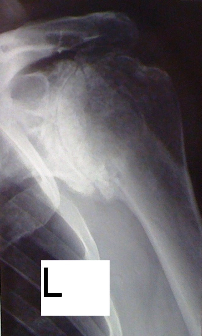
Preoperatively, osteoarthritis of the glenohumeral joint was present on X-rays in all cases. Moderate osteoarthritis was detected in three shoulders and severe osteoarthritis in ten (Fig. 1). The distribution of fatty infiltration of the rotator cuff muscles is shown in Table 2.
Fig. 1.
AP X-ray of a 74-year-old man who underwent a Trillat procedure for recurrent anterior instability in 1957. This X-ray was performed immediately before shoulder replacement surgery. The screw for fixation of the coracoid was removed in 1975
Table 2.
Degree of fatty infiltration of the subscapularis, supraspinatus and infraspinatus muscles for each patient
| Patient | Subscapularis muscle | Supraspinatus muscle | Infraspinatus muscle |
|---|---|---|---|
| 1 | 4 | 4 | 3 |
| 2 | 3 | 1 | 2 |
| 3 | 4 | 3 | 2 |
| 4 | 3 | 0 | 0 |
| 5 | 4 | 3 | 3 |
| 6 | 2 | 4 | 4 |
| 7 | 2 | 2 | 3 |
| 8 | 2 | 1 | 1 |
| 9 | 4 | 4 | 4 |
| 10 | 4 | 3 | 4 |
| 11 | 2 | 1 | 2 |
| 12 | 3 | 2 | 3 |
| 13 | 4 | 3 | 2 |
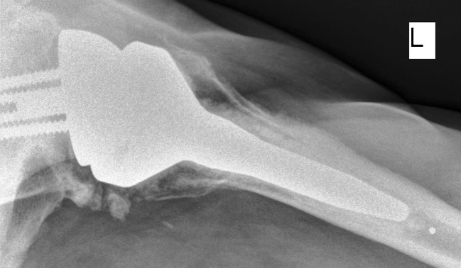
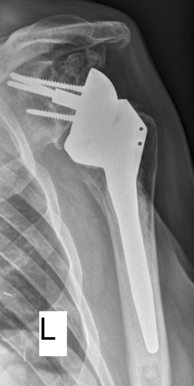
At the final follow-up, none of the glenoid or humeral implants was found to have loosened. Subsidence or tilting of the humeral or glenoid components were not found. Scapular notching was detected in five cases (38 %), with grade 1 in three shoulders and grade 2 in two (Figs. 2 and 3). An inferior spur at the glenoid/scapula neck was observed in four cases.
Fig. 2.
AP X-ray of the same patient as in Fig. 1 8 years after reverse shoulder arthroplasty. Grade 2 notching according to Sirveaux et al. and a small inferior spur are visible. No signs of component loosening were detected
Fig. 3.

Axillary X-ray of the same patient as in Figs. 1 and 2 8 years after reverse shoulder arthroplasty
Complications and revisions
Overall, one complication occurred (8 %). This patient with severe Parkinson’s disease had a fall six months post-operatively and suffered a humeral diaphyseal fracture distal to the implant. This fracture was treated at another centre with open reduction and internal plate fixation, which healed uneventfully. At the final follow-up (3.5 years) the patient was satisfied with the arthroplasty result.
Discussion
The occurrence of osteoarthritis of the glenohumeral joint following surgical stabilisation procedures is a disabling condition. It has been demonstrated that the development of osteoarthritis seems to be related to the pathology itself rather than to the chosen surgical procedure [8–10]. Risk factors for the development of osteoarthritis are a higher age at the time of initial dislocation, a longer time delay between the first dislocation and the stabilisation procedure and the presence of osseous lesions of the glenoid rim [9]. The rate of osteoarthritis may be higher in cases of technical failures related to the surgery (e.g. intra-articular screw placement, fracture of the bone block, recurrent dislocation). Anatomic shoulder arthroplasty has been described as a viable treatment option for patients with arthritis following shoulder stabilisation surgery [11–15].
An improvement of function and pain relief has been demonstrated in several studies addressing this topic. Bigliani et al. reported in 1995 the results of 17 patients with a mean age of 43 years treated with either hemiarthroplasty (n = 5) or total shoulder arthroplasty (n = 12) after various previous stabilisation procedures [12]; 76 % of patients were pain-free after arthroplasty and forward shoulder flexion increased from 111° to 159° and external rotation from −2° to 58°. No cuff deficiencies were reported in this series.
In 2001, Green and Norris reported a series of 17 cases with the same condition treated with hemiarthroplasty (n = 2) or total shoulder arthroplasty (n = 15) [13]. They also found an improvement in pain scores in 94 % of patients and an increase in forward elevation from 99° to 120°. They did not report on any cuff deficiencies in their series.
Sperling et al. published results of the largest existing cohort of 31 patients who underwent shoulder replacement surgery (hemiarthroplasty in ten and total shoulder arthroplasty in 21) with a mean clinical follow-up of seven years [15]. Significant pain relief and an increase in abduction (from 94° to 141°) and external rotation (from 4° to 43°) was demonstrated. They also did not report on any cuff deficiencies in their study.
Two other studies from Matsoukis et al. [14] and Lehmann et al. [11] analysed the results of total shoulder arthroplasty in patients previously managed operatively and non-operatively for instability. Comparing the post-operative clinical outcome, they did not find a difference between the two groups. A significant increase of the Constant score and the range of motion were observed at the final follow-up.
Matsoukis et al. showed an improvement in forward elevation (from 82° to 139°) and external rotation (from 4° to 39°). Moreover, they found a significant improvement in the Constant score from 31 to 66 points [14]. Overall, four tears of the rotator cuff were found in the operatively treated group. Lehmann et al. reported an increase of shoulder flexion from 114° to 140° and also an improvement in the Constant score (from 41 to 66 points) [11]. They found a partial tear of the supraspinatus tendon in ten cases. Both series did not report about fatty infiltration of the rotator cuff muscles.
Although all of the above-mentioned studies showed a significant improvement in the clinical outcome, there was a relatively high rate of patients dissatisfied with their result, complications and revisions when compared with those of unconstrained shoulder replacement for primary osteoarthritis [11, 12, 15, 24–26].
A high complication rate of 40 % was described in the study by Lehmann et al. with 20 % of patients requiring a revision procedure [11]. Sperling et al. reported about 55 % of unsatisfactory results using a modified rating system of Neer. In their series, complications occurred in 52 % of cases (n = 16) and 11 patients underwent revision surgery (35 %) [15]. The study by Bigliani et al. reported unsatisfactory outcomes in 23 % of patients and a revision rate of 18 % [12]. The same rate of revisions and complications was documented in the study by Green and Norris [13].
The overall complication rate of 8 % in this study was acceptable compared with these other studies [13, 11, 12, 15]. One of our patients with severe Parkinson’s disease had a fall six months post-operatively following an uncomplicated period of rehabilitation and suffered a humeral fracture. At the 40-month follow-up (32 months after open reduction and internal fixation) she was satisfied with the outcome. There were no other complications detected during the study period. Moreover, 92 % of patients were either satisfied or very satisfied in this study and this rate is clearly higher than those that were reported in studies for unconstrained implants [15, 11, 12, 24].
The clinical results in this series were slightly inferior to those of the above-mentioned studies, though we did demonstrate a significant increase in the Constant score and range of motion. Compared to other studies describing the clinical results of reverse shoulder arthroplasty, our results seem to be comparable [23, 27, 28]. All of our patients had undergone stabilisation procedures prior to arthroplasty; however, there are some important differences between our patients and the cohorts in the previously mentioned arthroplasty post-instability studies. Some of our patients already had rotator cuff tears at the time of the initial stabilisation procedure; in these cases one of the senior authors systematically performed a Trillat procedure. Moreover, a rupture of the subscapularis tendon and fatty infiltration of the muscle was found in all shoulders at the time of arthroplasty, and in most cases other rotator cuff tendons were also ruptured or had a fatty infiltration. This led us to perform a reverse shoulder arthroplasty rather than to use an unconstrained arthroplasty. In cases of intact rotator cuffs, the authors of this article recommend performing anatomic shoulder arthroplasty. However, the problem becomes more challenging if there is isolated fatty infiltration of the subscapularis muscle. Due to the promising short-term results of this study and the reported high complication rates in studies dealing with anatomic shoulder replacements, we recommend reverse shoulder arthroplasty even in cases with isolated severe fatty infiltration (grades 3 and 4 according to Goutallier et al. [21]) of the subscapularis muscle. In very young patients, however, other treatment options like hemiarthroplasty or arthrodesis should be kept in mind.
The mean age at the time of surgery in this study (70 years) was also higher compared to studies describing the results of conventional shoulder arthroplasty after instability surgery, ranging from 43 to 56 years. In contrast, the mean time delay of 15 years in this study between stabilisation and arthroplasty seems to be in line with most of the studies published [12–15].
This study has weaknesses. Although all patients were recorded in prospective databases, the analysis of the data was performed retrospectively, and the number of cases included is relatively low. However, we did present all of the cases from two specialised shoulder centres from a ten year period. In each of those centres more than 100 shoulder arthroplasties per year were performed during this time, underlining the fact that those who underwent reverse shoulder arthroplasty represent a rare patient cohort. To our knowledge, this is the first study reporting on the results of this implant concept after previous stabilisation surgeries.
Conclusion
Reverse arthroplasty seems to be a viable treatment option with high satisfaction rates for patients with previous shoulder stabilisation procedures who have osteoarthritis and rotator cuff tears. The improvements in clinical outcome as well as the radiographic results seem to be comparable with those of other studies reporting on the outcome of reverse shoulder arthroplasty for other conditions. The complication and revision rates were acceptable in this study.
Acknowledgments
The authors would like to thank the non-profit foundation Stiftung Endoprothetik, Hamburg, Germany, for supporting this study.
Footnotes
The study was approved by the Review Boards of both institutions: Centre Orthopedique Santy Lyon: Ref. Study 43, University of Heidelberg: S-305/2007
References
- 1.Gerber C, Terrier F, Ganz R. The Trillat procedure for recurrent anterior instability of the shoulder. J Bone Joint Surg Br. 1988;70(1):130–134. doi: 10.1302/0301-620X.70B1.3339045. [DOI] [PubMed] [Google Scholar]
- 2.Trillat A, Dejour H, Roullet J. Recurrent luxation of the shoulder and glenoid labrum lesions. Rev Chir Orthop Reparatrice Appar Mot. 1965;51(6):525–544. [PubMed] [Google Scholar]
- 3.Latarjet M. Treatment of recurrent dislocation of the shoulder. Lyon Chir. 1954;49(8):994–997. [PubMed] [Google Scholar]
- 4.Rowe CR, Patel D, Southmayd WW. The Bankart procedure: a long-term end-result study. J Bone Joint Surg Am. 1978;60(1):1–16. [PubMed] [Google Scholar]
- 5.Eden R. Zur Operation der habituellen Schulterluxation unter Mitteilung eines neuen Verfahrens bei Abriss am inneren Pfannenrande. Dtsch Z Chir. 1918;144:269. doi: 10.1007/BF02803861. [DOI] [Google Scholar]
- 6.Young AA, Maia R, Berhouet J, Walch G. Open Latarjet procedure for management of bone loss in anterior instability of the glenohumeral joint. J Shoulder Elbow Surg. 2011;20(2 Suppl):S61–S69. doi: 10.1016/j.jse.2010.07.022. [DOI] [PubMed] [Google Scholar]
- 7.Hybinette S. De la transplantation d'un fragment osseux pour remedier aux luxations recidivantes de l'epaule. Acta Chir Scand. 1932;71:411–445. [Google Scholar]
- 8.Hovelius L, Saeboe M. Neer Award 2008: arthropathy after primary anterior shoulder dislocation–223 shoulders prospectively followed up for twenty-five years. J Shoulder Elbow Surg. 2009;18(3):339–347. doi: 10.1016/j.jse.2008.11.004. [DOI] [PubMed] [Google Scholar]
- 9.Buscayret F, Edwards TB, Szabo I, Adeleine P, Coudane H, Walch G. Glenohumeral arthrosis in anterior instability before and after surgical treatment: incidence and contributing factors. Am J Sports Med. 2004;32(5):1165–1172. doi: 10.1177/0363546503262686. [DOI] [PubMed] [Google Scholar]
- 10.Marx RG, McCarty EC, Montemurno TD, Altchek DW, Craig EV, Warren RF. Development of arthrosis following dislocation of the shoulder: a case-control study. J Shoulder Elbow Surg. 2002;11(1):1–5. doi: 10.1067/mse.2002.119388. [DOI] [PubMed] [Google Scholar]
- 11.Lehmann L, Magosch P, Mauermann E, Lichtenberg S, Habermeyer P. Total shoulder arthroplasty in dislocation arthropathy. Int Orthop. 2010;34(8):1219–1225. doi: 10.1007/s00264-009-0928-5. [DOI] [PMC free article] [PubMed] [Google Scholar]
- 12.Bigliani LU, Weinstein DM, Glasgow MT, Pollock RG, Flatow EL. Glenohumeral arthroplasty for arthritis after instability surgery. J Shoulder Elbow Surg. 1995;4(2):87–94. doi: 10.1016/S1058-2746(05)80060-6. [DOI] [PubMed] [Google Scholar]
- 13.Green A, Norris TR. Shoulder arthroplasty for advanced glenohumeral arthritis after anterior instability repair. J Shoulder Elbow Surg. 2001;10(6):539–545. doi: 10.1067/mse.2001.118007. [DOI] [PubMed] [Google Scholar]
- 14.Matsoukis J, Tabib W, Guiffault P, Mandelbaum A, Walch G, Némoz C, Edwards TB. Shoulder arthroplasty in patients with a prior anterior shoulder dislocation. Results of a multicenter study. J Bone Joint Surg Am. 2003;85-A(8):1417–1424. doi: 10.2106/00004623-200308000-00001. [DOI] [PubMed] [Google Scholar]
- 15.Sperling JW, Antuna SA, Sanchez-Sotelo J, Schleck C, Cofield RH. Shoulder arthroplasty for arthritis after instability surgery. J Bone Joint Surg Am. 2002;84-A(10):1775–1781. doi: 10.2106/00004623-200210000-00006. [DOI] [PubMed] [Google Scholar]
- 16.Scarlat MM. Complications with reverse total shoulder arthroplasty and recent evolutions. Int Orthop. 2013;37(5):843–851. doi: 10.1007/s00264-013-1832-6. [DOI] [PMC free article] [PubMed] [Google Scholar]
- 17.Leung B, Horodyski M, Struk AM, Wright TW. Functional outcome of hemiarthroplasty compared with reverse total shoulder arthroplasty in the treatment of rotator cuff tear arthropathy. J Shoulder Elbow Surg. 2012;21(3):319–323. doi: 10.1016/j.jse.2011.05.023. [DOI] [PubMed] [Google Scholar]
- 18.Warner JJ, Shah A. Shoulder arthroplasty for the treatment of rotator cuff insufficiency. Instr Course Lect. 2011;60:113–121. [PubMed] [Google Scholar]
- 19.Constant CR, Murley AH. A clinical method of functional assessment of the shoulder. Clin Orthop Relat Res. 1987;214:160–164. [PubMed] [Google Scholar]
- 20.Samilson RL, Prieto V. Dislocation arthropathy of the shoulder. J Bone Joint Surg Am. 1983;65(4):456–460. [PubMed] [Google Scholar]
- 21.Goutallier D, Postel JM, Bernageau J, Lavau L, Voisin MC (1994) Fatty muscle degeneration in cuff ruptures. Pre- and postoperative evaluation by CT scan. Clin Orthop Relat Res (304):78–83 [PubMed]
- 22.Melis B, Defranco M, Lädermann A, Molé D, Favard L, Nérot C, Maynou C, Walch G. An evaluation of the radiological changes around the Grammont reverse geometry shoulder arthroplasty after eight to 12 years. J Bone Joint Surg Br. 2011;93(9):1240–1246. doi: 10.1302/0301-620X.93B9.25926. [DOI] [PubMed] [Google Scholar]
- 23.Sirveaux F, Favard L, Oudet D, Huquet D, Walch G, Molé D. Grammont inverted total shoulder arthroplasty in the treatment of glenohumeral osteoarthritis with massive rupture of the cuff. Results of a multicentre study of 80 shoulders. J Bone Joint Surg Br. 2004;86(3):388–395. doi: 10.1302/0301-620X.86B3.14024. [DOI] [PubMed] [Google Scholar]
- 24.Matsoukis J, Tabib W, Guiffault P, Mandelbaum A, Walch G, Némoz C, Cortés ZE, Edwards TB. Primary unconstrained shoulder arthroplasty in patients with a fixed anterior glenohumeral dislocation. J Bone Joint Surg Am. 2006;88(3):547–552. doi: 10.2106/JBJS.E.00368. [DOI] [PubMed] [Google Scholar]
- 25.Raiss P, Aldinger PR, Kasten P, Rickert M, Loew M. Total shoulder replacement in young and middle-aged patients with glenohumeral osteoarthritis. J Bone Joint Surg Br. 2008;90(6):764–769. doi: 10.1302/0301-620X.90B6.20387. [DOI] [PubMed] [Google Scholar]
- 26.Walch G, Young AA, Melis B, Gazielly D, Loew M, Boileau P (2011) Results of a convex-back cemented keeled glenoid component in primary osteoarthritis: multicenter study with a follow-up greater than 5 years. J Shoulder Elbow Surg 20:385–394 [DOI] [PubMed]
- 27.Wall B, Nové-Josserand L, O’Connor DP, Edwards TB, Walch G. Reverse total shoulder arthroplasty: a review of results according to etiology. J Bone Joint Surg Am. 2007;89(7):1476–1485. doi: 10.2106/JBJS.F.00666. [DOI] [PubMed] [Google Scholar]
- 28.Nolan BM, Ankerson E, Wiater JM. Reverse total shoulder arthroplasty improves function in cuff tear arthropathy. Clin Orthop Relat Res. 2011;469(9):2476–2482. doi: 10.1007/s11999-010-1683-z. [DOI] [PMC free article] [PubMed] [Google Scholar]


