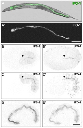
Fig. 3.

IFO-1 is expressed exclusively in the intestine and localizes to the adlumenal domain together with IFB-2. Fluorescence and DIC pictures of IFO-1::CFP-expressing adult worm (A,A′) and embryos (B-D′) of strain BJ155. (A-A′) IFO-1 fluorescence (green) is detected in adult worms only in the intestine where it localizes apically (overlay of DIC and fluorescence image in A). (B-D′) Embryos co-stained with anti-IFB-2 antibodies show that IFO-1 fluorescence is first detectable during polarization of the intestinal primordium, where it accumulates at the future apical pole together with IFB-2 (B,B′). In mid-morphogenesis, IFO-1 is also present at the lateral cortex that is negative for IFB-2. By contrast, IFB-2 localizes diffusely to the cytoplasm, which is negative for IFO-1 (C,C′). During further development, both proteins continue to accumulate at the apical domain whereas the respective lateral and cytoplasmic fluorescence signals diminish (3-fold embryo in D,D′). Note the absence of anti-IFB-2 signal (arrowheads) in some apical membrane regions that already stain positive for IFO-1::CFP (arrowheads). Scale bars: 100 μm in A′; 10 μm in D’ (for B-D′).
