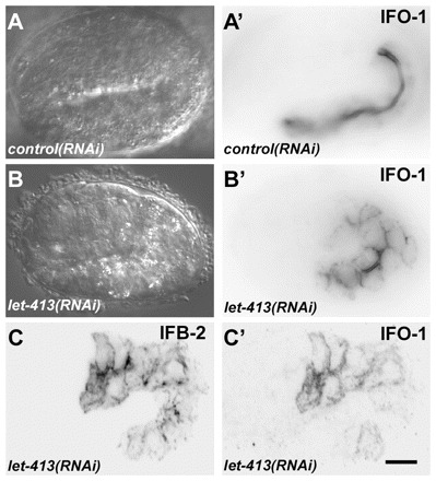
Fig. 8:

let-413 RNAi disturbs proper IFO-1 and IFB-2 localization. (A-B′) Embryos of reporter strain BJ186 kcEX28[ifo-1::yfp,myo-3p::mCherry] were treated with dsRNA directed against either mtm-6 (A,A′) or let-413 (B,B′). DIC images (A,B) and corresponding fluorescence micrographs (A′,B′) are shown. Note the typical exclusive apical IFO-1 distribution in the control that contrasts with the circumferential labeling of apical and basolateral cortical domains upon let-413 RNAi treatment. (C,C′) Fluorescence micrographs of 1.75-fold embryo of strain BJ155 kcEx29[ifo-1::cfp, myo-3p::mcherry] depicting antibody staining of anti-IFB-2 in C and IFO-1::CFP fluorescence in C′ upon let-413 RNAi. Note the non-polarized distribution of both. Scale bar: 10 μm.
