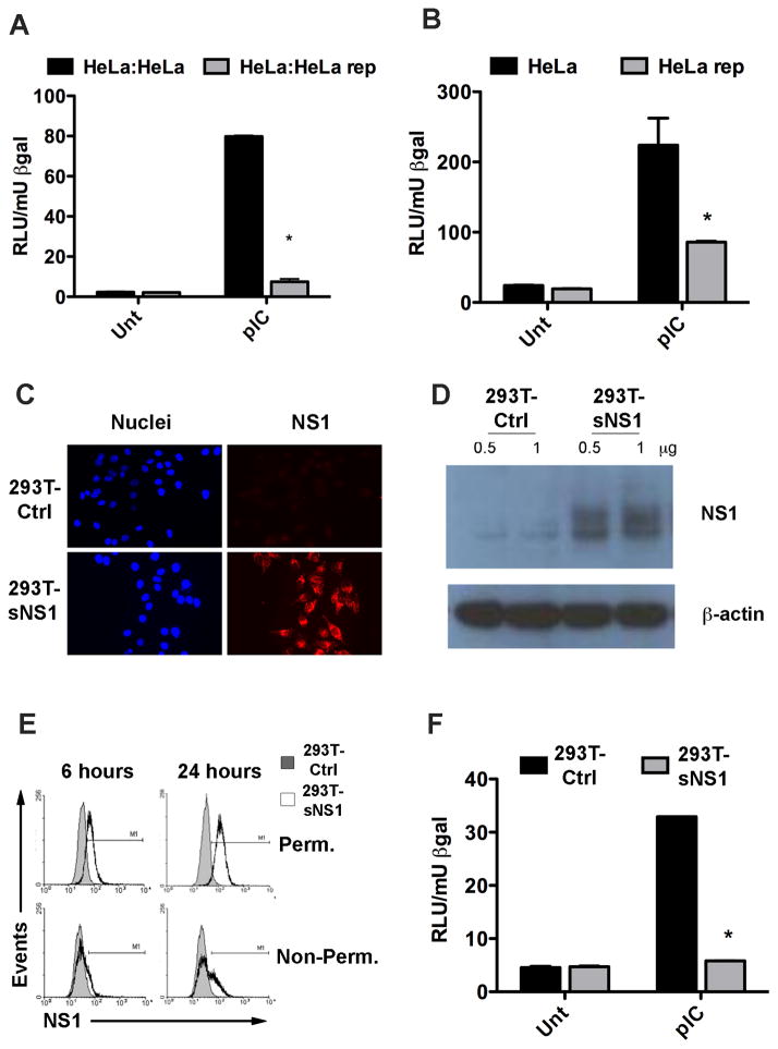
Figure 1.
NS1 containing cell culture supernatants inhibit the TLR3 response in HeLa cells. (A) HeLa cells were transfected with an IFNβ-Luc reporter construct and a β-gal expressing plasmid and subsequently co-cultured with either regular HeLa cells or HeLa cells harboring a WNV replicon (HeLa rep). Luc activity was determined 4h after the addition of pIC to the culture media and normalized to β-gal activity. Asterisks represent statistically significant differences, determined by student’s t-test, comparing the pIC response of HeLa:HeLa rep samples to that of HeLa:HeLa samples. (B) HeLa cells transfected with IFNβ reporter and β-gal as above were incubated for 16h with cell culture supernatants from either regular HeLa cells or HeLa rep cells then stimulated with pIC before Luc activity was determined. Asterisks represent statistically significant differences, determined by student’s t-test comparing the pIC response of HeLa rep cells to that of HeLa cells. (C) HeLa cells were incubated with cell culture supernatants from either 293T-Ctrl or 293T-NS1 cells for 16h. NS1 association with the cells was detected by immunofluorescence of permeabilized cells using WNV MHIAF. (D) HeLa cells were incubated with control or sNS1-containing supernatants for 16h and extensively washed before lysis. Whole cell lysates were analyzed by immunoblot with WNV MHIAF followed by HRP-labeled mouse antibody. (E) NS1 association with naïve cells by flow cytometry. HeLa cells were incubated with control or sNS1-containing supernatants for 6h and 24h either left unpermeabilized to detect NS1 on the cell surface or permeabilized prior to staining to detect internalized NS1. NS1 was detected with an NS1-specific antibody. (F) Secreted NS1-containing cell culture supernatants inhibit the TLR3 response in HeLa cells. HeLa cells transfected with IFNβ reporter construct and β-gal were incubated with supernatants from either 293T-Ctrl or 293T-NS1 cells for 16h and stimulated as above. Asterisks represent statistically significant differences, determined by student’s t-test of sNS1-containing supernatant treated samples compared to control treated samples

