Abstract
Introduction and Objectives:
Hypospadias is the most common congenital abnormality of the penis, and is most commonly diagnosed during the postnatal physical examination. However, milder forms of the condition can be difficult to detect, leading to delayed referral to specialist teams. We aim to determine whether there is an association between hypospadias and the position of the penoscrotal raphe.
Materials and Methods:
A case — control study was performed where clinical photographs from children undergoing hypospadias correction were compared with a control group of children without the condition. The position of the penoscrotal raphe was documented as midline, left or right. Pearson's chi squared test was used to determine significance.
Results:
Images for 80 children undergoing hypospadias correction were compared with 80 normal children in the maternity ward. 88.8% of the children with hypospadias had a penoscrotal raphe deviated from the midline compared with only 13.8% in the control group (P < 0.0003).
Conclusions:
Our study demonstrates a significant association between hypospadias and deviation of the penoscrotal raphe from the midline. Consideration should be given to whether to include this finding within the spectrum of abnormalities seen in hypospadias. Examination of the penoscrotal raphe is simple to perform and could aid in the early diagnosis in children with milder forms of the condition.
KEY WORDS: Deviation, hypospadias, penoscrotal raphe
INTRODUCTION
Hypospadias is the most common congenital abnormality of the penis, with a reported incidence of 0.4-8.2 in 1000 live births,[1] and studies suggest that the condition is becoming increasingly common.[2] One of the reasons for this is likely due to better diagnosis and increased reporting of the condition. Hypospadias is most commonly diagnosed by paediatricians during the postnatal physical examination. However, milder forms may be missed as nonretractile infant foreskin can mask the ectopic meatus.
Classically, hypospadias has been characterised by a triad of defects, namely an ectopic external urethral meatus positioned on the ventral surface of the penis, proximal to its normal location at the tip, chordee and a dorsally hooded prepuce. However, while these signs aid in the diagnosis of the condition, they may be absent or difficult to detect.
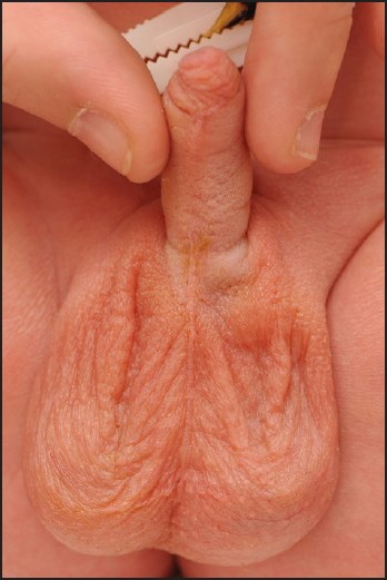
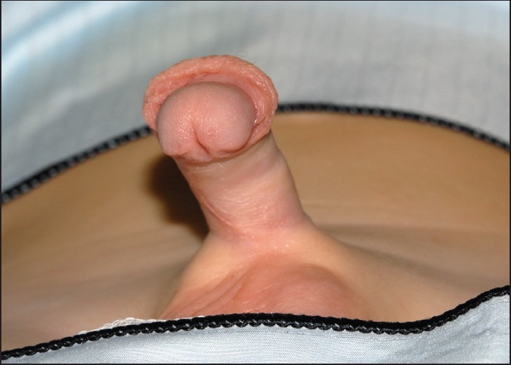
The raphe of the penis is a narrow, dark streak or ridge continuous posteriorly with the raphe of the scrotum and extending forward along the midline on the ventral surface of the penis. Our clinical observations have suggested that a high proportion of children with hypospadias have deviation of the penoscrotal raphe from the normal midline position [Figures 1 and 2]. This may represent abnormal fusion of the urethral folds during development. We therefore performed a study to determine whether this finding is more common in children with hypospadias. If our hypothesis showed a strong correlation, then this feature could be used to aid in the diagnosis of hypospadias.
Figure 1.

Normal child
Figure 2.

Hypospadias
MATERIALS AND METHODS
A case — control study was performed at the Royal Preston Hospital. Preoperative clinical photographs of children undergoing hypospadias correction were retrospectively analysed to determine the position of the penile raphe. These were compared with newborn, healthy infants who were prospectively recruited from the maternity ward. Infants were recruited to the control group following routine postnatal examination. Children with significant medical illness (requiring admission to the neonatal unit) and those with urogenital abnormalities were excluded.
Ethical approval was sought from the Research and Development Directorate. Following this, parental consent for photography was obtained by and following explanation of the study by a member of the Plastic Surgery team. Photographs were taken by the hospital clinical photography department and stored on their secure database.
Photographs of the ventral surface of the penis were analysed to determine whether the penoscrotal median raphe was positioned in the midline, to the left or to the right. The raphe was defined as deviated when its entire length was not confined to the midline.
RESULTS
Records were analysed from 95 children who had undergone hypospadias correction. Photographic records were not available for 15 children. These were compared with a control group of tx80 children without the condition. The results are shown in Table 1.
Table 1.
Results

The majority of children (88.8%) with hypospadias had a penile raphe deviated from the midline, whereas the majority of unaffected children (86.3%) had a centrally positioned raphe. The data were analysed using Pearson's chi squared test to determine the significance between the two variables. Significance was defined as P <0.05. A chi squared statistic of 12.6889 with one degree of freedom corresponds to P <0.0003 thus confirming an association between hypospadias and a deviated penile raphe.
DISCUSSION
Penoscrotal raphe deviation from the midline is an occasional finding in infants. However, there is no published evidence to determine whether this occurs more frequently in association with certain conditions. The aim of this study was to determine whether deviation is significantly more common in children with hypospadias. Our data strongly support the null hypothesis, with 88.1% of the children with the condition demonstrating raphe deviation.
Although a larger proportion of children with hypospadias (59.2%) demonstrated raphe deviation to the right (40.8%), this was not significant. Interestingly, however, 90.9% of the normal children with raphe deviation deviated to the left. This correlates with the results from a study by Sarkis et al.,[3] which demonstrated that 99% of the patients with isolated penile torsion rotated to the left.
Although severe forms of hypospadias can be diagnosed on foetal ultrasound scan, the majority of children are diagnosed by paediatricians at their postnatal check. However, milder variants can be missed. Consideration should be given to whether to include deviation of the penile raphe as part of the spectrum of anomalies seen in hypospadias. Our study suggests a strong correlation between children with the condition of raphe deviation. This, in combination with the low incidence of raphe deviation in normal children, indicates that this finding could prove to be particularly useful as a predictor of hypospadias in infants with nonretractile foreskin. Examination of the penile raphe is simple to perform and could prove to be a useful and safe indicator for the presence of hypospadias.
Footnotes
Source of Support: Nil
Conflict of Interest: None declared.
REFERENCES
- 1.Leung AK, Robson WL. Hypospadias: An update. Asian J Androl. 2007;9:16–22. doi: 10.1111/j.1745-7262.2007.00243.x. [DOI] [PubMed] [Google Scholar]
- 2.Borer J, Retik A. Hypospadias. In: Wein AJ, Kavoussi LR, Novick AC, Partin AW, Peters CA, editors. Campbell-Walsh Urology. 9th ed. Vol. 4. Philadelphia (PA): Saunders Elsevier; 2007. pp. 3703–43. [Google Scholar]
- 3.Sarkis PE, Sadasivam M. Incidence and predictive factors of isolated neonatal penile glandular torsion. J PediatrUrol. 2007;3:495–9. doi: 10.1016/j.jpurol.2007.03.002. [DOI] [PubMed] [Google Scholar]


