Abstract
Anorectal disorders result in many visits to healthcare specialists. These disorders include benign conditions such as hemorrhoids to more serious conditions such as malignancy; thus, it is important for the clinician to be familiar with these disorders as well as know how to conduct an appropriate history and physical examination. This article reviews the most common anorectal disorders, including hemorrhoids, anal fissures, fecal incontinence, proctalgia fugax, excessive perineal descent, and pruritus ani, and provides guidelines on comprehensive evaluation and management.
Keywords: Hemorrhoid, anal fissure, fecal incontinence, pruritus ani, proctalgia fugax, rectoanal abscess, fistula, excessive perineal descent
Anorectal disorders are a common reason for visits to both primary care physicians and gastroenterologists. These disorders are varied and include benign conditions such as hemorrhoids to more serious conditions such as malignancy; thus, it is important for the clinician to be familiar with these disorders as well as know how to conduct an appropriate history and physical examination. This article reviews the most common anorectal disorders, including hemorrhoids, anal fissures, fecal incontinence (FI), proctalgia fugax, excessive perineal descent, and pruritus ani, and provides guidelines on comprehensive evaluation and management.
Hemorrhoids
Hemorrhoids are an extremely common condition, affecting approximately 10 million persons per year. One study estimated that more than 50% of the US population over age 50 years has experienced hemorrhoids.1,2
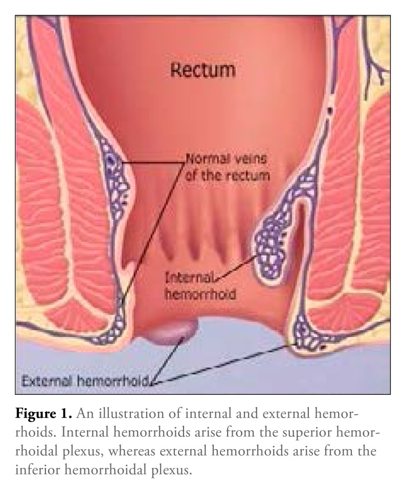
Hemorrhoids represent normal, submucosal, venous structures in the lower rectum and anal canal that may be internal or external depending on their relationship to the dentate line: internal hemorrhoids are located above the dentate line, and external hemorrhoids originate below the dentate line.
Internal hemorrhoids arise from the superior hemorrhoidal plexus. They are viscerally innervated with overlying rectal mucosa and are thus painless. External hemorrhoids arise from the inferior hem-orrhoidal plexus, have somatic innervation that contains numerous pain receptors, and are covered by squamous epithelium (Figure 1).
Figure 1.
An illustration of internal and external hemorrhoids. Internal hemorrhoids arise from the superior hemor-rhoidal plexus, whereas external hemorrhoids arise from the inferior hemorrhoidal plexus.
External skin tags are not hemorrhoids but residual excess tissue. These occur from prior thrombosis of external hemorrhoids or from inflammatory conditions such as perianal Crohn’s disease or anal fissures. As shown in Figure 1, the hemorrhoidal plexuses communicate and then drain to inferior pudendal veins and finally to the inferior vena cava.
Internal hemorrhoids are graded from 1 to 4. Grade 1 hemorrhoids bulge into the lumen but do not extend distal to the dentate line. Grade 2 hemorrhoids prolapse out of the anal canal with straining but reduce spontaneously. Grade hemorrhoids prolapse out of the anal canal with straining and require manual reduction into normal position. Grade hemorrhoids are not able to be reduced and are at risk for strangulation. There is no conventionally used system for grading external hemorrhoids.
The pathogenesis of symptomatic hemorrhoids is not completely understood but likely involves weakening of the anchoring connective tissue, which can then cause prolapse of internal hemorrhoids into the anal canal and protrusion of external hemorrhoids below the anal sphincter. Swelling and engorgement of the hemorrhoidal plexi occur due to factors that increase intra-abdominal pressure, such as straining, constipation, pregnancy, and prolonged sitting.3
The most common clinical presentations of symptomatic hemorrhoids include painless rectal bleeding, pruritus, fecal soilage, perianal irritation, or mucus discharge. Internal hemorrhoidal bleeding typically presents as bright red blood on toilet paper, drips in the toilet bowl, or on the surface of stool; however, blood loss can be substantial, leading to iron-deficient anemia. A patient complaining of bleeding associated with pain should bring to mind an alternative explanation, such as acute or chronic anal fissure, abscess, Crohn’s disease, irritated external hemorrhoids, perineal excoriation, or anal cancer. Thrombosed or engorged external hemorrhoids may present as pain without bleeding.
Treatment of hemorrhoids can be divided into operative and nonoperative therapies. In the setting of acute thrombosis of external hemorrhoids, the mainstay of therapy is pain control. This can be done by conservative measures such as sitz baths and analgesia or surgical excision of the thrombosis, which is most effective during the first 48 to 72 hours after onset of symptoms.4
All patients with symptomatic hemorrhoids should be counseled to avoid constipation and straining. This is most often achieved with fiber supplementation and a mild laxative, with the goal of bulking the stool, minimizing straining, and decreasing the time spent on the com-mode.2 The treatment of internal hemorrhoids depends on the degree of disease. Typically, patients with grades 1, 2, and 3 internal hemorrhoids can be treated nonopera-tively, whereas grade 4 disease or symptoms that do not respond to in-office management should be referred for surgical intervention. The goal of office procedures is to decrease the amount of redundant tissue, increase fixation of the hemorrhoid tissue to the wall of the rectum, and decrease vascularity.2 This can be achieved by rubber band ligation, sclerotherapy, or infrared coagulation, with band ligation being the most commonly performed procedure.
Rubber band ligation has been shown in a meta-analysis to be superior to sclerotherapy and infrared coagu-lation.5 Complications are minimal, with less than 2% of patients experiencing a significant complication such as large-volume bleeding or sepsis.6 The recurrence rate can be up to 13% at 5 years.7 Sclerotherapy involves injection of a caustic agent into the hemorrhoid, which results in fibrosis; however, recurrence is estimated in up to 30% of patients at 4 years.8 Infrared coagulation transforms infrared light into heat as it penetrates the hemorrhoid and results in sclerosis and fibrosis of the tissue. Although this therapy reportedly causes less discomfort than band ligation, the recurrence rate is also higher, and more treatments are required to resolve symptoms.9
Surgical hemorrhoidectomy is typically reserved for those patients with either grade 4 disease or those in whom in-office procedures have failed. The 2 most common surgical procedures are excisional hemorrhoid-ectomy and stapled hemorrhoidopexy. Hemorrhoidopexy uses a circular stapler to fix the anal cushions in place and resect the tissue, whereas, in hemorrhoidectomy, the hem-orrhoid cushions are surgically dissected away from the sphincter muscles and resected. Although hemorrhoido-pexy has been associated with less postoperative pain than hemorrhoidectomy, some studies have shown a slightly higher risk of recurrence.10-12
Anal Fissures
An anal fissure is a tear in the anoderm distal to the dentate line and can be acute or chronic. Acute fissures are those that have been present less than 2 to 3 months and heal with local management. Chronic anal fissures, due to scarring and poor blood flow, often require surgical intervention due to failed conservative management. Most commonly, anal fissures occur in the posterior midline; however, in up to 25% of women and 8% of men, a fissure can be located in the anterior midline. In patients who have lateral fissures, the clinician should consider an alternative etiology such as Crohn’s disease, malignancy, tuberculosis, or HIV infection.13,14
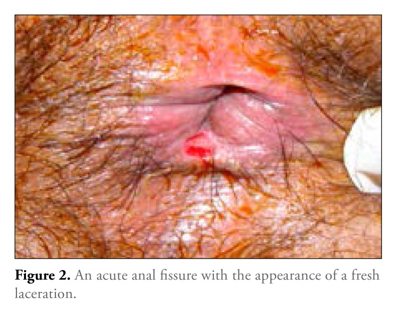
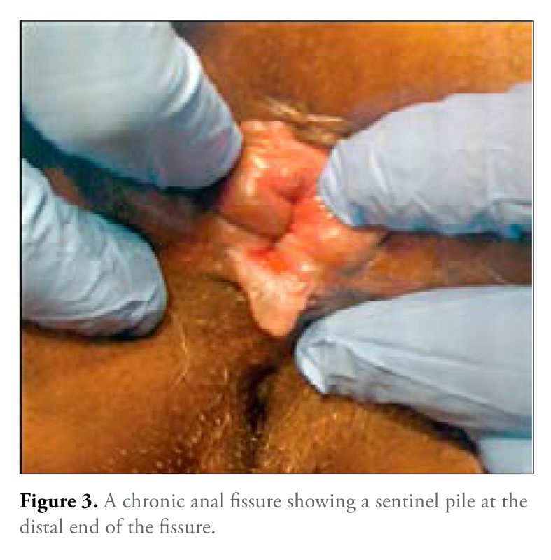
Patients presenting with an anal fissure complain of pain during and after passage of stool. Anal fissure pain is described as sharp, tearing, “like passing knives,” or “shards of glass.” If bright red blood per rectum occurs, it is usually low volume, although large-volume hema-tochezia can occur. An acute fissure appears similar to a fresh laceration (Figure 2), whereas a chronic fissure is frequently associated with skin tags (sentinel pile) at the distal end of the fissure (Figure 3). On digital examination, a chronic fissure feels rough, raised, or fibrotic in the mid-distal anal canal.
Figure 2.
An acute anal fissure with the appearance of a fresh laceration.
Figure 3.
A chronic anal fissure showing a sentinel pile at the distal end of the fissure.
The pathogenesis of anal fissures is thought to be 3-fold and includes trauma, ischemia, and elevated anal pressure. In the posterior midline, which is the site of most fissures, the blood flow is less than half of that seen in other quadrants of the anal canal, and this, in turn, likely contributes to a decreased ability to heal.15 Additionally, blood flow at the fissure site has been demonstrated to be lower than that at the posterior anal midline of control groups.16 Typically, there is elevated anal canal pressure in patients with an anal fissure, which is thought to be due to increased internal anal sphincter tone as well as spasm of the muscle beneath the tear, which, in turn, is due to pain from the initial trauma.17
Treatment of an anal fissure can be achieved medically or, in the case of refractory fissures, surgically. The gold-standard treatment for chronic, refractory anal fissures is a lateral internal sphincterotomy, which is effective and has a low rate of disease recurrence (<10%). A Cochrane database review found that medical management is less effective than surgical management in the treatment of refractory fissures; however, no medical treatments caused FI, which is a known complication of sphincterotomy.18,19 Additionally, sphincterotomy involves anesthesia, inpatient care, higher costs, and greater morbidity, which is why medical management is tried first.
The goal of medical treatment of the anal fissure is to relax the anal sphincter as well as to halt the cycle of sphincter spasm and tearing. Ultimately, this promotes increased blood flow to the area and healing of the fissure. A cornerstone of therapy is to soften the stool and regulate bowel habits in an effort to minimize trauma to the area. Additional topical therapies include nitroglyc-erin ointment (available in multiple concentrations from 0.2% to 0.4%) and topical nifedipine or diltiazem cream. Topical nitroglycerin has been shown in multiple trials to be better than placebo in the healing of anal fissures, and, although it can cause headaches transiently, this effect usually quickly diminishes with continued use of the medication.20-22 Although multiple studies have compared the efficacy of topical calcium channel blockers (CCBs) to nitroglycerin, the results are mixed.23-27 Injection of botulinum toxin into the internal anal sphincter is also an effective therapy. Although it is more invasive than topical therapies, it is less invasive than surgery and has shown promising results. Recent studies have found a healing rate of 83% to 92% after 25 to 30 units of botulinum toxin were injected into the internal anal sphincter. Additionally, botulinum toxin has been shown to be slightly more effective than topical nitroglycerin.28,29
Anorectal Abscesses and Fistulae
Anorectal abscesses and fistulae are anorectal disorders that are thought to be a spectrum of the same disease. Perianal abscess is the initial manifestation of infection that may then be followed by a more chronic, suppura-tive process, leading to perianal fistula. The conversion of abscess to fistula occurs in approximately 40% to 50% of cases.3,30 The prevalence of these disorders is difficult to calculate, as many patients with anorectal symptoms do not often come to medical attention, and reported data reflect single-institution experience; however, it is estimated that the incidence of anorectal abscess is 100,000 cases per year in the United States. The mean age of presentation is 40 years, with a male predominance of 2:1.30,31
Anorectal abscesses are thought to originate from an infected anorectal gland. The infection can then track through the perianal tissues to form a perianal fistula, which is a connection between the infected anal crypt gland and the perineum.3 Although the most common etiology for perianal fistula is an anorectal abscess, other etiologies include Crohn’s disease, radiation proctitis, foreign body, prior anal surgery, infections (such as HIV, tuberculosis, or actinomycosis), and malignancy.
Patients presenting with an anal abscess typically have an area of persistent pain and swelling that can be visualized and palpated. If the abscess is in the intersphincteric space, the clinician may not appreciate an abnormality externally but be able to palpate a boggy area on rectal examination. Perianal fistulae often present as drainage of blood, pus, or stool from an external opening in the perianal region. Fistulae are intermittently painful, a finding that may help distinguish them from abscesses, which evoke a constant throbbing pain. Patients with fistulae can experience perianal itching.
Treatment of an anorectal abscess requires incision and drainage to prevent spread, recurrence, and, hopefully, subsequent fistulization. The American Society of Colon and Rectal Surgeons states that antibiotics are not routinely required in the management of an anorectal abscess except in cases of immunosuppression, diabetes, extensive cellulitis, or prosthetic devices.32
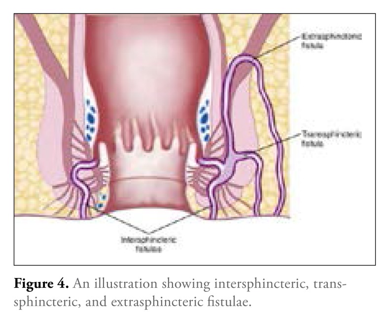
Treatment for a perianal fistula is determined by the anatomy of the fistula. There are 4 types of fistulae: intersphincteric, transsphincteric, suprasphincteric, and extrasphincteric (Figure 4). The diagnostic approach to examining a patient with a fistula is with magnetic resonance imaging (MRI) or endoscopic ultrasound before he or she undergoes examination by a surgeon under anesthesia. Asymptomatic Crohn’s fistulae usually require no treatment; however, internal fistulae (eg, gastrocolic, duodenocolic, or enterovesicular) that cause severe or persistent symptoms should always be treated surgically. Surgical treatment is guided by the type of fistula, with the goal of primary healing.33
Figure 4.
An illustration showing intersphincteric, transsphincteric, and extrasphincteric fistulae.
Fecal Incontinence
FI is a debilitating, embarrassing, and potentially devastating disorder. It is common and affects up to 24% of the general population, although figures vary widely depending on the definition of FI and the age group studied.34 The prevalence of FI is even higher in institutionalized and nursing home patients.35,36 Complaints of FI can range from flatal incontinence to minor soiling with small amounts of liquid stool or stool pellets to frank, involuntary passage of a complete bowel movement. Subtypes of FI include passive incontinence, which is the involuntary discharge of stool or gas without awareness; urge incontinence, which is the discharge of fecal matter in spite of active attempts to retain bowel contents; and fecal seepage, which is the passive leakage of stool usually following an otherwise normal evacuation.37
The mechanism of fecal continence is complex and involves numerous anatomic and physiologic factors. These include sphincter function, anorectal sensation, colonic transit, stool consistency, and neurologic as well as cognitive elements. Disruption of any number of the above can predispose a patient to incontinence. Etiologies of FI include obstetric injuries such as sphincter tear or nerve injury, surgical trauma, neuropathy, altered bowel habit (either constipation or diarrhea), rectal prolapse, and decreased rectal compliance (as can be seen in radiation proctitis or ulcerative colitis). Many patients have sphincter injuries that remain asymptomatic for years until they experience age- or hormone-related changes, such as muscle or tissue atrophy, that reduce the ability to compensate for their remote injury.
In evaluating a patient with FI, a detailed medical, surgical, and obstetric history, particularly of the incontinence, bowel symptoms, and medication use, should be obtained. Physical examination is vital to an accurate and comprehensive diagnosis and should include careful peri-anal and perineal examinations that look for an obvious pathology as well as perianal sensation via elicitation of the anocutaneous reflex (anal wink). Digital examination will reveal resting tone, anal sphincter length, and symmetry. Patients should be asked to bear down to assess for pelvic floor descent and rectal prolapse as well as asked to squeeze to assess squeezing pressure at rest and while bearing down.
In addition to the physical examination, there are a few diagnostic tests that are helpful in evaluating FI. Anorectal manometry quantifies internal and external anal sphincter function, rectal sensation, and compliance. There are several systems in use. The water-perfused probe is the least expensive and has been traditionally used. Solid-state probes with microtransducers have become common. High-resolution manometry systems with over 250 pressure sensors are available for the evaluation of pressure profiles and topographic changes in 3 dimensions. This method may increase diagnostic yield.38 Anal resting and squeeze pressure as well as rectoanal inhibitory reflex should always be measured. Anal resting and squeeze pressures are often low in FI, suggesting weak internal and external sphincters.39 Endoanal ultrasound remains the standard for identifying sphincter injury.40 It provides excellent resolution of the internal sphincter but is less accurate with the external sphincter. Dynamic pelvic floor MRI consists of a defecogram imaging technique that achieves better visualization of the sphincter complex as well as more comprehensive evaluation of the pelvic floor without using radiation.41,42 Gadolinium paste is injected into the rectum, after which a dynamic assessment of evacuation is recorded, with the patient in the supine position. Standard defecography involves instillation of barium paste into the rectum followed by dynamic recorded images as the patient sits on a commode. It is less expensive than MRI but exposes the patient to radiation and does not clearly show the relationship of surrounding pelvic floor organs to evacuation. Defecography allows visualization and measurement of perineal descent, of anorectal angles at rest, with bearing down, and during squeeze. It can also demonstrate the presence of rectoceles, enteroceles, and processes that influence evacuation and continence. MR defecography best demonstrates orientation of organs after pelvic surgery.
Patients may need endoscopic evaluation of the distal colon and rectum, primarily to exclude inflammation or other pathology that could contribute to incontinence. In patients with bowel habit changes, a full colonoscopy may be more appropriate as well as a comprehensive workup to identify the cause of diarrhea or constipation.
Treatment of FI begins with lifestyle modifications. Reducing medications that have a diarrheal adverse effect or that increase intestinal transit may result in significant improvement in FI. Diets high in artificial sugars and caffeine or low in fiber can reduce stool consistency while increasing episodes of stool loss and leakage. Limited mobility in elderly or physically impaired patients can play a role in FI. Scheduling toileting times, having ready access to assist devices, and shortening travel distances allow these patients better access to the toilet. If diarrhea is the cause for the FI, initiation of supplemental fiber bulking agents is often effective. Pharmacotherapy for diarrhea with agents such as loperamide, diphenoxylate/atropine, alosetron (Lotronex, Prometheus), clonidine, cholestyramine, colestipol, probiotics, tincture of opium, and amitriptyline is usually reserved for patients with more refractory symptoms that do not respond to bulk fiber supplements. In patients with overflow incontinence, a strict bowel regimen should be used to prevent constipation. A trial of fiber therapy should be first-line treatment for overflow incontinence as well as incontinence due to diarrhea. Although stool softeners are often used initially in clinical practice, a large study comparing the efficacy of psyllium vs docusate found that psyllium was significantly superior to docusate in providing relief of constipation.43
Biofeedback therapy is a nonoperative technique that is widely used when conservative dietary or bowel management interventions are insufficient. Biofeedback works by improving muscular strength and control of the pelvic floor, enhancing sensory perception of rectal distension and coordinating both aspects to improve continence. Although multiple studies have been performed to evaluate the effect of biofeedback on FI, many are retrospective, small, and without control groups. This has led to a wide estimate of success, ranging between 38% and 90%. Despite the varying estimates of efficacy, biofeedback has been shown to have a durable effect on symptoms.44-49 In patients in whom biofeedback and other conservative options have failed, another new but promising intervention is an injectable bulking agent.
Injection of hyaluronic acid/dextranomer (Solesta, Salix) can augment the anal sphincter area and increase anal sphincter pressures at rest and during squeeze, thereby improving continence primarily in those patients with low anal sphincter pressures. The material is injected into the submucosa in 4 quadrants just proximal to the dentate line. Studies have shown a decrease of up to 75% in the number of incontinence episodes.50,51 In patients who have failed conservative management with a bulking agent and biofeedback, injection therapy is first-line intervention and a safe office-based procedure typically performed by a gastroenterologist or colorectal surgeon. Sacral nerve stimulation, or direct repair of the anal sphincter, should be considered if symptoms are severe and if the anal sphincter damage is not amenable to injection therapy. MRI images will show hyaluronic acid/ dextranomer in situ, whereas CT does not. Sacral nerve stimulation is thought to work by improving squeeze pressures of the anal sphincter and improving rectal sensation and has shown promising results.52-55 Surgery may be necessary in women with evidence of a significant sphincter tear, often occurring from obstetric trauma.
Proctalgia Fugax
Proctalgia fugax is a functional gastrointestinal disorder characterized by severe and self-limiting anorectal pain. Attacks last from 5 seconds to 90 minutes, occur any time of the day, and sometimes wake patients from sleep.56 Attacks tend to be infrequent, averaging 1 episode monthly.57 Based on Rome III criteria, proctalgia fugax is defined as recurrent episodes of pain localized to the anus or lower rectum.57 Although symptoms of proctalgia fugax are unique and characteristic, conditions in the differential for recurrent anorectal pain include painful hemorrhoids, anal fissures, inflammatory conditions, fissures, and malignancy. The prevalence of proctalgia fugax is estimated to be 4% to 18% of the general population, with a female predominance.58,59 Patients cannot typically identify a trigger for the onset of pain, which does not radiate and occurs without concomitant symptoms.56
The pathophysiology of proctalgia fugax is incompletely understood but is thought to be due, in part, to spasm of the internal anal sphincter and/or pudendal nerve compression. Anorectal manometry has shown increased resting pressures and higher internal anal sphincter thick-ness.60,61 Pharmacologic treatment of proctalgia fugax is often unnecessary due to the fleeting, infrequent nature of the symptoms; however, in patients with more severe symptoms, treatment is aimed at the underlying proposed pathology and can include topical treatments with nitroglycerin or diltiazem, biofeedback, tricyclic antide-pressants, and more invasive therapy such as botulinum toxin injection and nerve blocks. Biofeedback therapy has shown promise in this disorder, is noninvasive, and has no documented adverse effects.60,62
Pruritus Ani
Pruritus ani affects between 1% and 5% of the population and has a male predominance.63 The condition is characterized by itching or burning in the perianal area. The causes of pruritus ani are numerous and include topical irritants such as soaps or laundry solutions; some foods such as coffee, chocolate, and citrus; certain sexually transmitted diseases; medications such as colchicine or neomycin; mechanical factors such as fecal soiling or hemorrhoids; infections; dermatologic disorders, including psoriasis or atopic dermatitis; systemic disorders; or malignancy. Obtaining a detailed patient history is critical for an accurate and timely diagnosis and should include an examination of the perianal area. The underlying pathophysiology of pruritus ani is such that there is an initial, localized irritation that is followed by the development of an inflammatory response. Scratching further augments and irritates the inflammatory response, and a self-propagating cycle develops. In patients with chronic pruritus ani, the clinician may see lichenification of the perianal skin due to repeated scratching.
Treatment of pruritus ani should be directed at the underlying cause, if identified. The clinician must educate patients about keeping the area dry and that persistent scratching will often lead to persistent itching due to aggravation of inflammation. The clinician can try an antihistamine or witch hazel to reduce the itching. Patients who have FI and soiling should be instructed to minimize aggressive wiping and cleansing and to use fragrance-free soaps or, ideally, only water.
Topical capsaicin 0.006% cream has been found effective in a randomized, placebo-controlled trial, as has topical hydrocortisone.64,65 Topical hydrocortisone, however, should not be used for more than 2 weeks due to its potential to cause perianal skin thinning. If symptoms do not improve, the clinician needs to consider dermatology evaluation with biopsy of the perianal skin.
Summary
Anorectal disorders are common and can significantly impair a person’s quality of life. Diagnosis is made by a comprehensive history of symptoms, visual inspection and digital rectal examination, along with selective tests. Diet, bowel habit, and lifestyle changes are often first-line therapy for hemorrhoids, minor irritation, and FI. When conservative therapy is not effective, in-office ligation, sclerotherapy, or infrared coagulation for hemorrhoids should be considered. Surgery is reserved for those with persistent symptoms or grade 4 disease.
The goal of medical treatment for chronic fissures is to reduce the cycle of spasm and tearing. Softening stool and regulating movements minimize trauma. Topical therapy such as nitroglycerin or nifedipine can be effective, although injection of botulinum toxin or surgery may be needed for refractory fissures.
Painful swelling characterizes the presentation of an anorectal abscess. Treatment of an abscess requires incision and drainage of the lesion, usually without antibiotics unless the patient is immunocompromised. Asymptomatic Crohn’s fistulae can be watched, whereas symptomatic fistulae require surgical treatment.
FI often responds to dietary or supplemental fiber with a timed bowel regimen. Injectable agents such as hyaluronic acid create a barrier, effectively reducing the number of incontinence episodes. Sacral nerve stimulation for FI has also shown promising results. Surgery for FI is reserved for refractory cases.
Proctalgia fugax is a functional anorectal pain condition that responds poorly to medical therapy, yet biofeed-back has shown some benefit and has no ill effects. Pruritus ani has many potential causes, with primary treatment directed at the underlying cause as well as reduction of perianal irritation. Topical hydrocortisone therapy can be effective but should be limited to 2 weeks due to its potential for thinning of the perianal skin.
Footnotes
Dr Foxx-Orenstein has received honoraria from Salix Pharmaceutical. Dr Umar and Dr Crowell have no relevant conflicts of interest to disclose.
References
- 1.Sanchez C, Chinn BT. Hemorrhoids. Clin Colon Rectal Surg. 2011;24(1):5–13. doi: 10.1055/s-0031-1272818. [DOI] [PMC free article] [PubMed] [Google Scholar]
- 2.Rivadeneira DE, Steele SR, Ternent C, Chalasani S, Buie WD, Rafferty JL. Standards Practice Task Force of The American Society of Colon and Rectal Surgeons. Practice parameters for the management of hemorrhoids (revised 2010) Dis Colon Rectum. 2011;54(9):1059–1064. doi: 10.1097/DCR.0b013e318225513d. [DOI] [PubMed] [Google Scholar]
- 3.Feldman M, Friedman LS, Brandt LJ, editors. Sleisenger andFordtrans Gastrointestinal and Liver Disease. 9th ed. Philadelphia, PA: Saunders Elsevier; 2010. [Google Scholar]
- 4.Greenspon J, Williams SB, Young HA, Orkin BA. Thrombosed external hemorrhoids: outcome after conservative or surgical management. Dis Colon Rectum. 2004;47(9):1493–1498. doi: 10.1007/s10350-004-0607-y. [DOI] [PubMed] [Google Scholar]
- 5.MacRae HM, McLeod RS. Comparison of hemorrhoidal treatment modalities. A meta-analysis. Dis Colon Rectum. 1995;38(7):687–694. doi: 10.1007/BF02048023. [DOI] [PubMed] [Google Scholar]
- 6.Bat L, Melzer E, Koler M, Dreznick Z, Shemesh E. Complications of rubber band ligation of symptomatic internal hemorrhoids. Dis Colon Rectum. 1993;36(3):287–290. doi: 10.1007/BF02053512. [DOI] [PubMed] [Google Scholar]
- 7.Su MY, Chiu CT, Lin WP, Hsu CM, Chen PC. Long-term outcome and efficacy of endoscopic hemorrhoid ligation for symptomatic internal hemorrhoids. World J Gastroenterol. 2011;17(19):2431–2436. doi: 10.3748/wjg.v17.i19.2431. [DOI] [PMC free article] [PubMed] [Google Scholar]
- 8.Fleshman J, Madoff R. Hemorrhoids. In: Cameron J, editor. Current Surgical Therapy. 8th ed. Philadelphia, PA: Elsevier; 2004. pp. 245–252. [Google Scholar]
- 9.MacRae HM, McLeod RS. Comparison of hemorrhoidal treatments: a meta-analysis. Can J Surg. 1997;40(1):14–17. [PMC free article] [PubMed] [Google Scholar]
- 10.Jayaraman S, Colquhoun PH, Malthaner RA. Stapled hemorrhoidopexy is associated with a higher long-term recurrence rate of internal hemorrhoids compared with conventional excisional hemorrhoid surgery. Dis Colon Rectum. 2007;50(9):1297–1305. doi: 10.1007/s10350-007-0308-4. [DOI] [PubMed] [Google Scholar]
- 11.Gravie JF, Lehur PA, Huten N, et al. Stapled hemorrhoidopexy versus milli-gan-morgan hemorrhoidectomy: a prospective, randomized, multicenter trial with 2-year postoperative follow up. Ann Surg. 2005;242(1):29–35. doi: 10.1097/01.sla.0000169570.64579.31. [DOI] [PMC free article] [PubMed] [Google Scholar]
- 12.Giordano P, Gravante G, Sorge R, Ovens L, Nastro P. Long-term outcomes of stapled hemorrhoidopexy vs conventional hemorrhoidectomy: a meta-analysis of randomized controlled trials. Arch Surg. 2009;144(3):266–272. doi: 10.1001/archsurg.2008.591. [DOI] [PubMed] [Google Scholar]
- 13.Oh C, Divino CM, Steinhagen RM. Anal fissure. 20-year experience. Dis Colon Rectum. 1995;38(4):378–382. doi: 10.1007/BF02054225. [DOI] [PubMed] [Google Scholar]
- 14.Dykes SL, Madoff RD. Benign anorectal: anal fissure. In: Wolff BG, Fleshman JW, Beck DE, Pemberton JH, Wexner SD, editors. The ASCRS Textbook of Colon and Rectal Surgery. New York, NY: Springer Science + Business Media, LLC; 2007. pp. 178–191. [Google Scholar]
- 15.Schouten WR, Briel JW, Auwerda JJ. Relationship between anal pressure and anodermal blood flow. The vascular pathogenesis of anal fissures. Dis Colon Rectum. 1994;37(7):664–669. doi: 10.1007/BF02054409. [DOI] [PubMed] [Google Scholar]
- 16.Schouten WR, Briel JW, Auwerda JJ, De Graaf EJ. Ischaemic nature of anal fissure. Br J Surg. 1996;83(1):63–65. doi: 10.1002/bjs.1800830120. [DOI] [PubMed] [Google Scholar]
- 17.Keck JO, Staniunas RJ, Coller JA, Barrett RC, Oster ME. Computer-generated profiles of the anal canal in patients with anal fissure. Dis Colon Rectum. 1995;38(1):72–79. doi: 10.1007/BF02053863. [DOI] [PubMed] [Google Scholar]
- 18.Nelson RL, Thomas K, Morgan J, Jones A. Nonsurgical therapy for anal fissure. Cochrane Database Syst Rev. 2012;2 doi: 10.1002/14651858.CD003431.pub3. CD003431. [DOI] [PMC free article] [PubMed] [Google Scholar]
- 19.Nyam DC, Pemberton JH. Long-term results of lateral internal sphincter-otomy for chronic anal fissure with particular reference to incidence of fecal incontinence. Dis Colon Rectum. 1999;42(10):1306–1310. doi: 10.1007/BF02234220. [DOI] [PubMed] [Google Scholar]
- 20.Lund JN, Scholefield JH. A randomised, prospective, double-blind, placebo-controlled trial of glyceryl trinitrate ointment in treatment of anal fissure. Lancet. 1997;349(9044):11–14. doi: 10.1016/S0140-6736(96)06090-4. [DOI] [PubMed] [Google Scholar]
- 21.Kennedy ML, Sowter S, Nguyen H, Lubowski DZ. Glyceryl trinitrate ointment for the treatment of chronic anal fissure: results of a placebo-controlled trial and long-term follow-up. Dis Colon Rectum. 1999;42(8):1000–1006. doi: 10.1007/BF02236691. [DOI] [PubMed] [Google Scholar]
- 22.Thornton MJ, Kennedy ML, King DW. Manometric effect of topical glyceryl trinitrate and its impact on chronic anal fissure healing. Dis Colon Rectum. 2005;48(6):1207–1212. doi: 10.1007/s10350-004-0916-1. [DOI] [PubMed] [Google Scholar]
- 23.Ezri T, Susmallian S. Topical nifedipine vs. topical glyceryl trinitrate for treatment of chronic anal fissure. Dis Colon Rectum. 2003;46(6):805–808. doi: 10.1007/s10350-004-6660-8. [DOI] [PubMed] [Google Scholar]
- 24.Cevik M, Boleken ME, Koruk I, et al. A prospective, randomized, double-blind study comparing the efficacy of diltiazem, glyceryl trinitrate, and lidocaine for the treatment of anal fissure in children. Pediatr Surg Int. 2012;28(4):411–416. doi: 10.1007/s00383-011-3048-4. [DOI] [PubMed] [Google Scholar]
- 25.Sajid MS, Vijaynagar B, Desai M, Cheek E, Baig MK. Botulinum toxin vs glyceryltrinitrate for the medical management of chronic anal fissure: a meta-analysis. Colorectal Dis. 2008;10(6):541–546. doi: 10.1111/j.1463-1318.2007.01387.x. [DOI] [PubMed] [Google Scholar]
- 26.Sanei B, Mahmoodieh M, Masoudpour H. Comparison of topical glyceryl trinitrate with diltiazem ointment for the treatment of chronic anal fissure: a randomized clinical trial. Acta Chir Belg. 2009;109(6):727–730. doi: 10.1080/00015458.2009.11680524. [DOI] [PubMed] [Google Scholar]
- 27.Perry WB, Dykes SL, Buie WD, Rafferty JF. Standards Practice Task Force of the American Society of Colon and Rectal Surgeons. Practice parameters for the management of anal fissures (3rd revision) Dis Colon Rectum. 2010;53(8):1110–1115. doi: 10.1007/DCR.0b013e3181e23dfe. [DOI] [PubMed] [Google Scholar]
- 28.Brisinda G, Cadeddu F, Brandara F, Marniga G, Maria G. Randomized clinical trial comparing botulinum toxin injections with 0.2 percent nitroglycerin ointment for chronic anal fissure. Br J Surg. 2007;94(2):162–167. doi: 10.1002/bjs.5514. [DOI] [PubMed] [Google Scholar]
- 29.Sileri P, Stolfi VM, Franceschilli L, et al. Conservative and surgical treatment of chronic anal fissure: prospective longer term results. J Gastrointest Surg. 2010;14(5):773–780. doi: 10.1007/s11605-010-1154-6. [DOI] [PubMed] [Google Scholar]
- 30.Abcarian H. Anorectal infection: abscess-fistula. Clin Colon Rectal Surg. 2011;24(1):14–21. doi: 10.1055/s-0031-1272819. [DOI] [PMC free article] [PubMed] [Google Scholar]
- 31.Sainio P. Fistula-in-ano in a defined population. Incidence and epidemiological aspects. Ann Chir Gynaecol. 1984;73(4):219–224. [PubMed] [Google Scholar]
- 32.Whiteford MH, Kilkenny J, 3rd, Hyman N, et al. Standards Practice Task Force; American Society of Colon and Rectal Surgeons. Practice parameters for the treatment of perianal abscess and fistula-in-ano (revised) Dis Colon Rectum. 2005;48(7):1337–1342. doi: 10.1007/s10350-005-0055-3. [DOI] [PubMed] [Google Scholar]
- 33.Siddiqui MR, Ashrafian H, Tozer P, et al. A diagnostic accuracy meta-analysis of endoanal ultrasound and MRI for perianal fistula assessment. Dis Colon Rectum. 2012;55(5):576–585. doi: 10.1097/DCR.0b013e318249d26c. [DOI] [PubMed] [Google Scholar]
- 34.Halland M, Talley NJ. Fecal incontinence: mechanisms and management. Curr Opin Gastroenterol. 2012;28(1):57–62. doi: 10.1097/MOG.0b013e32834d2e8b. [DOI] [PubMed] [Google Scholar]
- 35.Varma MG, Brown JS, Creasman JM, et al. Fecal incontinence in females older than aged 40 years: who is at risk? Dis Colon Rectum. 2006;49(6):841–851. doi: 10.1007/s10350-006-0535-0. [DOI] [PMC free article] [PubMed] [Google Scholar]
- 36.Nelson R, Furner S, Jesudason V. Fecal incontinence in Wisconsin nursing homes: prevalence and associations. Dis Colon Rectum. 1998;41(10):1226–1229. doi: 10.1007/BF02258218. [DOI] [PubMed] [Google Scholar]
- 37.Rao SS. Pathophysiology of adult fecal incontinence. Gastroenterology. 2004;126(1 suppl 1):S14–S22. doi: 10.1053/j.gastro.2003.10.013. [DOI] [PubMed] [Google Scholar]
- 38.Rao SS. Advances in diagnostic assessment of fecal incontinence and dyssyner-gic defecation. Clin Gastroenterol Hepatol. 2010;8(11):910–919. doi: 10.1016/j.cgh.2010.06.004. [DOI] [PMC free article] [PubMed] [Google Scholar]
- 39.Diamant NE, Kamm MA, Wald A, Whitehead WE. AGA technical review on anorectal testing techniques. Gastroenterology. 1999;116(3):735–760. doi: 10.1016/s0016-5085(99)70195-2. [DOI] [PubMed] [Google Scholar]
- 40.Bharucha AE. Update of tests of colon and rectal structure and function. J Clin Gastroenterol. 2006;40(2):96–103. doi: 10.1097/01.mcg.0000196190.42296.a9. [DOI] [PubMed] [Google Scholar]
- 41.Maccioni F. Functional disorders of the ano-rectal compartment of the pelvic floor: clinical and diagnostic value of dynamic MRI. Abdom Imaging. 2013;38(5):930–951. doi: 10.1007/s00261-012-9955-6. [DOI] [PubMed] [Google Scholar]
- 42.Rentsch M, Paetzel C, Lenhart M, Feuerbach S, Jauch KW, Furst A. Dynamic magnetic resonance imaging defecography: a diagnostic alternative in the assessment of pelvic floor disorders in proctology. Dis Colon Rectum. 2001;44(7):999–1007. doi: 10.1007/BF02235489. [DOI] [PubMed] [Google Scholar]
- 43.McRorie JW, Daggy BP, Morel JG, Diersing PS, Miner PB, Robinson M. Psyl-lium is superior to docusate sodium for treatment of chronic constipation. Aliment Pharmacol Ther. 1998;12(5):491–497. doi: 10.1046/j.1365-2036.1998.00336.x. [DOI] [PubMed] [Google Scholar]
- 44.Boselli AS, Pinna F, Cecchini S, et al. Biofeedback therapy plus anal elec-trostimulation for fecal incontinence: prognostic factors and effects on anorectal physiology. World J Surg. 2010;34(4):815–821. doi: 10.1007/s00268-010-0392-9. [DOI] [PubMed] [Google Scholar]
- 45.Byrne CM, Solomon MJ, Young JM, Rex J, Merlino CL. Biofeedback for fecal incontinence: short-term outcomes of 513 consecutive patients and predictors of successful treatment. Dis Colon Rectum. 2007;50(4):417–427. doi: 10.1007/s10350-006-0846-1. [DOI] [PubMed] [Google Scholar]
- 46.Heymen S, Scarlett Y, Jones K, Ringel Y, Drossman D, Whitehead WE. Randomized controlled trial shows biofeedback to be superior to pelvic floor exercises for fecal incontinence. Dis Colon Rectum. 2009;52(10):1730–1737. doi: 10.1007/DCR.0b013e3181b55455. [DOI] [PMC free article] [PubMed] [Google Scholar]
- 47.Lee BH, Kim N, Kang SB, et al. The long-term clinical efficacy of biofeedback therapy for patients with constipation or fecal incontinence. J Neurogastroenterol Motil. 2010;16(2):177–185. doi: 10.5056/jnm.2010.16.2.177. [DOI] [PMC free article] [PubMed] [Google Scholar]
- 48.Madoff RD, Parker SC, Varma MG, Lowry AC. Faecal incontinence in adults. Lancet. 2004;364(9434):621–632. doi: 10.1016/S0140-6736(04)16856-6. [DOI] [PubMed] [Google Scholar]
- 49.Pager CK, Solomon MJ, Rex J, Roberts RA. Long-term outcomes of pelvic floor exercise and biofeedback treatment for patients with fecal incontinence. Dis Colon Rectum. 2002;45(8):997–1003. doi: 10.1007/s10350-004-6350-6. [DOI] [PubMed] [Google Scholar]
- 50.Danielson J, Karlbom U, Wester T, Graf W. Efficacy and quality of life 2 years after treatment for faecal incontinence with injectable bulking agents. Tech Coloproctol. 2013;17(4):389–395. doi: 10.1007/s10151-012-0949-8. [DOI] [PubMed] [Google Scholar]
- 51.Graf W, Mellgren A, Matzel KE, Hull T, Johansson C, Bernstein M. Efficacy of dextranomer in stabilised hyaluronic acid for treatment of faecal incontinence: a randomised, sham-controlled trial. Lancet. 2011;377(9770):997–1003. doi: 10.1016/S0140-6736(10)62297-0. [DOI] [PubMed] [Google Scholar]
- 52.Malouf AJ, Vaizey CJ, Nicholls RJ, Kamm MA. Permanent sacral nerve stimulation for fecal incontinence. Ann Surg. 2000;232(1):143–148. doi: 10.1097/00000658-200007000-00020. [DOI] [PMC free article] [PubMed] [Google Scholar]
- 53.Matzel KE, Stadelmaier U, Hohenfellner M, Gall FP. Electrical stimulation of sacral spinal nerves for treatment of faecal incontinence. Lancet. 1995;346(8983):1124–1127. doi: 10.1016/s0140-6736(95)91799-3. [DOI] [PubMed] [Google Scholar]
- 54.Faucheron JL, Chodez M, Boillot B. Neuromodulation for fecal and urinary incontinence: functional results in 57 consecutive patients from a single institution. Dis Colon Rectum. 2012;55(12):1278–1283. doi: 10.1097/DCR.0b013e31826c7789. [DOI] [PubMed] [Google Scholar]
- 55.Hull T, Giese C, Wexner SD, et al. Long-term durability of sacral nerve stimulation therapy for chronic fecal incontinence. Dis Colon Rectum. 2013;56(2):234–245. doi: 10.1097/DCR.0b013e318276b24c. [DOI] [PubMed] [Google Scholar]
- 56.de Parades V, Etienney I, Bauer P, Taouk M, Atienza P. Proctalgia fugax: demographic and clinical characteristics. What every doctor should know from a prospective study of 54 patients. Dis Colon Rectum. 2007;50(6):893–898. doi: 10.1007/s10350-006-0754-4. [DOI] [PubMed] [Google Scholar]
- 57.Bharucha AE, Wald A, Enck P, Rao S. Functional anorectal disorders. Gastroenterology. 2006;130(5):1510–1518. doi: 10.1053/j.gastro.2005.11.064. [DOI] [PubMed] [Google Scholar]
- 58.Boyce PM, Talley NJ, Burke C, Koloski NA. Epidemiology of the functional gastrointestinal disorders diagnosed according to Rome II criteria: an Australian population-based study. Intern Med J. 2006;36(1):28–36. doi: 10.1111/j.1445-5994.2006.01006.x. [DOI] [PubMed] [Google Scholar]
- 59.Whitehead WE, Wald A, Diamant NE, Enck P, Pemberton JH, Rao SS. Functional disorders of the anus and rectum. Gut. 1999;45(suppl 2):II55–II59. doi: 10.1136/gut.45.2008.ii55. [DOI] [PMC free article] [PubMed] [Google Scholar]
- 60.Atkin GK, Suliman A, Vaizey CJ. Patient characteristics and treatment outcome in functional anorectal pain. Dis Colon Rectum. 2011;54(7):870–875. doi: 10.1007/DCR.0b013e318217586f. [DOI] [PubMed] [Google Scholar]
- 61.Eckardt VF, Dodt O, Kanzler G, Bernhard G. Anorectal function and morphology in patients with sporadic proctalgia fugax. Dis Colon Rectum. 1996;39(7):755–762. doi: 10.1007/BF02054440. [DOI] [PubMed] [Google Scholar]
- 62.Palsson OS, Heymen S, Whitehead WE. Biofeedback treatment for functional anorectal disorders: a comprehensive efficacy review. Appl Psychophysiol Biofeed-back. 2004;29(3):153–174. doi: 10.1023/b:apbi.0000039055.18609.64. [DOI] [PubMed] [Google Scholar]
- 63.Lacy BE, Weiser K. Common anorectal disorders: diagnosis and treatment. Curr Gastroenterol Rep. 2009;11(5):413–419. doi: 10.1007/s11894-009-0062-y. [DOI] [PubMed] [Google Scholar]
- 64.Lysy J, Sistiery-Ittah M, Israelit Y, et al. Topical capsaicin—a novel and effective treatment for idiopathic intractable pruritus ani: a randomised, placebo controlled, crossover study. Gut. 2003;52(9):1323–1326. doi: 10.1136/gut.52.9.1323. [DOI] [PMC free article] [PubMed] [Google Scholar]
- 65.Al-Ghnaniem R, Short K, Pullen A, Fuller LC, Rennie JA, Leather AJ. 1% hydrocortisone ointment is an effective treatment of pruritus ani: a pilot randomized controlled crossover trial. Int J Colorectal Dis. 2007;22(12):1463–1467. doi: 10.1007/s00384-007-0325-8. [DOI] [PubMed] [Google Scholar]


