Abstract
Dexamethasone could be more effective than prednisolone at similar anti-inflammatory doses in the treatment of childhood acute lymphoblastic leukemia. In order to check if this “superiority” of dexamethasone might be dose-dependent, we conducted a randomized phase III trial comparing dexamethasone (6 mg/m2/day) to prednisolone (60 mg/m2/day) in induction therapy. All newly diagnosed children and adolescents with acute lymphoblastic leukemia in the 58951 EORTC trial were randomized on prephase day 1 or day 8. The main endpoint was event-free survival; secondary endpoints were overall survival and toxicity. A total of 1947 patients with acute lymphoblastic leukemia were randomized. At a median follow-up of 6.9 years, the 8-year event-free survival rate was 81.5% in the dexamethasone arm and 81.2% in the prednisolone arm; the 8-year overall survival rates were 87.2% and 89.0% respectively. The 8-year incidences of isolated or combined central nervous system relapse were 2.9% and 4.5% in the dexamethasone and prednisolone arms, respectively. The incidence of grade 3–4 toxicities during induction and the frequency of osteonecrosis were similar in the two arms. In conclusion, dexamethasone and prednisolone, used respectively at the doses of 6 and 60 mg/m2/day during induction, were equally effective and had a similar toxicity profile. Dexamethasone decreased the 8-year central nervous system relapse incidence by 1.6%. This trial was registered at www.clinicaltrials.gov as #NCT00003728.
Introduction
Acute lymphoblastic leukemia (ALL) is the most frequent malignant disease in childhood. The 5-year event-free survival rate in patients treated with contemporary treatment schedules currently ranges from 80 to 85%1 and corticosteroids are a critical component of these intensive multidrug chemotherapeutic regimens. Dexamethasone is known to have a stronger in-vitro cytotoxic effect on lymphoblasts than prednisolone.2,3 It has been suggested previously that dexamethasone penetrates the central nervous system (CNS) better than prednisolone does, resulting in a lower rate of meningeal leukemia in children treated with dexamethasone.4,5 In the 1990s, the DCLSG ALL VI, a non-randomized Dutch study of standard-risk patients used dexamethasone during induction and frequent vincristine + dexamethasone reinduction pulses throughout continuation, resulting in a 6-year event-free survival rate of 86%.6
Subsequently, randomized trials7,8 demonstrated that substituting dexamethasone for prednisolone could decrease the risk of bone marrow and CNS relapse. However, the reported incidence of septic episodes or aseptic osteonecrosis was higher in the dexamethasone arm than in the prednisolone arm.6–10
In 1998, the European Organisation for Research and Treatment of Cancer – Children’s Leukemia Group (EORTC-CLG) started a randomized trial in ALL and lymphoblastic non-Hodgkin lymphoma patients. The trial had three main objectives: (i) to assess the value of dexamethasone (6 mg/m2/day) versus prednisolone (60 mg/m2/day) administered in induction for all patients; (ii) to assess the value of an increased number of administrations of L-asparaginase throughout consolidation and late intensification in patients not at very high risk; and (iii) to assess the value of vincristine and corticosteroid pulses added to continuation therapy in average-risk patients.11 Here, we report the results of the dexamethasone versus prednisolone comparison in childhood ALL.
Methods
Patients
Patients under 18 years of age with previously untreated ALL were eligible for the trial. Patients with ALL with FAB L3 morphology were excluded, as were patients previously treated with corticosteroids for more than 7 days.
Minimal residual disease monitoring was based on quantitative detection of leukemic clone-specific T-cell-receptor/immunoglobulin gene rearrangements, as previously described.12 Immunophenotypes, cytogenetics and minimal residual disease were centrally reviewed. As described in the Online Supplementary Appendix, patients were assigned to different risk groups: very low risk, average risk and very high risk.11
Informed consent from the parents or legal guardians was provided before entry into the study, which was conducted according to the Declaration of Helsinki. The protocol was approved by the EORTC Protocol Review Committee and by the local institutional ethical committees.
Definitions
CNS disease was graded according to the classification proposed by Pui.15,16 Complete remission, remission failure and relapse were defined previously11 (Online Supplementary Appendix). Toxicity was graded according to World Health Organization (WHO) criteria.17
Treatment
The protocol was a Berlin-Frankfurt-Munster (BFM)-like protocol, without cranial or local irradiation. The general scheme of the protocol is shown in Figure 1. Concerning the randomization dexamethasone versus prednisolone, the patients could be randomly assigned either before the beginning of the prephase (day 1), or at the beginning of protocol IA (day 8), at the investigator’s discretion. In the latter case, prednisolone was used throughout the prephase.
Figure 1.
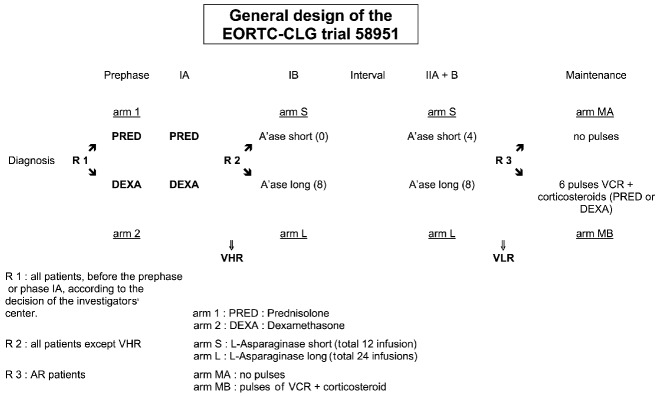
General scheme of the EORTC-CLG 58951 trial. IA: induction phase; IB: consolidation phase; II A+B: re-induction and re-consolidation phase; VCR: vincristine; VLR: very low risk (group); AR: average risk (group); VHR: very high risk (group).
All patients had to receive dexamethasone (6 mg/m2/day) or prednisolone (60 mg/m2/day), orally, in two divided doses throughout prephase (day 1 to day 7) and induction therapy (day 8 to day 35, including a tapering down period of 8 days). During protocol IIA, all patients received dexamethasone 6 mg/m2. Treatment details for the different risk groups are summarized in Online Supplementary Tables S1, S2 and S3.11 If they had an HLA identical donor, all very high-risk patients were eligible for hematopoietic stem cell transplantation except those whose only very high-risk criterion was a poor corticosteroid response on day 8, without T-cell immunophenotype or early precursor B-ALL or white blood cell count ≥100×109/L. Otherwise, the patients continued chemotherapy for a total treatment duration of 2 years.
Statistical analysis
The primary endpoint was the event-free survival from the date of first complete remission, achieved after induction IA or consolidation IB, for all patients who started induction therapy (whether this was according to protocol or not and whether the patient was eligible or not) until relapse or death in first complete remission. Patients who did not enter complete remission after these treatment steps or who died during induction therapy were considered as having had events at time 0.
The secondary endpoints were the overall duration of survival from date of randomization until death, whatever the cause; and the response to the prephase treatment (blasts <1×10⁹/L versus ≥1×10⁹/L).
The trial was powered to detect a treatment hazard ratio (HR) of 0.70. Further information on sample size computations, randomization technique, stratification factors, and statistical analysis methods18 are included in the Online Supplementary Appendix.
Results
Patients’ characteristics
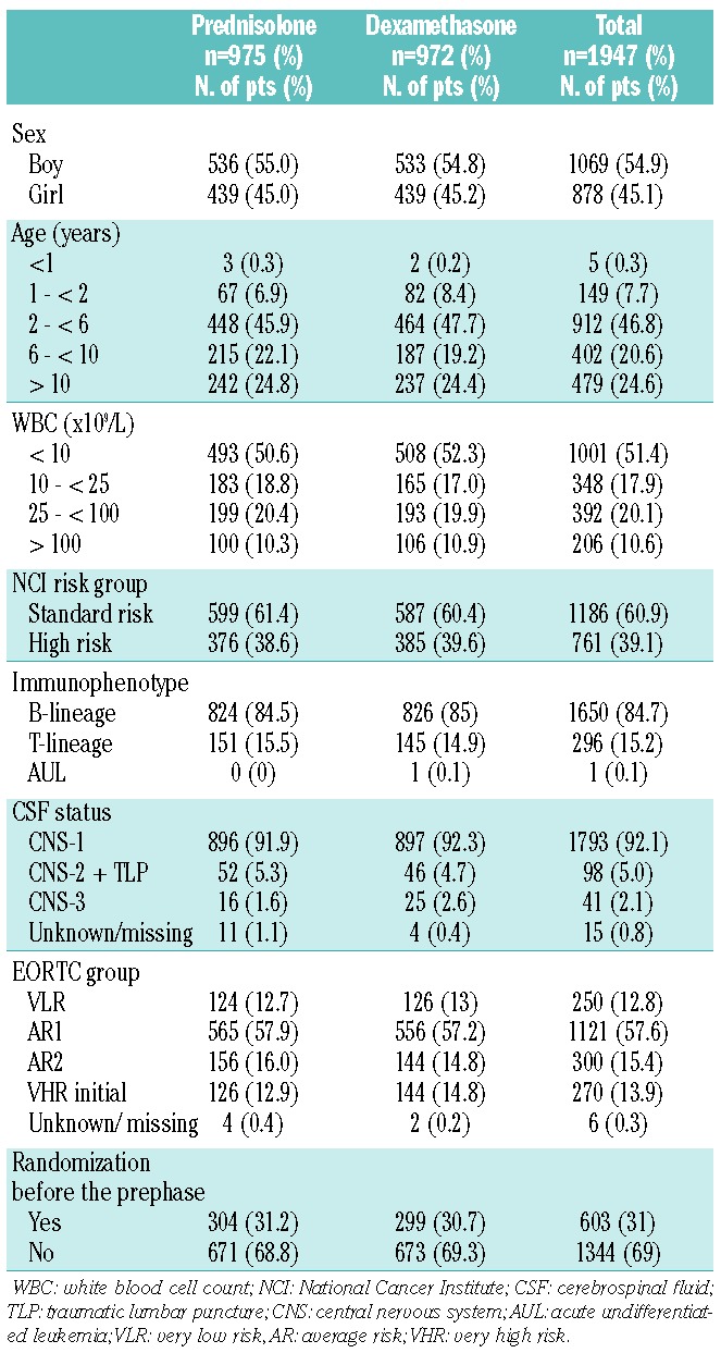
Between December 1998 and August 2008, 1947 patients with newly diagnosed ALL were enrolled in the EORTC 58951 trial and randomized to receive either dexamethasone (972 patients) or prednisolone (975 patients) during induction. Nine were considered ineligible (Table 1). However, these nine patients were included in the overall survival analyses in order to comply with the intent-to-treat principle,19,20 while six were excluded from the event-free survival analysis, as induction was not started. The distribution of the patients and disease characteristics was well balanced in the two treatment groups (Table 2). A total of 603 patients were randomized before day 1 of prephase and 1344 at the end of the prephase.
Table 1.
Flow chart (CONSORT statement).
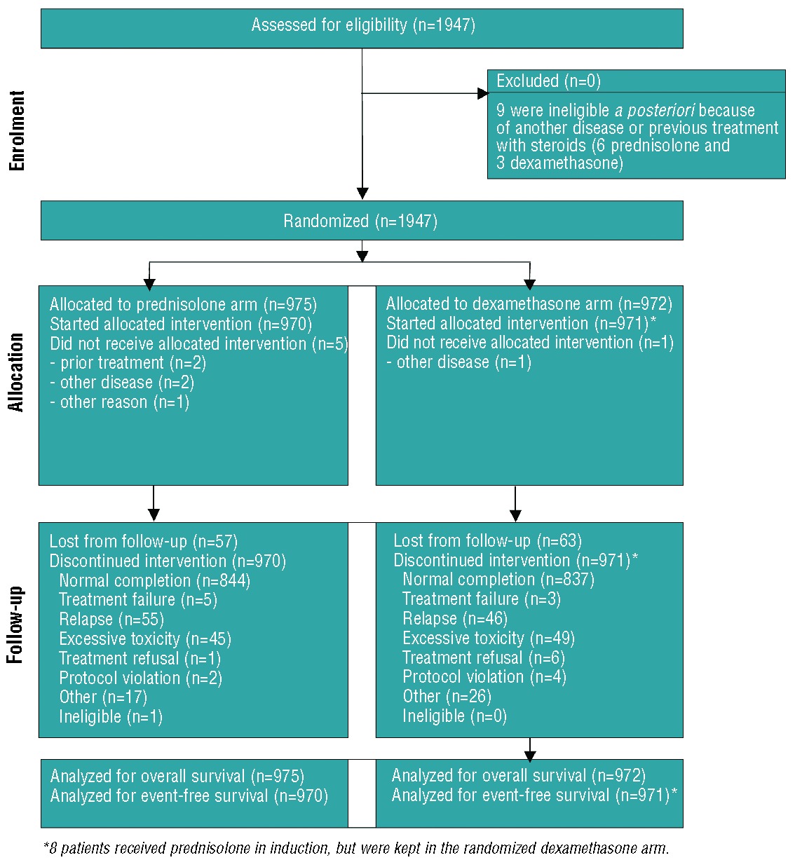
Table 2.
Patient and disease characteristics according to randomization arm.
Overall treatment outcome
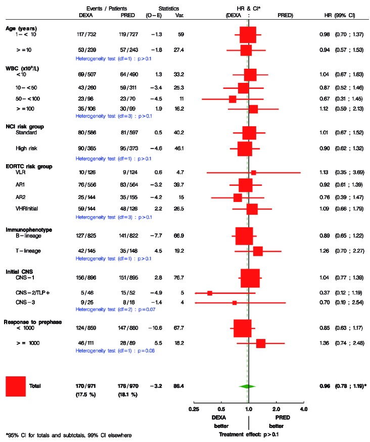
For the whole cohort, the 5-year and 8-year event-free survival rates were 82.6% (SE: 0.9%) and 81.3% (SE: 0.9%), respectively. The 5- and 8-year overall survival rates were 89.7% (SE: 0.7%) and 88.1% (SE: 0.8%), respectively. The event-free survival was similar in the two treatment groups (HR: 0.96; 95%CI 0.78–1.19, P=0.73) (Figure 2A) as was the overall survival (HR: 1.12; 95%CI 0.85–1.46, P=0.42) (Figure 2B).
Figure 2.
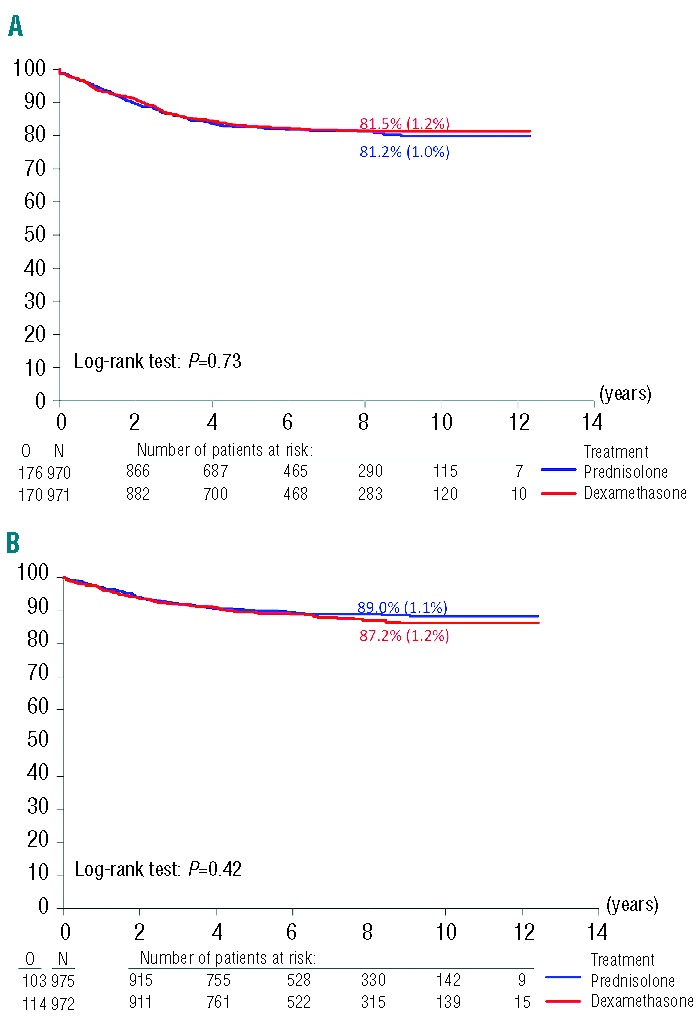
Event-free survival (A) and overall survival (B) according to randomization arm. O: observed number of events; N: number of patients randomized; %: 8-year event-free or overall survival estimation according to the Kaplan-Meier technique, followed by the standard error (SE); (A) Treatment comparison adjusted for sex in a Cox model stratified by EORTC and NCI risk groups: HR, 0.94 (95% CI 0.76–1.16); P=0.57.
At the end of induction (phase IA), the rate of complete remission was similar in the two treatment groups (Table 3). Induction failures were due to refractory leukemia in 28 patients, to fatal infections in ten patients (6 patients received dexamethasone and 4 patients received prednisolone), to hemorrhage in two patients (both received dexamethasone) and other causes in two patients (both received dexamethasone).
Table 3.
Outcome according to randomization arm.
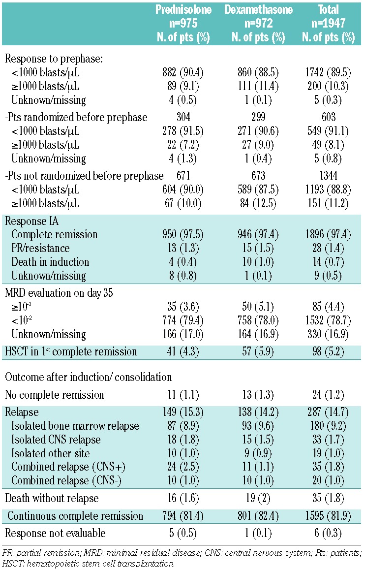
Minimal residual disease was evaluated at the end of induction (day 35) in 1617 patients (83%), of whom 85 (5.2%) had detectable disease above 10−2 (Table 3): 50 out of 808 patients in the dexamethasone arm and 35 out of 809 patients in the prednisolone arm. Twenty patients in each group acquired a very high risk status only on day 35 on the basis of this minimal residual disease result.
The 180 patients who had an isolated bone marrow relapse were well balanced in the two treatment groups (Table 3). CNS relapses (isolated or combined) were less frequent with dexamethasone (26 versus 42 for prednisolone). Thus, the 8-year cumulative incidence of isolated or combined CNS relapse was lower in the dexamethasone arm than in the prednisolone arm: 2.9% (SE: 0.6%) versus 4.5% (SE: 0.7%), respectively (P=0.048, Online Supplementary Figure S1). The difference remained statistically significant after adjustment for EORTC and NCI risk groups and sex. The 8-year cumulative incidence of non-CNS relapses was similar in the two groups (11.9% versus 12.5%; P=0.70). Thirty-five patients died in first complete remission: 19 in the dexamethasone arm versus 16 in the prednisolone arm (Table 3).
Treatment outcome according to immunophenotype and risk group
The 8-year event-free survival rate of patients with precursor B-cell ALL was 83.4% in the dexamethasone arm versus 82.0% in prednisolone arm whereas for T-cell ALL it was 71.3% versus 76.7%, respectively. The 8-year overall survival rate of patients with precursor B-cell ALL was 89.6% in the dexamethasone arm versus 90.2% in the prednisolone arm, whereas for patients with T-cell ALL it was 74.2% versus 82.1%, respectively (Online Supplementary Figures S2 and S3).
With regards to the different risk groups, for precursor B-cell ALL, the 8-year event-free and overall survival rates were 92.2% and 97.3% for very low risk patients; 84.7% and 91.6% for the average risk-1 group, 73.0% and 82.5% for the average risk-2 group, and 60.5% and 71.8% for very high risk patients (Online Supplementary Figure S4). For T-cell ALL patients, the patients in the average risk-2 group had an 8-year event-free survival rate of 82.4% (SE: 9.2%) and an 8-year overall survival rate of 86.8% (SE: 3.6%) whereas the patients in the initial very high risk group had an event-free survival rate of 60.0% (SE: 4.9%) and an overall survival rate of 63.6% (SE: 7.0%).
For precursor B-cell ALL, NCI risk did not have a significant impact on treatment difference with regards to either event-free survival (Figure 3) or overall survival. In the NCI standard-risk group, the 8-year event-free survival rate was 85.8% in both groups; the 8-year overall survival rate was 91.3% in the dexamethasone arm and 94.2% in the prednisolone arm. In the NCI high-risk group, the 8-year event-free survival rate was 79.5% versus 74.4% (HR: 0.74) in the dexamethasone and prednisolone arms, respectively, while the 8-year overall survival rate was 85.9% versus 82.1% (HR: 0.72), respectively (Online Supplementary Figure S5).
Figure 3.
Forest plots for event-free survival according to randomization arm. *NCI risk groups are described in Table 2. DEXA: dexamethasone; PRED: prednisolone; WBC: white blood cells; VLR: very low risk; AR: average risk; VHR: very high risk.
Response to prephase treatment in randomized patients
A subgroup of patients (n=603) was randomly assigned to dexamethasone (n=299) or prednisolone (n=304) before the start of the prephase. The rate of “good responders” was 90.6% in the dexamethasone arm versus 91.5% in the prednisolone arm (Table 3).
Treatment outcome according to other factors
Forest plot analysis (Figure 3) did not reveal heterogeneity of treatment effect regarding event-free survival according to stratification and other baselines characteristics factors, except for CNS status (P=0.07) and prephase response (P=0.08). We found better results with dexamethasone than prednisolone in CNS-2 and CNS-3 groups (HR: 0.37 and 0.7 respectively), but not in the CNS-1 subgroup. In patients with a “good response” to prephase, treatment results were similar, but in “poor responders” to prephase, there was a trend to worse results with dexamethasone than with prednisolone, both in B-cell ALL patients (8-year event-free survival: 60.8% versus 72.5%; HR: 1.41) and in T-cell ALL patients (8-year event-free survival: 55.6% versus 67.6%; HR: 1.47).
The treatment difference regarding disease-free survival was small, and remained practically unchanged, whether patients were randomized to receive a short course of asparaginase (HR: 0.90) or a long course (HR: 0.93).
Steroid toxicity
The incidences of grade 3–4 toxicities reported during induction were similar in the two randomized steroid groups (Table 4). Infections were responsible for six of the ten deaths in the dexamethasone group and for four toxic deaths in the prednisolone group. Furthermore, during consolidation, in the long-course asparaginase arm, the incidence of grade 3–4 infections was higher among patients who received dexamethasone during induction therapy (26.7%) than among those who received prednisolone (22.4%).
Table 4.
Grade 3/4 toxicity during induction, according to randomization arm.
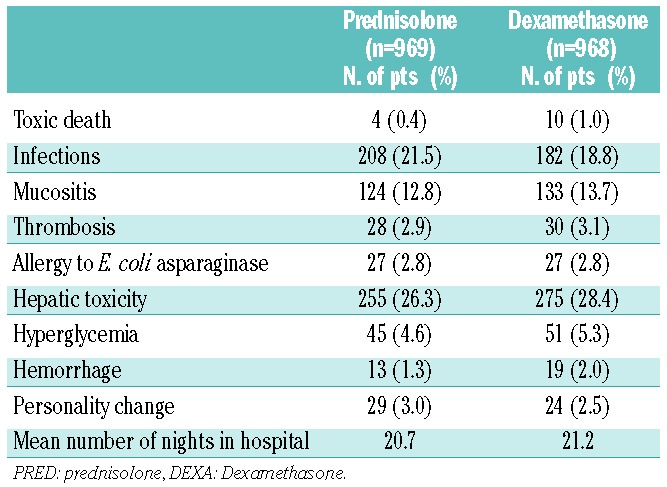
The number of cases of aseptic osteonecrosis was similar in the two arms: dexamethasone, 24 patients (2.5%) and prednisolone, 25 patients (2.6%).
Discussion
With an 8-year event-free survival rate of 81.3% and an 8-year overall survival rate of 88%, the EORTC 58951 trial demonstrated an improvement of approximately 10% compared to rates in the previous EORTC 58881 study.14 Furthermore, similar results for event-free and overall survival were obtained with dexamethasone 6 mg/m2/day and prednisolone 60 mg/m2/day in induction, so the dexamethasone:prednisolone ratio (6:60) could be considered as equivalent from the point of view of clinical efficacy.
Corticosteroids are a major component of the induction therapy for ALL in childhood.21,22 Dexamethasone, an alternative to prednisolone, is known to penetrate the CNS better than prednisolone.4 It was previously reported that the rate of isolated or combined CNS relapse in children was lower in those randomly assigned to dexamethasone (versus prednisolone), whatever the dose of dexamethasone used.5–9,23–25 This is in line with our findings of a decreased cumulative incidence of CNS relapse with dexamethasone.
Dexamethasone is also known to have a greater in vitro cytotoxic effect on lymphoblasts than prednisolone.2,3 In their studies, Bostrom7 and Mitchell8, who used a dose ratio of 6:40, reported a 6-year event-free survival rate of 85% with dexamethasone versus 77% with prednisolone7 and a 5-year event-free survival rate of 84.2% versus 75.6%,8 respectively. In the BFM trial, with a 10:60 ratio, the 6-year event-free survival rate was 84.1% in the dexamethasone arm and 79.1% in the prednisolone arm and the incidence of any type of relapse was decreased in the dexamethasone arm.5 However, all these groups also reported more toxicities and more toxic deaths with dexamethasone. The EORTC 58951 is the only protocol with a dexamethasone:prednisolone ratio of 6:60.26 With this ratio, we obtained similar treatment outcomes. Dexamethasone did not increase the incidence of complications or toxic deaths. This equivalence could be explained by various factors. In the dexamethasone group, we observed a trend towards more bone marrow relapses and more patients with minimal residual disease (≥10−2) at the end of induction. In the dexamethasone arm, we also observed a non-significant trend towards a higher “poor response” rate to the prephase. These parameters are not significant by themselves but might cooperate in favor of prednisolone, except for the prevention of CNS relapse.
In NCI standard-risk B-ALL, we obtained better results with prednisolone than those reported by Bostrom et al.7, whereas their results with dexamethasone were comparable with ours. This is most likely attributable to the lower dosage of prednisolone (40 mg/m2) applied in their study. In our trial, the 6:60 ratio resulted in similar outcomes in the whole population, although we found worse results for the poor prephase responders (B and T-ALL) treated with dexamethasone versus prednisolone, maybe because corticosteroids are not the major drug in the treatment of these “poor responders”. Similarly, the reduction of relapse was most pronounced in T-ALL patients with good prednisone response after the prephase in the BFM study.25 For patients with a prednisone “poor response”, who will undergo heavy treatment, prednisolone is probably more appropriate since it avoids excessive toxicity.
Toxic effects, including toxic deaths, have also been reported.8,10,26,27 In our study, we did not observe more toxicity in the dexamethasone group. Thirty-five patients died in complete remission and half of these children had been randomly assigned to dexamethasone. No differences were found in duration of hospitalization or supportive care interventions. There was no influence of the administration of dexamethasone on patients’ behavior, nor in the occurrence of grade 3–4 infections. So, in contrast to the results reported by Hurwitz et al.,27 the substitution of dexamethasone for prednisolone in our study did not compromise remission induction and did not result in a higher incidence of septic episodes or death from toxicity. An increased rate of toxic deaths was reported with a dexamethasone dosage of 10 mg/m2.25 Consequently, the dosage of dexamethasone during induction therapy may play a role in the occurrence of toxicity.
In our study, we observed more toxic complications in patients in complete remission treated in the arm given a long course of asparaginase who received dexamethasone in induction therapy. These data suggest that cumulative toxicity of the dexamethasone/asparaginase association is probably responsible for a longer and deeper neutropenia, even with a dosage of dexamethasone of 6 mg/m2. This is in accordance with the asparaginase-associated myelosuppression recently reported by the Dana Farber group.28
Corticosteroids are also known for their skeletal complications such as aseptic osteonecrosis.29 In our current analysis, we found more osteonecrosis in patients older than 10 years, with a similar incidence rate in the dexamethasone and prednisolone arms, using dexamethasone 6 mg/m2/day during induction (IA) and re-induction (IIA). We even noted a trend towards a higher incidence of aseptic osteonecrosis in females older than 10 years in the prednisolone group.
In conclusion, for the whole childhood ALL population, we observed similar event-free and overall survival results with dexamethasone and prednisolone, with no benefit, at the dosages tested, from replacing prednisolone by dexamethasone, except for the CNS relapse incidence. Furthermore, at the dosage used in this trial, prednisolone seemed to have greater efficiency than dexamethasone on bone marrow blasts.
For the ongoing treatment of ALL in childhood, we recommend the use of dexamethasone during induction therapy for patients with involvement of the CNS (CNS2 and CNS3 status). For T-ALL patients not at very high risk, in order to decrease the relapse risk we propose dexamethasone at a dosage of 10 mg/m2/day (i.e. a 1:6 ratio) based on recent BFM data,25 but special attention should be paid to potential toxicities currently observed with the use of a higher dosage of dexamethasone.25,26 For the other groups, including very high-risk patients who already receive intensive chemotherapy, we recommend prednisolone at a dose of 60 mg/m2.
Acknowledgments
The authors would like to thank the EORTC-CLG study group members for their participation in the study and the EORTC HQ Data Management Department members (Séraphine Rossi, Lies Meirlaen, Liv Meert, Aurélie Dubois, Christine Waterkeyn, Alessandra Busato, Isabel VandeVelde and Gabriel Solbu) for their support of this trial as well as Drs. Francisco Bautista (EORTC-CLG fellow) and Matthias Karrasch (former EORTC Clinical Research Physician).
Footnotes
The online version of this article has a Supplementary Appendix.
Funding
This study was supported by the EORTC Charitable Trust Foundation, Vlaamse Liga Tegen Kanker, Belgian Federation against Cancer, non-profit organization, TéléVie and Kinderkankerfonds from Belgium.
Authorship and Disclosures
Information on authorship, contributions, and financial & other disclosures was provided by the authors and is available with the online version of this article at www.haematologica.org.
References
- 1.Pui CH, Mullighan CG, Evans WE, Relling MV. Pediatric acute lymphoblastic leukemia: where are we going and how do we get there¿ Blood. 2012;120(6):1165–74 [DOI] [PMC free article] [PubMed] [Google Scholar]
- 2.Ito C, Evans WE, McNinch L, Coustan-Smith E, Mahmoud H, Pui CH, et al. Comparative cytotoxicity of dexamethasone and prednisolone in childhood acute lymphoblastic leukemia. J Clin Oncol. 1996;14(8):2370–6 [DOI] [PubMed] [Google Scholar]
- 3.Kaspers GJ, Veerman AJ, Popp-Snijders C, Lomecky M, Van Zantwijk CH, Swinkels LM, et al. Comparison of the antileukemic activity in vitro of dexamethasone and prednisolone in childhood acute lymphoblastic leukemia. Med Pediatr Oncol. 1996;27(2):114–21 [DOI] [PubMed] [Google Scholar]
- 4.Balis FM, Lester CM, Chrousos GP, Heideman RL, Poplack DG. Differences in cerebrospinal fluid penetration of corticosteroids: possible relationship to the prevention of meningeal leukemia. J Clin Oncol. 1987;5(2):202–7 [DOI] [PubMed] [Google Scholar]
- 5.Jones B, Freeman AI, Shuster JJ, Jacquillat C, Weil M, Pochedly C, et al. Lower incidence of meningeal leukemia when prednisone is replaced by dexamethasone in the treatment of acute lymphocytic leukemia. Med Pediatr Oncol. 1991;19(4):269–75 [DOI] [PubMed] [Google Scholar]
- 6.Veerman AJP, Hählen K, Kamps WA, Van Leeuwen EF, De Vaan GA, Solbu G, et al. High cure rate with a moderately intensive treatment regimen in non high-dose risk childhood acute lymphoblastic leukemia: results of protocol ALL VI from the Dutch Childhood Leukemia Study Group. J Clin Oncol. 1996;14(3):911–8 [DOI] [PubMed] [Google Scholar]
- 7.Bostrom BC, Sensel MR, Sather HN, Gaynon PS, La MK, Johnston K, et al. Dexamethasone versus prednisone and daily oral versus weekly intravenous mercaptopurine for patients with standard-risk acute lymphoblastic leukemia: a report from the Children’s Cancer Group. Blood. 2003;101:3809–17 [DOI] [PubMed] [Google Scholar]
- 8.Mitchell CD, Richards SM, Kinsey SE, Lilleyman J, Vora A, Eden TO. Benefit of dexamethasone compared with prednisolone for childhood acute lymphoblastic leukaemia: results of the UK Medical Research Council ALL97 randomized trial. Br J Haematol. 2005;129(6):734–45 [DOI] [PubMed] [Google Scholar]
- 9.Silverman LB, Declerck L, Gelber RD, Dalton VK, Asselin BL, Barr RD, et al. Results of Dana-Farber Cancer Institute Consortium protocols for children with newly diagnosed acute lymphoblastic leukemia (1981–1995). Leukemia. 2000;14(12):2247–56 [DOI] [PubMed] [Google Scholar]
- 10.Igarashi S, Manabe A, Ohara A, Kumagai M, Saito T, Okimoto Y, et al. No advantage of dexamethasone over prednisolone for the outcome of standard- and intermediate-risk childhood acute lymphoblastic leukemia in the Tokyo Children’s Cancer Study Group L95-14 protocol. J Clin Oncol. 2005;23(27):6489–98 [DOI] [PubMed] [Google Scholar]
- 11.De Moerloose B, Suciu S, Bertrand Y, Mazingue F, Robert A, Uyttebroeck A, et al. Children’s Leukemia Group of the European Organisation for Research and Treatment of Cancer (EORTC). Improved outcome with pulses of vincristine and corticosteroids in continuation therapy of children with average risk acute lymphoblastic leukemia (ALL) and lymphoblastic non-Hodgkin lymphoma (NHL): report of the EORTC randomized phase 3 trial 58951. Blood. 2010;116(1):36–44 [DOI] [PMC free article] [PubMed] [Google Scholar]
- 12.Guidal C, Vilmer E, Grandchamp B, Cavé H. A competitive PCR-based method using TCRD, TCRG and IGH rearrangements for rapid detection of patients with high levels of minimal residual disease in acute lymphoblastic leukemia. Leukemia. 2002;16(4):762–4 [DOI] [PubMed] [Google Scholar]
- 13.Cavé H, Van Der Werff Ten Bosch J, Suciu S, Guidal C, Waterkeyn C, Otten J, et al. Clinical significance of minimal residual disease in childhood acute lymphoblastic leukemia. New Engl J Med. 1998;339:591–8 [DOI] [PubMed] [Google Scholar]
- 14.Vilmer E, Suciu S, Ferster A, Bertrand Y, Cavé H, Thyss A, et al. Long-term results of three randomized trials (58831, 58832, 58881) in childhood acute lymphoblastic leukemia: a CLCG-EORTC report. Leukemia. 2000;14(12):2257–66 [DOI] [PubMed] [Google Scholar]
- 15.Pui CH, Evans WE. Acute lymphoblastic leukemia. N Engl J Med. 1998;339(9):605–15 [DOI] [PubMed] [Google Scholar]
- 16.Pui CH, Howard SC. Current management and challenges of malignant disease in the CNS in paediatric leukaemia. Lancet Oncol. 2008;9(3):257–68 [DOI] [PubMed] [Google Scholar]
- 17.Miller AB, Hoogstraten B, Staquet M, Winkler A. Reporting results of cancer treatment. Cancer. 1981;47(1):207–14 [DOI] [PubMed] [Google Scholar]
- 18.Kalbfleisch JD, Prentice RL. The clinical analysis of failure time date. 2nd ed New-Jersey: Wiley Inter-Science; 2002 [Google Scholar]
- 19.Schulz KF, Altman DG, Moher D, CONSORT Group. CONSORT 2010 Statement: updated guidelines for reporting parallel group randomised trials. Trials. 2010;11:3220334632 [Google Scholar]
- 20.Moher D, Hopewell S, Schulz KF, Montori V, Gøtzsche PC, Devereaux PJ, et al. CONSORT 2010 explanation and elaboration: updated guidelines for reporting parallel group randomised trial. BMJ. 2010;340:c869. [DOI] [PMC free article] [PubMed] [Google Scholar]
- 21.Gaynon PS, Lustig RH. The use of glucocorticoids in acute lymphoblastic leukemia of childhood. Molecular, cellular, and clinical considerations. J Pediatr Hematol Oncol. 1995;17(1):1–12 [DOI] [PubMed] [Google Scholar]
- 22.Gaynon PS, Carrel AL. Glucocorticosteroid therapy in childhood acute lymphoblastic leukemia. Adv Exp Med Biol. 1999;457:593–605 [DOI] [PubMed] [Google Scholar]
- 23.Veerman AJ, Hählen K, Kamps WA, Vanleeuwen EF, de Vaan GA, Vanwering ER, et al. Dutch Childhood Leukemia Study Group: early results of study ALL VI (1984–1988). Haematol Blood Transfus. 1990;33:473–7 [DOI] [PubMed] [Google Scholar]
- 24.Silverman LB, Gelber RD, Dalton VK, Asselin BL, Barr RD, Clavell LA, et al. Improved outcome for children with acute lymphoblastic leukemia: results of Dana-Farber Consortium Protocol 91-01. Blood. 2001;97(5):1211–8 [DOI] [PubMed] [Google Scholar]
- 25.Schrappe M, Zimmermann M, Moricke A, et al. Dexamethasone in induction can eliminate one third of all relapses in childhood acute lymphoblastic leukemia (ALL): results of an international randomized trial in 3655 patients (Trial AIEOP-BFM ALL 2000). Blood (ASH Annu Meet Abstr). 2008;112:117 [Google Scholar]
- 26.Teuffel O, Kuster SP, Hunger SP, Conter V, Hitzler J, Ethier MC, et al. Dexamethasone versus prednisone for induction therapy in childhood acute lymphoblastic leukemia: a systematic review and meta-analysis. Leukemia. 2011;25(8):1232–8 [DOI] [PubMed] [Google Scholar]
- 27.Hurwitz CA, Silverman LB, Schorin MA, Clavell LA, Dalton VK, Glick KM, et al. Substituting dexamethasone for prednisone complicates remission induction in children with acute lymphoblastic leukemia. Cancer. 2000;88(8):1964–9 [PubMed] [Google Scholar]
- 28.Merryman R, Stevenson KE, Gostic WJ, 2nd, Neuberg D, O’Brien J, Sallan SE, Silverman LB. Asparaginase-associated myelosuppression and effects on dosing of other chemotherapeutic agents in childhood acute lymphoblastic leukemia. Pediatr Blood Cancer. 2012;59(5):925–7 [DOI] [PMC free article] [PubMed] [Google Scholar]
- 29.Mattano LA, Jr, Sather HN, Trigg ME, Nachman JB. Osteonecrosis as a complication of treating acute lymphoblastic leukemia in children: a report from the Children’s Cancer Group. J Clin Oncol. 2000;18(18):3262–72 [DOI] [PubMed] [Google Scholar]


