Abstract
Ligands incorporating a tetraazamacrocycle receptor, a ‘click’- derived triazole and a 1,8-naphthalimide fluorophore have proven utility as probes for metal ions. Three new cyclam-based molecular probes are reported, in which a piperidinyl group has been introduced at the 4-position of the naphthalimide fluorophore. These compounds have been synthesized using the copper(I)-catalyzed azide-alkyne Huisgen cycloaddition and their photophysical properties studied in detail. The alkylamino group induces the expected red-shift in absorption and emission spectra relative to the simple naphthalimide derivatives and gives rise to extended fluorescence lifetimes in aqueous buffer. The photophysical properties of these systems are shown to be highly solvent-dependent. Screening the fluorescence responses of the new conjugates to a wide variety of metal ions reveals significant and selective fluorescence quenching in the presence of copper(II), yet no fluorescence enhancement with zinc(II) as observed previously for the simple naphthalimide derivatives. Reasons for this different behaviour are proposed. Cytotoxicity testing shows that these new cyclam-triazole-dye conjugates display little or no toxicity against either DLD-1 colon carcinoma cells or MDA-MB-231 breast carcinoma cells, suggesting a potential role for these and related systems in biological sensing applications.
Introduction
Given the various essential roles played by metal ions in biological systems and environmental processes, the development of fluorescent probes with high selectivity and sensitivity for these species is of great importance. [1]–[8] Due to their tunable photophysical properties and ease of preparation, 1,8-naphthalimide derivatives are commonly used as fluorophores in metal ion probes. [9]–[22] Structural modifications of the 1,8-naphthalimide are readily accommodated on either the aromatic naphthalene moiety or the imide-NH site.
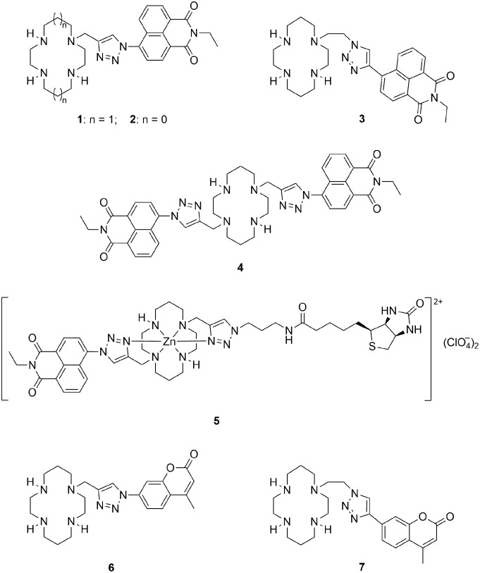
We have recently developed a novel class of fluorescent probes for Zn2+ (Figure 1) by attaching the 1,8-naphthalimide fluorophore to a tetraazamacrocycle scaffold via copper(I)-catalyzed azide-alkyne Huisgen cycloaddition (colloquially known as the click reaction). [19]–[22] The click-generated triazole is a linker but also acts as a coordination site, thus playing a role in the metal ion binding and detection. Compounds 1–4 signal the binding of Zn2+ to the tetraazamacrocycle-triazole moiety with a multifold increase in fluorescence emission of the pendant 1,8-naphthalimide. Reversing the triazole topology in the cyclam-triazole-naphthalimide system (3 vs. 1) gives a 10-fold brighter fluorescence response to Zn2+ in HEPES buffer (10 mM, pH 7.4). [22] Furthermore, the cyclam-based probe 1 has been used to detect the cellular Zn2+ flux during apoptosis in vitro, [21] and the cyclen-based probe 2 has been applied in vivo to image Zn2+ in zebrafish. [19] In a related approach, tethering a second pendant group (biotin) to the zinc(II) complex of compound 1 afforded a fluorescent ‘allosteric scorpionand’ probe 5 that visualizes the binding of the pendant biotin to the cognate biomolecule avidin. [23] Replacement of the 1,8-naphthalimide dye in compounds 1 and 3 with the coumarin fluorophore provided probes 6 and 7 (Figure 1) that respond selectively to Cu2+ and Hg2+ [22], [24].
Figure 1. Fluorescent probes 1–7 used in previous studies.
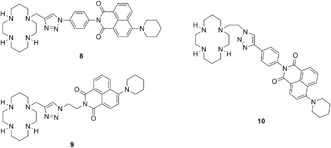
To minimize cell damage and interference from background autofluorescence in cell-based assays, the absorption and emission spectra of the fluorescent probe should be as close as possible to the red end of the visible spectrum. [25] In this regard the spectral characteristics of probes 1–7 are sub-optimal (λabs∼320–360 nm, λem∼380–460 nm in aqueous buffer). [19]–[24] Previous studies have shown that introducing alkylamino groups at the naphthalene moiety of 1,8-naphthalimide induces such a bathochromic shift. [11], [26]–[29] To this end, we designed three new cyclam-piperidinylnaphthalimide conjugates 8–10 (Figure 2). A phenyl linker was used in compounds 8 and 10 to connect the cyclam-triazole moiety to the piperidinylnaphthalimide fluorophore, while compound 9, containing a flexible ethylene chain, was designed as a control to verify the importance of conjugation. The metal-ion responsiveness, fluorescence quantum yields and decay times, and cytotoxicity of these new conjugates were investigated to explore their potential for application as metal ion probes in vitro and in vivo.
Figure 2. Cyclam-piperidinylnaphthalimide conjugates 8–10 studied in this work.
Results and Discussion
(a) Synthesis
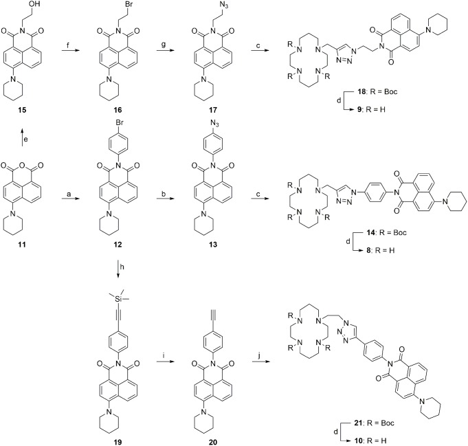
Synthesis of the cyclam-piperidinylnaphthalimide conjugates 8–10 required the preparation of precursors 13, 17 and 20 (Figure 3). Azide 17 [30], [31] and alkyne 20 [29], [30], [32] were successfully synthesized according to literature procedures, whereas the preparation of azide 13 proved challenging. Conversion of bromide 12 to the corresponding azide 13 was initially attempted with sodium azide in the presence of sodium ascorbate, copper(I) iodide and N, N′-dimethylethylenediamine (DMEDA) at reflux in either an ordinary round-bottomed flask or a pressure tube. [33]–[35] A solvent screen including methanol/water, ethanol/water or dimethyl sulfoxide (DMSO)/water (7∶3 in all cases) failed to afford the desired azide 13, giving instead full recovery of starting material 12; this outcome may be attributed to the extraordinarily low solubility of bromide 12 in these solvent combinations. Switching to tetrahydrofuran (THF)/water (7∶3), all reactants and reagents were dissolved at reflux in the pressure tube and reaction proceeded to give azide 13 in 50% yield. The corresponding amine was also detected by LCMS analysis of the reaction mixture, consistent with previous observations that both azide and amine may be generated through a copper-assisted aromatic substitution reaction with sodium azide. [33] Reacting each of the three precursors 13, 17 and 20 individually with the complementary propargyl-tri-Boc cyclam [23], [36] or azidoethyl-tri-Boc cyclam [24] under standard click conditions [24], [36] yielded the Boc-protected cyclam-piperidinylnaphthalimide conjugates 14, 18 and 21 respectively in good to excellent yields. Removal of Boc groups from these conjugates was effected in a mixture of TFA/DCM/H2O (90∶5∶5), [23], [36], [37] followed by basification to recover the corresponding free amines 8–10. However, the outcome of the basification step was contingent on the base used. Addition of 2 M sodium hydroxide solution [36], [38] or saturated sodium carbonate solution [36] resulted in decomposition of the desired free amines or incomplete removal of trifluoroacetate counter ions respectively (indicated by analysis with 1H and 13C NMR spectroscopy). Successful isolation of the pure amines 8–10 was achieved using excess Ambersep 900 (hydroxide form) in methanol.
Figure 3. Synthesis of the cyclam-piperidinylnaphthalimide conjugates 8–10.
Reagents and conditions: (a) 4-bromoaniline, piperidine, 2-methoxyethanol, reflux, 72 h, 90%; (b) NaN3, CuI, sodium ascorbate, DMEDA, THF/H2O (7∶3), 12 h, 50%; (c) propargyl-tri-Boc cyclam, CuSO4·5H2O, sodium ascorbate, THF/H2O (7∶3), rt for 13 and 50°C for 17, 12 h, 14: 96%, 18: 92%; (d) (i) TFA/DCM/H2O (90∶5∶5), rt, 6 h; (ii) Ambersep 900 hydroxide form, CH3OH, rt, 15 min, 8: 96%, 9: 99%, 10: 99%; (e) 2-aminoethanol, EtOH, reflux, 22 h, 92%; (f) PBr3, pyridine, THF, 50°C, 16 h, 60%; (g) NaN3, EtOH, reflux, 6 h, 80%; (h) trimethylsilylacetylene, CuI, triphenylphosphine, Pd(PPh3)4, Et3N, pyridine, 85°C, o/n, 94%; (i) K2CO3, CH3OH, rt, o/n, 97%; (j) 2-azidoethyl-tri-Boc cyclam, CuSO4·5H2O, sodium ascorbate, THF/H2O (7∶3), 12 h, 66%.
(b) Photophysical Properties
i) Steady-state photophysical properties
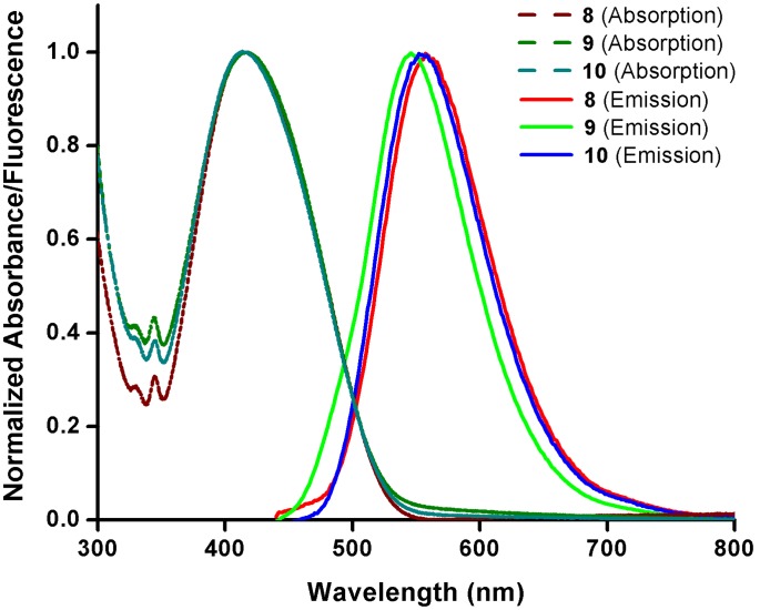
The steady state photophysical properties of cyclam-piperidinylnaphthalimide conjugates 8–10 were investigated using both UV-Vis and fluorescence spectroscopy. The UV-Vis absorption spectra of 8–10 in HEPES buffer (10 mM, pH 7.4) are almost identical, with the lowest-energy absorption (λabs) centered at 415±2 nm and stretching out to 500 nm (Figure 4). The fluorescence emission spectra of 8–10 are only slightly shifted giving a broad emission band ranging from 500 to 700 nm, centered around 545–558 nm (λem) (Figure 4). Introduction of the piperidine to the naphthalimide fluorophore not only leads to a red-shifted emission maximum but also to a broadening of both the absorption and emission bands. The similarity of these spectra in aqueous buffer is remarkable, and implies i) the role of the linker (phenyl 8 vs ethyl 9) exerts minimal influence and ii) the triazole connectivity (8 vs 10) does not have a significant impact on the UV-Vis absorption and fluorescence emission of these conjugates. The fact that the π-system is not extended in 8 or 10 by conjugation of the phenyl group with the 1,8-naphthalimide core can be rationalized by considering a twisting of the two aromatic planes to minimize adverse steric interactions. This effect may be enhanced after excitation of the probe, giving rise to charge separated states and significant solvent-dependent variation in spectral properties.
Figure 4. Normalized UV-Vis and fluorescence spectra of 8–10.
Experiments were carried out in HEPES buffer (10 mM, pH 7.4) at 25°C.
Screening the spectral properties of 8–10 in various solvents spanning a wide range of polarities revealed a solvent-dependent shift in absorption, and – to a much larger extent – emission maxima (Table 1). Comparing measurements made in aqueous buffer versus non-polar toluene shows that the impact of solvent polarity is less in the case of ethyl-linked 9, where the emission in HEPES buffer (545 nm) shifts less than 40 nm in toluene (507 nm). In the emissions of 8 and 10, a blue-shift of nearly 60 nm is seen in toluene relative to HEPES buffer. The Stokes shifts ( ) (calculated from the difference of the absorption and emission maxima) allow easier comparison: the Stokes shifts of ligands 8 and 10 respond similarly throughout the solvent screen; the slight differences that are observed between the two ligands can be attributed to the effect of the different triazole connectivity (further evidence for the minor impact this structural change exerts on the spectral properties). More importantly, there is a distinct decrease in the Stokes shift of both compounds when moving from HEPES buffer into less polar solvents, suggesting that charge separation in the excited state is most likely linked to conformational changes. In the ethyl-linked analogue 9, the effect of the solvent is weaker, indicating that the excited state of 9 incorporates a much smaller charge separation. Lippert-Mataga plots [39]–[41] (Figures S1–S3 and Text S1 in File S1) were constructed to build a picture of solvent-fluorophore interactions. The Stokes shifts of all three conjugates in the hydrogen bonding solvents (e.g. alcohols) are typically greater than those in solvents that less readily form hydrogen bonds (e.g. toluene); such behavior can be attributed to protic solvent-fluorophore hydrogen bonding and has been observed for other fluorophores [42], [43].
Table 1. Photophysical properties of 8–10 in various solvents with decreasing polarity from aqueous (HEPES buffer) to toluene.
| λabs/nm | λem/nm | /cm−1 | |||||||
| Solvent | 8 | 9 | 10 | 8 | 9 | 10 | 8 | 9 | 10 |
| HEPES | 415 | 417 | 414 | 558 | 545 | 555 | 6175 | 5632 | 6137 |
| MeOH | 415 | 7 | 415 | 542 | 538 | 542 | 5646 | 5393 | 5646 |
| EtOH | 413 | 415 | 412 | 540 | 536 | 539 | 5695 | 5440 | 5719 |
| n-PrOH | 413 | 413 | 412 | 537 | 534 | 536 | 5591 | 5486 | 5615 |
| n-BuOH | 413 | 411 | 411 | 536 | 534 | 534 | 5556 | 5604 | 5604 |
| DMSO | 415 | 416 | 413 | 540 | 536 | 540 | 5578 | 5382 | 5695 |
| MeCN | 411 | 412 | 409 | 539 | 537 | 538 | 5778 | 5650 | 5863 |
| DMF | 410 | 412 | 409 | 538 | 535 | 537 | 5803 | 5580 | 5828 |
| Acetone | 406 | 409 | 405 | 531 | 530 | 530 | 5798 | 5582 | 5823 |
| EtOAc | 401 | 404 | 398 | 518 | 520 | 515 | 5633 | 5522 | 5708 |
| THF | 403 | 406 | 401 | 517 | 518 | 514 | 5472 | 5326 | 5482 |
| DCM | 418 | 421 | 415 | 523 | 524 | 521 | 4803 | 4669 | 4903 |
| CHCl3 | 418 | 418 | 415 | 513 | 516 | 512 | 4430 | 4544 | 4565 |
| Toluene | 404 | 406 | 402 | 500 | 507 | 499 | 4752 | 4907 | 4836 |
λabs: wavelength of maximum UV-Vis absorbance; λem: wavelength of maximum emission intensity; : Stokes shift.
ii) Response to metal ions
The UV-Vis and fluorescence responses of conjugates 8–10 to a wide variety of metal ions (Ag+, Ba2+, Ca2+, Cd2+, Co2+, Cu2+, Fe2+, Fe3+, Hg2+, K+, Li+, Mg2+, Mn2+, Na+, Ni2+, Pb2+, Rb+ and Zn2+) were assessed in HEPES buffer (10 mM, pH 7.4 – see File S1). Of the metals tested, only Cu2+ triggered a significant response, quenching the fluorescence of all three conjugates (Figures S4–S6 in File S1). This response is consistent with previous observations that Cu2+ quenches the fluorescence of derivatives 1, 3, 6 and 7, [20]–[22] and may be due to paramagnetic or heavy atom effects; [44]–[47] work is underway to determine the mechanism of Cu2+- mediated fluorescence quenching in these systems. However none of the cyclam-piperidinylnaphthalimide conjugates 8–10 show any meaningful response to either Zn2+ or Hg2+, in contrast to the previously-reported cyclam-naphthalimide conjugates 1, 3 and 4 which exhibited fluorescence increases in the presence of Zn2+ and quenching in response to Hg2+ respectively. [20]–[22] Addition of Co2+, Fe2+ and Fe3+ each triggered a small to moderate reduction in the fluorescence of 10, but had no effect on the fluorescence of 8 or 9. Taken together, these fluorescence results imply that both the nature of the pendant fluorophore and the connectivity between the fluorophore and metal-cyclam complex play a role in the metal-ion responsiveness of these conjugates. The addition of these metal ions had no significant effect on the UV-Vis absorption spectra of all three conjugates (Figures S7–S9 in File S1).
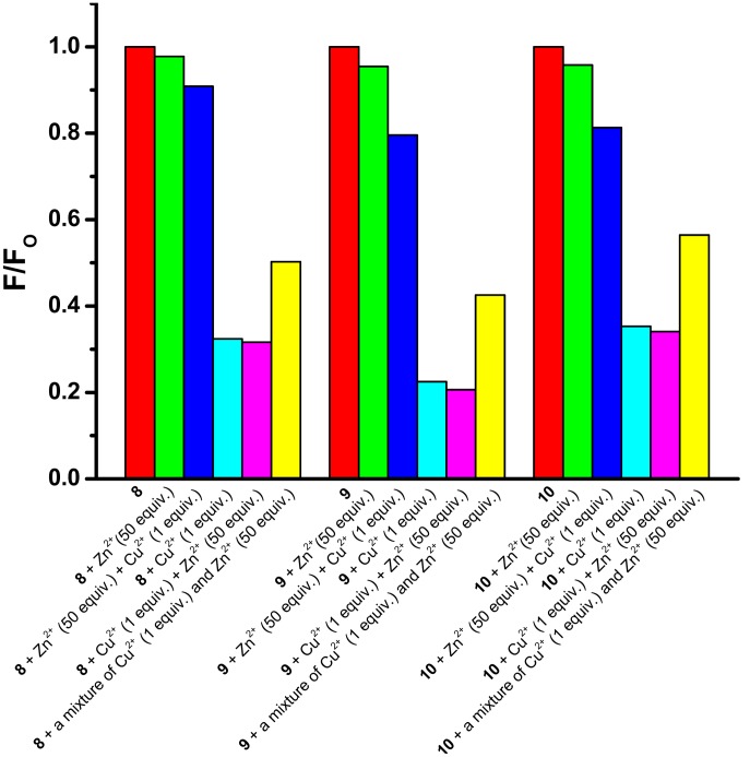
To investigate the effectiveness of 8–10 as probes for Cu2+ in the presence of competing metal ions, competitive binding experiments were conducted using Zn2+. Thus a 10 µM solution of 8–10 in HEPES buffer (10 mM, pH 7.4) was combined with 50 equivalents of Zn2+, followed after approximately 3 minutes by 1 equivalent of Cu2+. In all cases, much weaker fluorescence quenching was observed than in the experiments in which the two metal ions were added in the reverse order, or a premixed Cu2+/Zn2+ (1∶50) solution was added (Figure 5). These results show the effectiveness of 8–10 as Cu2+- probes but indicate a limitation in the presence of high Zn2+ concentrations.
Figure 5. Competitive binding experiments.
Experiments were carried out to investigate the effectiveness of Cu2+-induced (1 equiv.) quenching of the fluorescence of probes 8–10 (10 µM) in HEPES buffer (10 mM, pH 7.4) at 25°C in the presence of excess Zn2+ (50 equiv.).
The fluorescence responses of probes 8–10 were evaluated over a wide pH range, both in the absence and presence of Cu2+ (Figures S10–S12 in File S1). These experiments indicate optimum responsiveness to Cu2+ at neutral pH. At low pH, the fluorescence responses of the free ligands 8–10 change little in the presence of Cu2+, presumably due to inhibition of metal coordination when the cyclam amine groups are protonated. The fluorescence of the free ligands 8–10 is diminished at high pH, as previously observed with probe 1. [21] However, it is the absence of any protonation-induced fluorescence enhancement with probes 8–10 that is more significant. This indicates that the photoinduced electron transfer (PET) from the cyclam-triazole moiety to naphthalimide observed with probes 1–3 [22] does not occur with the piperidinylnaphthalimide fluorophore incumbent. This in turn means that the full fluorescence response of 8–10 is turned ‘on’ in the free ligands, eliminating the possibility of a fluorescence ‘turn-on’ pathway upon protonation or metal binding. The fact that PET is not favoured with probes 8–10 can be rationalized by considering the push-pull-character of the 4-aminonaphthalimide, where the electron acceptor is located at the amine and the electron donor at the imide. In the excited state, the resultant negative charge density on the imide inhibits acceptance of an additional electron via PET when the electron donor is connected at this position on the fluorophore [48].
iii) Time resolved photophysical properties and fluorescence quantum yields
Fluorescence quantum yields were acquired in three representative solvents (HEPES buffer, ethyl acetate and acetonitrile) to investigate the intrinsic photophysical properties in more detail (Table 2). In HEPES-buffer and acetonitrile, the quantum yields and the fluorescence decay times of the free ligands 8–10 are generally low, although ligand 9 gives a significantly longer decay time (<τ> = 4.86 ns) in buffer compared to ligands 8 and 10 (<τ> = 2.47 and 2.42 ns respectively). The quantum yields in ethyl acetate (0.25–0.50) are at least one order of magnitude higher than in acetonitrile (0.036–0.052) and about twice as high as in buffer (0.005–0.009). Clearly solvent has a strong influence on the photophysical properties of these probes. Strong solvent-dependence was also observed in the decay time profile of all three ligands. In ethyl acetate, ligands 8 and 10 decay with a single exponential profile, ligand 9 with a bis-exponential profile. In acetonitrile, decay times for all three ligands are fitted with two exponentials. In buffer, three exponentials give the best fit in all three cases. These multi-exponential fits indicate the presence of multiple excited species in these solvents. The additional components observed in aqueous buffer over the organic solvents can be rationalized by considering changes in ligand protonation, which give rise to new species that are absent in the aprotic organic solvents. Interestingly, the photophysical properties of 8, 9 and 10 change relative to each other with changes in solvent: while similar values for quantum yields and decay times are observed for all three ligands in acetonitrile, only 8 and 10 afford similar data in ethyl acetate and buffer, and the values recorded for 9 are appreciably different. In ethyl acetate, longer decay times for 8 and 10 (5.73 and 6.36 ns respectively) and higher quantum yields (0.44 and 0.50) are found compared to ligand 9, but in buffer ligand 9 gives higher values than ligands 8 and 10. Notably, the averaged decay of 9 is particularly long (∼5 ns) in the aqueous solvent. The long decay times may render these new probes suitable for time-correlated assays, e.g. fluorescence lifetime imaging (FLIM) techniques in biological samples.
Table 2. Fluorescence quantum yields (Φ F) and decay times (τ) of 8–10 in HEPES buffer (10 mM, pH 7.4), ethyl acetate and acetonitrile.
| Solvent | Compound | Φ F | τ/ns | <τ>/ns | ||
| HEPES | 8 | 0.005 | 0.12 (28%) | 2.32 (46%) | 5.28 (26%) | 2.47 |
| 9 | 0.009 | 0.10 (9%) | 4.03 (45%) | 6.60 (46%) | 4.86 | |
| 10 | 0.005 | 0.17 (26%) | 1.83 (37%) | 4.58 (37%) | 2.42 | |
| EtOAc | 8 | 0.44 | 5.73 | 5.73 | ||
| 9 | 0.25 | 2.36 (29%) | 5.13 (71%) | 4.33 | ||
| 10 | 0.50 | 6.36 | 6.36 | |||
| MeCN | 8 | 0.043 | 0.85 (94%) | 8.38 (6%) | 1.30 | |
| 9 | 0.036 | 0.80 (94%) | 8.58 (6%) | 1.27 | ||
| 10 | 0.052 | 1.04 (95%) | 7.18 (5%) | 1.35 | ||
<τ> is the averaged time from the multi-exponential decay profiles.
(c) Biological Evaluation
The cytotoxicity of cyclam-piperidinylnaphthalimide conjugates 8–10 and Boc-protected precursors 14, 18, and 21 was assayed against DLD-1 colon carcinoma cells and MDA-MB-231 breast carcinoma cells (Table 3). Cisplatin was used as a positive control in this cell viability study; its cytotoxicity against DLD-1 cells was found to be 11.2±0.3 µM, consistent with literature value of 11.8±1.2 µM. [49] Five of the six cyclam-piperidinylnaphthalimide compounds did not display any significant cytotoxicity against either cell line, with a safe dosage level of 20 µM. The single exception was conjugate 10, which showed moderate activity against both carcinoma cell lines. In general, IC50 values for all cyclam-piperidinylnaphthalimide conjugates 8–10 were lower than those for the corresponding Boc-protected counterparts 14, 18 and 21.
Table 3. Cytotoxicity of 8–10, 14, 18 and 21 against DLD-1 colon carcinoma cells and MDA-MB-231 breast carcinoma cells after incubation for 72 h.
| IC50/µM | ||
| Compound | DLD-1 | MDA-MB-231 |
| cisplatin | 11.2±0.3 | 22.0±0.6 |
| 8 | >100 | 54.1±0.8 |
| 9 | >200 | >100 |
| 10 | 53.8±1.3 | 37.4±0.6 |
| 14 | >200 | >200 |
| 18 | >200 | >100 |
| 21 | >200 | >200 |
IC50 values are expressed as mean ± standard error of mean of at least 3 independent experiments.
Conclusions
We have reported the synthesis of three cyclam-piperidinylnaphthalimide conjugates 8–10 which respond to the presence of copper(II) with a significant decrease in fluorescence. Despite the different triazole connectivities and the variation of the pendant alkyl arm length, these probes exhibit remarkably similar photophysical properties. However, these photophysical properties are highly dependent on solvent, as seen in the UV-Vis and fluorescence spectra, quantum yields and decay times of all three ligands. The influence of the flexible ethyl linker is reflected in the long averaged fluorescence decay time of compound 9 in HEPES buffer, which is twice as long as those of ligands 8 and 10. None of the probes display significant cytotoxicity to mammalian cells, supporting the potential suitability of this new probe class for sensing, labeling or imaging studies in biological systems.
Experimental
(a) General Materials
All reactions except azidation of 12 were carried out with continuous magnetic stirring in ordinary glassware; azidation of 12 was performed in a 15 mL Ace pressure tube, purchased from Sigma-Aldrich. Heating of reactions was conducted with a paraffin oil bath or a water bath. All reagents and solvents were purchased from Sigma-Aldrich, Alfa Acer, Merck, or Ajax Finechem. Reagents were used as received unless otherwise specified. Hexane and ethyl acetate were distilled before use. Dichloromethane and ethanol were distilled over calcium hydride and stored over activated 4 Å molecular sieves. Tetrahydrofuran was distilled over sodium wire/benzophenone. Methanol and acetonitrile were collected freshly from a PureSolv MD 7 solvent purification system having been passed through anhydrous alumina columns.
(b) Instrumentation and Methods
1H and 13C NMR spectra were recorded at 300 K on a Bruker AVANCE 300 spectrometer (1H at 300.13 MHz and 13C at 75.47 MHz) or a Bruker DRX 400 spectrometer (1H at 400.13 MHz and 13C at 100.61 MHz). 1H and 13C NMR spectra are referenced to 1H signals of residual nondeuterated solvents (or tetramethylsilane) and 13C signals of the deuterated solvents respectively. 1H NMR signals are reported with chemical shift values δ (ppm), multiplicity (s = singlet, d = doublet, t = triplet, q = quartet, dd = doublet of doublets, m = multiplet and br = broad), relative integral, coupling constants J (Hz) and assignments. Infrared spectra were recorded on a Bruker Alpha FT-IR spectrometer. Low resolution and high resolution mass spectra were recorded on a Finnigan LCQ mass spectrometer and a Bruker 7T Fourier Transform Ion Cyclotron Resonance (FT-ICR) Mass Spectrometer respectively. Ionisation of all samples was carried out using either ESI or APCI. Melting points were determined on an OptiMelt 100 automated melting point apparatus and are uncorrected. Elemental analyses were carried out by the Campbell Microanalytical Laboratory (University of Otago, New Zealand) on a Carlo Erba EA 1108 Elemental Analyser. HEPES buffer was sterile filtered before use and the pH values were determined by a Mettler Toledo S20 SevenEasy™ pH meter or Minilab ISFET pH meter. Analytical TLC was performed on Merck silica gel 60 F254 pre-coated aluminum plates (0.2 mm) and visualized under UV light (254 nm), followed by staining with ninhydrin. Flash column chromatography was carried out using Merck silica gel 60 (0.040–0.063 mm). UV-Vis spectra were recorded on a Varian Cary 4000 or Varian Cary 1E UV-visible spectrophotometer. Fluorescence spectra were recorded on a Varian Cary Eclipse fluorescence spectrophotometer. Temperature control for both UV-visible spectrophotometer and fluorescence spectrophotometer was provided by a Varian Cary PCB water peltier system. In the time-resolved measurements, the frequency doubled output of Titan sapphire laser (Tsunami 3960; Spectra Physics) was used for excitation. The repetition rate of 80.2 MHz was reduced to 3.8 MHz with a Pulse Picker (Pulse Select; APE). The luminescence was detected in a right angle configuration to the incomming beam. For the detection, a multichannel plate (ELDI EM1-132/300; europhoton GmbH) coupled to a FL920 fluorescence lifetime spectrometer (Edinburgh Instruments) was used. The time-resolved emission was recorded in time-correlated single photon counting mode. The FAST software package (Edinburgh Instruments) was used to analyse the fluorescence decays. The fluorescence quantum yields were measured on a PL Quantum Yield Measurement System C9920-02 with an integrating sphere (Hamamatsu).
(c) Synthesis
See the Supporting Information for synthetic experimental procedures of known compounds (Text S2 and Figures S13–S15 in File S1) and 1H and 13C NMR spectra of novel compounds (Figures S16–S33 in File S1).
General Synthetic Procedure A: The Copper(I)-Catalyzed Huisgen 1,3-Dipolar Cycloaddition of Azides and Alkynes
Alkyne (1.00 eq.) and azide (1.00 eq.) were dissolved in THF/H2O (7∶3, 50 mM in alkyne). A brown cloudy solution of CuSO4·5H2O (0.05 eq., 5 mol%) and sodium ascorbate (0.10 eq., 10 mol%) in H2O (25 mM in copper) was added. The reaction mixture was stirred at room temperature [23], [36], [37] or heated at 50°C under Ar for 12 h and quenched with saturated aqueous NH4Cl (100 L/mol copper). THF was evaporated under reduced pressure, and the remaining mixture was extracted with DCM (3×). The combined organic extracts were dried over Na2SO4 and concentrated under reduced pressure. The residue was purified by flash column chromatography (silica gel, EtOAc:petroleum benzine = 1∶1 ramping to EtOAc) to give the desired triazole.
General Synthetic Procedure B: TFA-Mediated Boc Removal [23], [36], [37] & Basification of Trifluoroacetates
Boc-protected amine (1.0 eq.) was dissolved in a mixture of TFA/DCM/H2O (90∶5∶5, 5 mM). The reaction mixture was stirred at room temperature for 6 h and concentrated under reduced pressure. The residue was dissolved in CH3OH (5 mL), and Ambersep 900 hydroxide form (pre-swelled with H2O for 30 min and CH3OH for 30 min) in CH3OH (10 mL) was added. The mixture was stirred at room temperature for 15 min and filtered, and the solid was washed with CH3OH (15 mL). The filtrate and washings were combined and concentrated under reduced pressure to give the desired N-functionalized cyclam.
2-(4-Azidophenyl)-6-(piperidin-1-yl)-1H-benzo[de]isoquinoline-1,3(2H)-dione (13)
To a solution of 12 (218 mg, 0.501 mmol) and sodium azide (65.0 mg, 1.00 mmol) in THF/H2O (7 mL/3 mL) in an Ace pressure tube were added CuI (19 mg, 0.10 mmol), sodium ascorbate (10 mg, 0.050 mmol) and DMEDA (22 µL, 0.20 mmol). The reaction mixture was heated at reflux under Ar for 12 h and cooled to room temperature. After addition of H2O (7 mL), the reaction mixture was extracted with DCM (3×25 mL). The combined organic extracts were concentrated under reduced pressure, and the residue was purified by flash column chromatography (silica gel, DCM) to give 13 as a yellow solid (100 mg, 50%). R F (DCM) 0.66. m.p. 190–191°C. IR ν max/cm−1 2936, 2853, 2794, 2112, 1705, 1657, 1589, 1506, 1452, 1375, 1287, 1234, 1195, 1143. 1H NMR (300 MHz, CDCl3) δ 1.60–1.84 (m, 2H, CH2CH2CH 2CH2CH2), 1.84–2.10 (m, 4H, CH2CH 2CH2CH 2CH2), 3.26 (t, 4H, J 5.1, CH2NCH2), 7.18 (d, 2H, J 8.4, Ph-H), 7.19 (d, 1H, J 7.5, naphthalene-H), 7.30 (d, 2H, J 8.7, Ph-H), 7.70 (t, 1H, J 7.8, naphthalene-H), 8.43 (d, 1H, J 8.4, naphthalene-H), 8.51 (d, 1H, J 8.1, naphthalene-H), 8.59 (d, 1H, J 7.5, naphthalene-H). 13C NMR (75 MHz, CDCl3) δ 24.4, 26.3, 54.6, 114.9, 115.7, 120.0, 123.2, 125.5, 126.4, 130.3, 130.4, 131.2, 131.6, 132.5, 133.3, 140.2, 157.8, 164.3, 164.8 (four carbon signals overlapping or obscured). HRMS (ESI) 420.14297 ([M+Na]+); calcd. for C23H19N5NaO2 ([M+Na]+) 420.14310.
Tri-tert-butyl 11-((1-(4-(1,3-dioxo-6-(piperidin-1-yl)-1H-benzo[de]isoquinolin-2(3H)-yl)phenyl)-1H-1,2,3-triazol-4-yl)methyl)-1,4,8,11-tetraazacyclotetradecane-1,4,8-tricarboxylate (14)
Propargyl-tri-Boc cyclam [23], [36] (84 mg, 0.16 mmol) and azide 13 (62 mg, 0.16 mmol) were reacted at room temperature using general synthetic procedure A to give 14 as a yellow foam (140 mg, 96%). R F (EtOAc:hexane = 1∶1) 0.19. IR ν max/cm−1 2973, 2934, 2857, 2816, 1670, 1584, 1517, 1460, 1413, 1365, 1322, 1298, 1234, 1160, 1075, 1039, 995, 915, 857, 832. 1H NMR (300 MHz, CDCl3) δ 1.44 (s, 9H, C(CH3)3), 1.46 (s, 18H, 2×C(CH3)3), 1.70–1.85 (m, 4H, CH2CH2CH 2CH2CH2 & NCH2CH 2CH2N), 1.85–2.02 (m, 6H, CH2CH 2CH2CH 2CH2 & NCH2CH 2CH2N), 2.45–2.60 (m, 2H, CH 2N(CH2-triazole)CH2), 2.62–2.78 (m, 2H, CH2N(CH2-triazole)CH 2), 3.28 (t, 4H, J 4.8, CH 2N(naphthalene)CH 2), 3.20–3.54 (m, 12H, 3×CH 2N(Boc)CH 2), 3.91 (s, 2H, NCH 2-triazole), 7.22 (d, 1H, J 8.1, naphthalene-H), 7.49 (d, 2H, J 8.7, Ph-H), 7.72 (t, 1H, J 7.8, naphthalene-H), 7.96 (d, 2H, J 8.4, Ph-H), 8.08 (br s, 1H, triazole-H), 8.46 (d, 1H, J 8.4, naphthalene-H), 8.54 (d, 1H, J 8.1, naphthalene-H), 8.62 (d, 1H, J 7.2, naphthalene-H). 13C NMR (75 MHz, CDCl3) δ 24.4, 26.3, 28.6, 45.7, 47.4, 48.9, 51.4, 54.6, 79.7, 114.9, 115.6, 121.2, 123.1, 125.5, 126.4, 130.5, 131.4, 131.6, 133.3, 136.0, 136.9, 144.4, 155.6, 155.9, 157.9, 164.1, 164.7 (twenty four carbon signals overlapping or obscured). MS (ESI) m/z 936.0 ([M+H]+, 48%), 958.1 ([M+Na]+, 100%). HRMS (ESI) 936.53398 ([M+H]+); calcd. for C51H70N9O8 ([M+H]+) 936.53419.
2-(4-(4-((1,4,8,11-Tetraazacyclotetradecan-1-yl)methyl)-1H-1,2,3-triazol-1-yl)phenyl)-6-(piperidin-1-yl)-1H-benzo[de]isoquinoline-1,3(2H)-dione (8)
Compound 14 (112 mg, 0.120 mmol) was deprotected using general synthetic procedure B to give 8 as a yellow glue (73 mg, 96%). IR ν max/cm−1 3384, 3287, 3123, 3058, 2926, 2850, 2820, 1701, 1660, 1584, 1517, 1457, 1367, 1233, 1189, 1135, 1114, 1078, 1044, 998, 833. 1H NMR (400 MHz, CDCl3) δ 1.56–1.81 (m, 4H, CH2CH2CH 2CH2CH2 & NCH2CH 2CH2N), 1.81–2.10 (m, 6H, CH2CH 2CH2CH 2CH2 & NCH2CH 2CH2N), 2.40–3.10 (m, 19H, 3×CH2NHCH2 & CH 2N(CH2-triazole)CH 2), 3.15–3.40 (m, 4H, CH 2N(naphthalene)CH 2), 3.93 (s, 2H, NCH 2-triazole), 7.20 (d, 1H, J 8.0, naphthalene-H), 7.47 (d, 2H, J 8.4, Ph-H), 7.70 (t, 1H, J 8.0, naphthalene-H), 7.92 (d, 2H, J 8.4, Ph-H), 8.11 (s, 1H, triazole-H), 8.44 (d, 1H, J 8.4, naphthalene-H), 8.51 (d, 1H, J 8.0, naphthalene-H), 8.58 (d, 1H, J 7.2, naphthalene-H). 13C NMR (100 MHz, CDCl3) δ 24.3, 26.1, 28.8, 46.9, 47.1, 48.0, 48.8, 49.3, 49.5, 50.7, 52.9, 54.5, 54.7, 114.7, 115.3, 120.8, 121.0, 122.9, 125.4, 126.3, 130.3, 130.4, 131.2, 131.5, 133.2, 135.8, 136.9, 145.0, 157.8, 163.9, 164.5 (five carbon signals overlapping or obscured). MS (ESI) m/z 636.3 ([M+H]+, 100%). HRMS (ESI) 636.37673 ([M+H]+); calcd. for C36H46N9O2 ([M+H]+) 636.37690.
Tri-tert-butyl 11-((1-(2-(1,3-dioxo-6-(piperidin-1-yl)-1H-benzo[de]isoquinolin-2(3H)-yl)ethyl)-1H-1,2,3-triazol-4-yl)methyl)-1,4,8,11-tetraazacyclotetradecane-1,4,8-tricarboxylate (18)
Propargyl-tri-Boc cyclam [23], [36] (298 mg, 0.553 mmol) and azide 17 (193 mg, 0.552 mmol) were reacted at room temperature using general synthetic procedure A to give 18 as a yellow foam (453 mg, 92%). R F (EtOAc:hexane = 1∶1) 0.19. IR ν max/cm−1 2973, 2934, 2859, 2815, 1689, 1659, 1585, 1459, 1363, 1240, 1162, 1035, 864. 1H NMR (400 MHz, CDCl3) δ 1.44 (s, 9H, C(CH3)3), 1.47 (s, 18H, 2×C(CH3)3), 1.63–1.78 (m, 4H, CH2CH2CH 2CH2CH2 & NCH2CH 2CH2N), 1.83–1.99 (m, 6H, CH2CH 2CH2CH 2CH2 & NCH2CH 2CH2N), 2.28–2.42 (m, 2H, CH 2N(CH2-triazole)CH2), 2.49–2.61 (m, 2H, CH2N(CH2-triazole)CH 2), 3.23 (t, 4H, J 5.2, CH 2N(naphthalene)CH 2), 3.18–3.52 (m, 12H, 3×CH 2N(Boc)CH 2), 3.78 (s, 2H, NCH 2-triazole), 4.64 (t, 2H, J 6.0), 4.75 (t, 2H, J 6.0) (total 4H, triazole-CH 2CH 2N), 7.15 (d, 1H, J 8.4, naphthalene-H), 7.55 (br s, 1H, triazole-H), 7.64 (dd, 1H, J 8.4 & 7.2, naphthalene-H), 8.37 (dd, 1H, J 8.4 & 1.2, naphthalene-H), 8.40 (d, 1H, J 8.0, naphthalene-H), 8.47 (dd, 1H, J 7.6 & 1.2, naphthalene-H). 13C NMR (100 MHz, CDCl3) δ 24.3, 26.2, 26.6, 28.5, 28.6, 39.5, 45.2, 47.0, 47.9, 50.7, 51.7, 53.0, 54.5, 79.5, 114.7, 115.1, 122.5, 123.1, 125.3, 126.3, 130.1, 131.1, 131.3, 133.0, 143.0, 155.5, 155.8, 157.7, 163.7, 164.3 (seventeen carbon signals overlapping or obscured). MS (ESI) m/z 588.2 ([M-3Boc+H]+, 22%), 688.0 ([M-2Boc+H]+, 16%), 788.2 ([M-Boc+H]+, 31%), 888.2 ([M+H]+, 100%), 910.3 ([M+Na]+, 46%). HRMS (ESI) 910.51580 ([M+Na]+); calcd. for C47H69N9NaO8 ([M+Na]+) 910.51613.
2-(2-(4-((1,4,8,11-Tetraazacyclotetradecan-1-yl)methyl)-1H-1,2,3-triazol-1-yl)ethyl)-6-(piperidin-1-yl)-1H-benzo[de]isoquinoline-1,3(2H)-dione (9)
Compound 18 (133 mg, 0.150 mmol) was deprotected using general synthetic procedure B to give 9 as a yellow glue (87 mg, 99%). IR ν max/cm−1 3269, 2934, 2846, 2814, 1692, 1653, 1584, 1516, 1458, 1384, 1356, 1239, 1118, 1079, 1028. 1H NMR (400 MHz, CDCl3) δ 1.50–2.00 (m, 10H, CH2CH 2CH 2CH 2CH2 & 2×NCH2CH 2CH2N), 2.20–2.84 (m, 16H, 3×CH 2NHCH 2 & CH 2N(CH2-triazole)CH 2), 3.95–3.50 (m, 7H, 3×CH2NHCH2 & CH 2N(naphthalene)CH 2), 3.69 (s, 2H, NCH 2-triazole), 4.54 (t, 2H, J 5.2), 4.65 (t, 2H, J 5.2) (total 4H, triazole-CH 2CH 2N), 7.04 (d, 1H, J 8.0, naphthalene-H), 7.54 (t, 1H, J 7.6, naphthalene-H), 7.60 (s, 1H, triazole-H), 8.26 (d, 1H, J 6.8, naphthalene-H), 8.28 (d, 1H, J 7.6, naphthalene-H), 8.36 (d, 1H, J 6.8, naphthalene-H). 13C NMR (100 MHz, CDCl3) δ 24.2, 25.7, 26.1, 28.4, 39.5, 46.7, 47.0, 47.6, 47.8, 48.6, 49.2, 50.7, 52.4, 54.0, 54.4, 114.6, 114.9, 122.3, 123.0, 125.2, 126.1, 129.9, 131.0, 131.1, 132.8, 143.9, 157.6, 163.5, 164.1 (three carbon signals overlapping or obscured). MS (ESI) m/z 588.2 ([M+H]+, 100%). HRMS (ESI) 588.37694 ([M+H]+); calcd. for C32H46N9O2 ([M+H]+) 588.37690.
6-(Piperidin-1-yl)-2-(4-((trimethylsilyl)ethynyl)phenyl)-1H-benzo[de]isoquinoline-1,3(2H)-dione (19) [32]
To a solution of 12 (1.31 g, 3.01 mmol), CuI (269 mg, 1.41 mmol), triphenylphosphine (1.06 g, 4.04 mmol) and Pd(PPh3)4 (263 mg, 0.228 mmol) in Et3N (18 mL) were added pyridine (9 mL) and trimethylsilylacetylene (4.25 mL, 30.1 mmol). The reaction mixture was heated at 85°C under Ar overnight and cooled to room temperature before addition of DCM (40 mL). The organic phase was washed with H2O (4×40 mL), dried over MgSO4 and concentrated under reduced pressure. The residue was purified by flash column chromatography (silica gel, DCM:hexane = 5∶1) to give 19 as a yellow solid (1.28 g, 94%). R F (DCM) 0.67. m.p. 258–259°C. IR ν max/cm−1 2959, 2926, 2854, 2794, 2364, 2332, 2156, 1707, 1658, 1590, 1508, 1453, 1401, 1376, 1237, 1196, 1148, 869, 838. 1H NMR (300 MHz, CDCl3) δ 0.27 (s, 9H, Si(CH3)3), 1.65–1.80 (m, 2H, CH2CH2CH 2CH2CH2), 1.84–1.98 (m, 4H, CH2CH 2CH2CH 2CH2), 3.25 (t, 4H, J 5.1, CH2NCH2), 7.18 (d, 1H, J 8.1, naphthalene-H), 7.24 (d, 2H, J 8.1, Ph-H), 7.61 (d, 2H, J 8.4, Ph-H), 7.69 (t, 1H, J 7.8, naphthalene-H), 8.43 (d, 1H, J 8.4, naphthalene-H), 8.50 (d, 1H, J 8.1, naphthalene-H), 8.58 (d, 1H, J 7.5, naphthalene-H). 13C NMR (75 MHz, CDCl3) δ 0.1, 24.4, 26.3, 54.6, 95.1, 104.7, 114.9, 115.8, 123.2, 123.6, 125.5, 126.4, 128.9, 130.4, 131.2, 131.5, 132.9, 133.2, 135.9, 157.8, 164.1, 164.6 (six carbon signals overlapping or obscured). HRMS (ESI) 453.19960 ([M+H]+); calcd. for C28H29N2O2Si ([M+H]+) 453.19928.
2-(4-Ethynylphenyl)-6-(piperidin-1-yl)-1H-benzo[de]isoquinoline-1,3(2H)-dione (20) [32]
To a solution of 19 (78 mg, 0.17 mmol) in CH3OH (5 mL) was added K2CO3 (95 mg, 0.69 mmol). The reaction mixture was stirred at room temperature overnight and filtered. The solids were washed with H2O (3×5 mL), dried and purified by flash column chromatography (silica gel, DCM:hexane = 1∶1 ramping to DCM) to give 20 as a yellow solid (63 mg, 97%). R F (DCM:hexane = 5∶1) 0.50. m.p. 263–264°C. IR ν max/cm−1 3254, 2943, 2917, 2850, 2807, 2361, 2331, 1696, 1647, 1585, 1508, 1449, 1372, 1233, 1187, 1138, 1077, 1034, 1000, 914, 832. 1H NMR (400 MHz, CDCl3) δ 1.70–1.77 (m, 2H, CH2CH2CH 2CH2CH2), 1.87–1.94 (m, 4H, CH2CH 2CH2CH 2CH2), 3.12 (s, 1H, C≡CH), 3.26 (t, 4H, J 5.2, CH2NCH2), 7.20 (d, 1H, J 8.0, naphthalene-H), 7.28 (d, 2H, J 8.4, Ph-H), 7.65 (d, 2H, J 8.4, Ph-H), 7.70 (t, 1H, J 8.0, naphthalene-H), 8.44 (d, 1H, J 8.4, naphthalene-H), 8.52 (d, 1H, J 8.0, naphthalene-H), 8.60 (d, 1H, J 7.2, naphthalene-H). 13C NMR (75 MHz, CDCl3) δ 24.5, 26.4, 54.7, 83.3, 114.9, 115.8, 122.6, 123.2, 125.6, 126.5, 129.1, 130.5, 131.3, 131.6, 133.2, 133.3, 136.3, 157.9, 164.2, 164.7 (five carbon signals overlapping or obscured). HRMS (ESI) 403.14150 ([M+Na]+); calcd. for C25H20N2NaO2 ([M+Na]+) 403.14170.
Tri-tert-butyl 11-(2-(4-(4-(1,3-dioxo-6-(piperidin-1-yl)-1H-benzo[de]isoquinolin-2(3H)-yl)phenyl)-1H-1,2,3-triazol-1-yl)ethyl)-1,4,8,11-tetraazacyclotetradecane-1,4,8-tricarboxylate (21)
Azide S3 (382 mg, 0.670 mmol) and alkyne 20 (255 mg, 0.670 mmol) were reacted at 50°C using general synthetic procedure A to give 21 as a yellow foam (423 mg, 66%). R F (EtOAc:hexane = 1∶1) 0.17. IR ν max/cm−1 2973, 2937, 2861, 2817, 1689, 1584, 1462, 1413, 1366, 1235, 1161, 1074. 1H NMR (300 MHz, CDCl3) δ 1.46 (s, 9H, C(CH3)3), 1.47 (s, 18H, 2×C(CH3)3), 1.62–1.85 (m, 6H, CH2CH2CH 2CH2CH2 & 2×NCH2CH 2CH2N), 1.85–2.00 (m, 4H, CH2CH 2CH2CH 2CH2), 2.46–2.62 (m, 2H, CH 2N(CH2CH2-triazole)CH2), 2.62–2.79 (m, 2H, CH2N(CH2CH2-triazole)CH 2), 2.92–3.10 (m, 2H, NCH 2CH2-triazole), 3.10–3.50 (m, 16H, 3×CH 2N(Boc)CH 2 & CH 2N(naphthalene)CH 2), 4.36–4.53 (m, 2H, NCH2CH 2-triazole), 7.21 (d, 1H, J 8.1, naphthalene-H), 7.38 (d, 2H, J 8.1, Ph-H), 7.71 (t, 1H, J 7.8, naphthalene-H), 7.91 (br s, 1H, triazole-H), 8.01 (d, 2H, J 8.1, Ph-H), 8.45 (d, 1H, J 8.4, naphthalene-H), 8.53 (d, 1H, J 8.1, naphthalene-H), 8.61 (d, 1H, J 7.2, naphthalene-H). 13C NMR (75 MHz, CDCl3) δ 24.4, 26.3, 28.6, 45.9, 47.0, 47.5, 47.9, 48.3, 52.4, 53.5, 54.6, 55.1, 79.8, 79.9, 114.8, 115.9, 120.7, 123.3, 125.5, 126.4, 126.5, 129.4, 130.4, 130.9, 131.1, 131.5, 133.2, 135.6, 147.1, 155.6, 155.8, 157.7, 164.2, 164.8 (eighteen carbon signals overlapping or obscured). MS (ESI) m/z 972.2 ([M+Na]+, 100%). HRMS (ESI) 972.53168 ([M+Na]+); calcd. for C52H71N9NaO8 ([M+Na]+) 972.53178.
2-(4-(1-(2-(1,4,8,11-Tetraazacyclotetradecan-1-yl)ethyl)-1H-1,2,3-triazol-4-yl)phenyl)-6-(piperidin-1-yl)-1H-benzo[de]isoquinoline-1,3(2H)-dione (10)
Compound 21 (143 mg, 0.150 mmol) was deprotected using general synthetic procedure B to give 10 as a yellow glue (97 mg, 99%). IR ν max/cm−1 3282, 3056, 2933, 2814, 1700, 1659, 1582, 1509, 1457, 1363, 1230, 1188, 1134, 1076, 1045, 1000, 912, 830. 1H NMR (400 MHz, CDCl3) δ 1.60–1.82 (m, 6H, CH2CH2CH 2CH2CH2 & 2×NCH2CH 2CH2N), 1.82–2.00 (m, 4H, CH2CH 2CH2CH 2CH2), 2.20–2.85 (m, 19H, 3×CH2NHCH2 & CH 2N(CH2CH2-triazole)CH 2), 2.94 (t, 2H, J 6.0, NCH 2CH2-triazole), 3.12–3.42 (m, 4H, CH 2N(naphthalene)CH 2), 4.57 (t, 2H, J 6.0, NCH2CH 2-triazole), 7.19 (d, 1H, J 8.0, naphthalene-H), 7.37 (d, 2H, J 8.4, Ph-H), 7.69 (t, 1H, J 8.0, naphthalene-H), 8.01 (d, 2H, J 8.0, Ph-H), 8.20 (s, 1H, triazole-H), 8.43 (d, 1H, J 8.4, naphthalene-H), 8.50 (d, 1H, J 8.0, naphthalene-H), 8.58 (d, 1H, J 6.8, naphthalene-H). 13C NMR (100 MHz, CDCl3) δ 24.3, 26.1, 28.5, 46.7, 47.2, 47.6, 48.3, 48.6, 50.7, 51.2, 52.9, 54.4, 54.6, 114.7, 115.7, 121.7, 123.1, 125.3, 126.3, 129.3, 130.3, 130.9, 131.0, 131.3, 132.9, 135.5, 146.5, 157.5, 164.0, 164.5 (seven carbon signals overlapping or obscured). MS (ESI) m/z 650.2 ([M+H]+, 100%), 1299.1 ([2M+H]+, 30%). HRMS (ESI) 650.39253 ([M+H]+); calcd. for C37H48N9O2 ([M+H]+) 650.39255.
(d) Photophysical Studies
All UV-Vis and fluorescence experiments were performed with a 1 cm fluorescence quartz cuvette. For metal ion binding studies, a small amount (2–10 µL) of a solution of metal perchlorate (2–10 mM in metal) in HEPES buffer (10 mM, pH 7.4) was added to a solution of 8–10 (10 µM, 2 mL) in HEPES buffer (10 mM, pH 7.4). For competitive binding studies, 50 equivalents of Zn2+ or Cu2+ were added to a 10 µM solution of 8–10 in HEPES buffer (10 mM, pH 7.4), followed after approximately 3 min by addition of 1 equivalent of Cu2+ or Zn2+ respectively; a premixed Cu2+/Zn2+ (1 equivalent/50 equivalents) solution was added to a 10 µM solution of 8–10 in HEPES buffer (10 mM, pH 7.4). For pH studies, the pH value of a solution of 8–10 (10 µM, 2 mL) in HEPES buffer (10 mM, pH 7.4) was adjusted with either 1 M HClO4 or 1 M NaOH prior to addition of Cu2+. For the solvent studies, solutions of 8–10 (1–2 µM, 2 mL) were prepared in different solvents. For the quantum yield and time resolved measurements in HEPES-buffer, ethyl acetate and MeCN, solutions were prepared freshly and the absorbance was adjusted to 0.1.
(e) Cell Viability Assay
The cytotoxicity of compounds 8–10, 14, 18 and 21 was evaluated using cell viability assay as described previously. [50] Compounds 8–10, 14, 18 and 21 were prepared as a 10 mM stock solution in DMSO and diluted with growth medium (2% FCS and 1% glutamine) to give rise to a range of concentrations (0–200 µM). DLD-1 colon carcinoma cells and MDA-MB-231 breast carcinoma cells were cultured as monolayers in Advanced DMEM, supplemented with 2% FCS, 1% glutamine and 1% antibiotic/antimycotic (A/A). Cells were incubated at 37°C with 5% CO2 in a humidified incubator, seeded at 1×10⁴ cells per well of a 96-well plate in 100 µL of growth medium, and allowed to adhere for 15 h. Growth medium was removed, and 100 µL of compounds at different concentrations were added in triplicate. The plates were incubated for 72 h. 3-(4,5-Dimethylthiazol-2-yl)-2,5-diphenyltetrazolium bromide (MTT, 20 µL), a water-aqueous soluble yellow tetrazole compound, was added to a final concentration of 1 mM per well and the plates were incubated for 4 h. Growth medium was removed, and DMSO (150 µL) was added to dissolve the water-insoluble purple formazan crystals. The plates were shaken until all the crystals were dissolved. The absorbances at 600 nm were read by a microplate reader (Victor, PerkinElmer) and averaged for each concentration. IC50 value was determined by the concentration of the compound at which the absorbance was half of that of the cells grown in only growth medium. The average of three independent IC50 values for each concentration was used to calculate the standard error of the mean.
Supporting Information
File S1 Contains the files: Text S1. Lippert-Mataga Equation. Text S2. Synthesis of Known Compounds. Figure S1. Stokes shift () of 8 versus orientation polarizability (Δ f ). The red, straight line represents the best linear fit to the 13 data points [coefficient of determination R 2 = 0.560, slope = (4.32±1.07)×103 cm−1, intercept = (4.41±0.26)×103 cm−1]. Figure S2. Stokes shift () of 9 versus orientation polarizability (Δ f ). The red, straight line represents the best linear fit to the 13 data points [coefficient of determination R 2 = 0.392, slope = (3.00±1.02)×103 cm−1, intercept = (4.61±0.25)×103 cm−1]. Figure S3. Stokes shift () of 10 versus orientation polarizability (Δ f ). The red, straight line represents the best linear fit to the 13 data points [coefficient of determination R 2 = 0.562, slope = (4.07±1.00)×103 cm−1, intercept = (4.53±0.25)×103 cm−1]. Figure S4. Fluorescence spectra of 8 (10 µM) in the presence of various metal ions. Experiments were carried out in HEPES buffer (10 mM, pH 7.4) at 25°C and the fluorescence emission spectra were recorded about 5 min after addition of various metal ions (1 equiv.). Figure S5. Fluorescence spectra of 9 (10 µM) in the presence of various metal ions. Experiments were carried out in HEPES buffer (10 mM, pH 7.4) at 25°C and the fluorescence emission spectra were recorded about 5 min after addition of various metal ions (1 equiv.). Figure S6. Fluorescence spectra of 10 (10 µM) in the presence of various metal ions. Experiments were carried out in HEPES buffer (10 mM, pH 7.4) at 25°C and the fluorescence emission spectra were recorded about 5 min after addition of various metal ions (1 equiv.). Figure S7. UV-Vis spectra of 8 (10 µM) in the presence of various metal ions. Experiments were carried out in HEPES buffer (10 mM, pH 7.4) at 25°C and the UV-Vis spectra were recorded about 5 min after addition of various metal ions (1 equiv.). Figure S8. UV-Vis spectra of 9 (10 µM) in the presence of various metal ions. Experiments were carried out in HEPES buffer (10 mM, pH 7.4) at 25°C and the UV-Vis spectra were recorded about 5 min after addition of various metal ions (1 equiv.). Figure S9. UV-Vis spectra of 10 (10 µM) in the presence of various metal ions. Experiments were carried out in HEPES buffer (10 mM, pH 7.4) at 25°C and the UV-Vis spectra were recorded about 5 min after addition of various metal ions (1 equiv.). Figure S10. Fluorescence emission of 8 (10 µM) in the absence and presence of Cu2+ (1 equiv.) over a range of pH values. Experiments were carried out in HEPES buffer (10 mM, pH 7.4) at 25°C. Figure S11. Fluorescence emission of 9 (10 µM) in the absence and presence of Cu2+ (1 equiv.) over a range of pH values. Experiments were carried out in HEPES buffer (10 mM, pH 7.4) at 25°C. Figure S12. Fluorescence emission of 10 (10 µM) in the absence and presence of Cu2+ (1 equiv.) over a range of pH values. Experiments were carried out in HEPES buffer (10 mM, pH 7.4) at 25°C. Figure S13. Synthesis of 2-azidoethyl-tri-Boc cyclam S3. Reagents and conditions: (a) (i) NaN3, H2O, reflux, o/n; (ii) p-toluenesulfonyl chloride, Et3N, rt, 6 h; (iii) glycine, rt, 2 h, 65%; (b) tri-Boc cyclam, Na2CO3, CH3CN, reflux, 96 h, 66%. Figure S14. Synthesis of compound 12. Reagents and conditions: (a) piperidine, 2-methoxyethanol, reflux, 36 h, 86%; (b) 4-bromoaniline, piperidine, 2-methoxyethanol, reflux, 72 h, 90%. Figure S15. Synthesis of compound 17. Reagents and conditions: (a) 2-aminoethanol, EtOH, reflux, 22 h, 92%; (b) PBr3, pyridine, THF, 50°C, 16 h, 60%; (c) NaN3, EtOH, reflux, 6 h, 80%. Figure S16. 1H NMR spectrum (300 MHz) of 13 in CDCl3. Figure S17. 13C NMR spectrum (75 MHz) of 13 in CDCl3. Figure S18. 1H NMR spectrum (300 MHz) of 14 in CDCl3. Figure S19. 13C NMR spectrum (75 MHz) of 14 in CDCl3. Figure S20. 1H NMR spectrum (400 MHz) of 8 in CDCl3. Figure S21. 13C NMR spectrum (100 MHz) of 8 in CDCl3. Figure S22. 1H NMR spectrum (400 MHz) of 18 in CDCl3. Figure S23. 13C NMR spectrum (100 MHz) of 18 in CDCl3. Figure S24. 1H NMR spectrum (400 MHz) of 9 in CDCl3. Figure S25. 13C NMR spectrum (100 MHz) of 9 in CDCl3. Figure S26. 1H NMR spectrum (300 MHz) of 19 in CDCl3. Figure S27. 13C NMR spectrum (75 MHz) of 19 in CDCl3. Figure S28. 1H NMR spectrum (400 MHz) of 20 in CDCl3. Figure S29. 13C NMR spectrum (75 MHz) of 20 in CDCl3. Figure S30. 1H NMR spectrum (300 MHz) of 21 in CDCl3. Figure S31. 13C NMR spectrum (75 MHz) of 21 in CDCl3. Figure S32. 1H NMR spectrum (400 MHz) of 10 in CDCl3. Figure S33. 13C NMR spectrum (100 MHz) of 10 in CDCl3.
(ZIP)
Funding Statement
This work was supported by the National Breast Cancer Fund (http://www.nbcf.org.au/) with Novel Concept Grant NC-10-69, the Australian Research Council (http://www.arc.gov.au/) with Discovery Project Grant DP120104035, and the University of Sydney, with a University of Sydney International Scholarship (USydIS) to Mingfeng Yu. The funders had no role in study design, data collection and analysis, decision to publish, or preparation of the manuscript.
References
- 1. Hyman LM, Franz KJ (2012) Probing oxidative stress: small molecule fluorescent sensors of metal ions, reactive oxygen species, and thiols. Coord Chem Rev 256: 2333–2356. [DOI] [PMC free article] [PubMed] [Google Scholar]
- 2. Yang Y, Zhao Q, Feng W, Li F (2013) Luminescent chemodosimeters for bioimaging. Chem Rev 113: 192–270. [DOI] [PubMed] [Google Scholar]
- 3. Grabchev I, Staneva D, Betcheva R (2012) Fluorescent dendrimers as sensors for biologically important metal cations. Curr Med Chem 19: 4976–4983. [DOI] [PubMed] [Google Scholar]
- 4. Mbatia HW, Burdette SC (2012) Photochemical tools for studying metal ion signaling and homeostasis. Biochemistry 51: 7212–7224. [DOI] [PubMed] [Google Scholar]
- 5. Dean KM, Qin Y, Palmer AE (2012) Visualizing metal ions in cells: an overview of analytical techniques, approaches, and probes. BBA-Mol Cell Res 1823: 1406–1415. [DOI] [PMC free article] [PubMed] [Google Scholar]
- 6. Schäferling M (2012) The art of fluorescence imaging with chemical sensors. Angew Chem Int Ed 51: 3532–3554. [DOI] [PubMed] [Google Scholar]
- 7. Jeong Y, Yoon J (2012) Recent progress on fluorescent chemosensors for metal ions. Inorg Chim Acta 381: 2–14. [Google Scholar]
- 8. Fegley MEA, Pinnock SS, Malele CN, Jones Jr WE (2012) Metal-containing conjugated polymers as fluorescent chemosensors in the detection of toxicants. Inorg Chim Acta 381: 78–84. [DOI] [PMC free article] [PubMed] [Google Scholar]
- 9. Xu Z, Xiao Y, Qian X, Cui J, Cui D (2005) Ratiometric and selective fluorescent sensor for Cu(II) based on internal charge transfer (ICT). Org Lett 7: 889–892. [DOI] [PubMed] [Google Scholar]
- 10. Xu Z, Qian X, Cui J (2005) Colorimetric and ratiometric fluorescent chemosensor with a large red-shift in emission: Cu(II)-only sensing by deprotonation of secondary amines as receptor conjugated to naphthalimide fluorophore. Org Lett 7: 3029–3032. [DOI] [PubMed] [Google Scholar]
- 11. Huang J, Xu Y, Qian X (2009) A red-shift colorimetric and fluorescent sensor for Cu2+ in aqueous solution: unsymmetrical 4,5-diaminonaphthalimide with N-H deprotonation induced by metal ions. Org Biomol Chem 7: 1299–1303. [DOI] [PubMed] [Google Scholar]
- 12. Singh N, Kaur N, McCaughan B, Callan JF (2010) Ratiometric fluorescent detection of Cu(II) in semi-aqueous solution using a two-fluorophore approach. Tetrahedron Lett 51: 3385–3387. [Google Scholar]
- 13. Xu Z, Pan J, Spring DR, Cui J, Yoon J (2010) Ratiometric fluorescent and colorimetric sensors for Cu2+ based on 4,5-disubstituted-1,8-naphthalimide and sensing cyanide via Cu2+ displacement approach. Tetrahedron 66: 1678–1683. [Google Scholar]
- 14. Xu Z, Kim S, Kim HN, Han SJ, Lee C, et al. (2007) A naphthalimide–calixarene as a two-faced and highly selective fluorescent chemosensor for Cu2+ or F− . Tetrahedron Lett 48: 9151–9154. [Google Scholar]
- 15. Xu H, Zeng X, Dai H (2011) A new fluorescent chemosensor for metal ions based upon 1,8-naphthalimide and 8-hydroxyquinoline. Chin J Chem 29: 2165–2168. [Google Scholar]
- 16. Dai H, Xu H (2012) Selective and sensitive fluorescent chemosensors for Cu2+ ion based upon bis-1,8-naphthalimide dyads. Chin J Chem 30: 267–272. [Google Scholar]
- 17. Dai H, Yan Y, Guo Y, Fan L, Che Z, et al. (2012) A selective and sensitive “turn-on” fluorescent chemosensor for recognition of Hg2+ ions in water. Chem Eur J 18: 11188–11191. [DOI] [PubMed] [Google Scholar]
- 18. Dai H, Xu H (2011) A water-soluble 1,8-naphthalimide-based ‘turn on’ fluorescent chemosensor for selective and sensitive recognition of mercury ion in water. Bioorg Med Chem Lett 21: 5141–5144. [DOI] [PubMed] [Google Scholar]
- 19. Jobe K, Brennan CH, Motevalli M, Goldup SM, Watkinson M (2011) Modular ‘click’ sensors for zinc and their application in vivo . Chem Commun 47: 6036–6038. [DOI] [PubMed] [Google Scholar]
- 20. Tamanini E, Flavin K, Motevalli M, Piperno S, Gheber LA, et al. (2010) Cyclam-based “clickates”: homogeneous and heterogeneous fluorescent sensors for Zn(II). Inorg Chem 49: 3789–3800. [DOI] [PubMed] [Google Scholar]
- 21. Tamanini E, Katewa A, Sedger LM, Todd MH, Watkinson M (2009) A synthetically simple, click-generated cyclam-based zinc(II) sensor. Inorg Chem 48: 319–324. [DOI] [PubMed] [Google Scholar]
- 22. Ast S, Rutledge PJ, Todd MH (2012) Reversing the triazole topology in a cyclam-triazole-dye ligand gives a 10-fold brighter signal response to Zn2+ in aqueous solution. Eur J Inorg Chem 2012: 5611–5615. [Google Scholar]
- 23. Yu M, Yu Q, Rutledge PJ, Todd MH (2013) A fluorescent “allosteric scorpionand” complex visualizes a biological recognition event. ChemBioChem 14: 224–229. [DOI] [PubMed] [Google Scholar]
- 24. Lau YH, Price JR, Todd MH, Rutledge PJ (2011) A click fluorophore sensor that can distinguish CuII and HgII via selective anion-induced demetallation. Chem Eur J 17: 2850–2858. [DOI] [PubMed] [Google Scholar]
- 25. Kronick MN, Grossman PD (1983) Immunoassay techniques with fluorescent phycobiliprotein conjugates. Clin Chem 29: 1582–1586. [PubMed] [Google Scholar]
- 26. Parkesh R, Clive Lee T, Gunnlaugsson T (2007) Highly selective 4-amino-1,8-naphthalimide based fluorescent photoinduced electron transfer (PET) chemosensors for Zn(II) under physiological pH conditions. Org Biomol Chem 5: 310–317. [DOI] [PubMed] [Google Scholar]
- 27. Gunnlaugsson T, Kruger PE, Lee TC, Parkesh R, Pfeffer FM, et al. (2003) Dual responsive chemosensors for anions: the combination of fluorescent PET (Photoinduced Electron Transfer) and colorimetric chemosensors in a single molecule. Tetrahedron Lett 44: 6575–6578. [Google Scholar]
- 28. Mednykh YA, Manaev YA, Volchkov VV, Uzhinov BM (2004) Influence of the configuration of the amine nitrogen atom on the efficiency of fluorescence of 4-aminonaphthalimide derivatives. Russ J Gen Chem 74: 1728–1733. [Google Scholar]
- 29. Ding G, Xu Z, Zhong G, Jing S, Li F, et al. (2008) Synthesis, photophysical and electroluminescent properties of novel naphthalimide derivatives containing an electron-transporting unit. Res Chem Intermed 34: 299–308. [Google Scholar]
- 30. Wang D, Zhang X, He C, Duan C (2010) Aminonaphthalimide-based imidazolium podands for turn-on fluorescence sensing of nucleoside polyphosphates. Org Biomol Chem 8: 2923–2925. [DOI] [PubMed] [Google Scholar]
- 31. Tagg T, McAdam CJ, Robinson BH, Simpson J (2008) N-(2-Bromoethyl)-4-piperidino-1,8-naphthalimide and N-(3-bromopropyl)-4-piperidino-1,8-naphthalimide. Acta Crystallogr C 64: o388–o391. [DOI] [PubMed] [Google Scholar]
- 32. Baier MC, Huber J, Mecking S (2009) Fluorescent conjugated polymer nanoparticles by polymerization in miniemulsion. J Am Chem Soc 131: 14267–14273. [DOI] [PubMed] [Google Scholar]
- 33. Markiewicz JT, Wiest O, Helquist P (2010) Synthesis of primary aryl amines through a copper-assisted aromatic substitution reaction with sodium azide. J Org Chem 75: 4887–4890. [DOI] [PubMed] [Google Scholar]
- 34.Zhu W, Ma D (2004) Synthesis of aryl azides and vinyl azides via proline-promoted CuI-catalyzed coupling reactions. Chem Commun: 888–889. [DOI] [PubMed]
- 35. Andersen J, Madsen U, Björkling F, Liang X (2005) Rapid synthesis of aryl azides from aryl halides under mild conditions. Synlett 2005: 2209–2213. [Google Scholar]
- 36. Yu M, Price JR, Jensen P, Lovitt CJ, Shelper T, et al. (2011) Copper, nickel, and zinc cyclam–amino acid and cyclam–peptide complexes may be synthesized with “click” chemistry and are noncytotoxic. Inorg Chem 50: 12823–12835. [DOI] [PubMed] [Google Scholar]
- 37. Yu M, Lim NH, Ellis S, Nagase H, Triccas JA, et al. (2013) Incorporation of bulky and cationic cyclam-triazole moieties into marimastat can generate potent MMP inhibitory activity without inducing cytotoxicity. ChemistryOpen 2: 99–105. [DOI] [PMC free article] [PubMed] [Google Scholar]
- 38. Tamanini E, Rigby SEJ, Motevalli M, Todd MH, Watkinson M (2009) Responsive metal complexes: a click-based “allosteric scorpionate” complex permits the detection of a biological recognition event by EPR/ENDOR spectroscopy. Chem Eur J 15: 3720–3728. [DOI] [PubMed] [Google Scholar]
- 39. Lippert E (1955) Dipolmoment und elektronenstruktur von angeregten molekülen. Z Naturforsch, A: Phys Sci 10: 541–545. [Google Scholar]
- 40. Mataga N, Kaifu Y, Koizumi M (1955) The solvent effect on fluorescence spectrum, change of solute-solvent interaction during the lifetime of excited solute molecule. Bull Chem Soc Jpn 28: 690–691. [Google Scholar]
- 41. Mataga N, Kaifu Y, Koizumi M (1956) Solvent effects upon fluorescence spectra and the dipolemoments of excited molecules. Bull Chem Soc Jpn 29: 465–470. [Google Scholar]
- 42. Filarowski A, Kluba M, Cieslik-Boczula K, Koll A, Kochel A, et al. (2010) Generalized solvent scales as a tool for investigating solvent dependence of spectroscopic and kinetic parameters. Application to fluorescent BODIPY dyes. Photochem Photobiol Sci 9: 996–1008. [DOI] [PubMed] [Google Scholar]
- 43. Werner TC, Hoffman RM (1973) Relation between an excited state geometry change and the solvent dependence of 9-methyl anthroate fluorescence. J Phys Chem 77: 1611–1615. [Google Scholar]
- 44. McClure DS (1952) Spin-orbit interaction in aromatic molecules. J Chem Phys 20: 682–686. [Google Scholar]
- 45. Varnes AW, Dodson RB, Wehry EL (1972) Interactions of transition-metal ions with photoexcited states of flavines. Fluorescence quenching studies. J Am Chem Soc 94: 946–950. [DOI] [PubMed] [Google Scholar]
- 46. Masuhara H, Shioyama H, Saito T, Hamada K, Yasoshima S, et al. (1984) Fluorescence quenching mechanism of aromatic hydrocarbons by closed-shell heavy metal ions in aqueous and organic solutions. J Phys Chem 88: 5868–5873. [Google Scholar]
- 47. De Costa MDP, Jayasinghe WAPA (2004) Detailed studies on complexation behaviour and mechanism of fluorescence quenching of naphthalene linked hydroxamic acid with transition metal ions by UV-visible and fluorescence spectra. J Photochem Photobiol, A 162: 591–598. [Google Scholar]
- 48. de Silva AP, Gunaratne HQN, Habib-Jiwan J-L, McCoy CP, Rice TE, et al. (1995) New fluorescent model compounds for the study of photoinduced electron transfer: the influence of a molecular electric field in the excited state. Angew Chem Int Ed 34: 1728–1731. [Google Scholar]
- 49. Zhang JZ, Bryce NS, Lanzirotti A, Chen CKJ, Paterson D, et al. (2012) Getting to the core of platinum drug bio-distributions: the penetration of anti-cancer platinum complexes into spheroid tumour models. Metallomics 4: 1209–1217. [DOI] [PubMed] [Google Scholar]
- 50. Kim BJ, Hambley TW, Bryce NS (2011) Visualising the hypoxia selectivity of cobalnIII) prodrugs. Chem Sci 2: 2135–2142. [Google Scholar]
Associated Data
This section collects any data citations, data availability statements, or supplementary materials included in this article.
Supplementary Materials
File S1 Contains the files: Text S1. Lippert-Mataga Equation. Text S2. Synthesis of Known Compounds. Figure S1. Stokes shift () of 8 versus orientation polarizability (Δ f ). The red, straight line represents the best linear fit to the 13 data points [coefficient of determination R 2 = 0.560, slope = (4.32±1.07)×103 cm−1, intercept = (4.41±0.26)×103 cm−1]. Figure S2. Stokes shift () of 9 versus orientation polarizability (Δ f ). The red, straight line represents the best linear fit to the 13 data points [coefficient of determination R 2 = 0.392, slope = (3.00±1.02)×103 cm−1, intercept = (4.61±0.25)×103 cm−1]. Figure S3. Stokes shift () of 10 versus orientation polarizability (Δ f ). The red, straight line represents the best linear fit to the 13 data points [coefficient of determination R 2 = 0.562, slope = (4.07±1.00)×103 cm−1, intercept = (4.53±0.25)×103 cm−1]. Figure S4. Fluorescence spectra of 8 (10 µM) in the presence of various metal ions. Experiments were carried out in HEPES buffer (10 mM, pH 7.4) at 25°C and the fluorescence emission spectra were recorded about 5 min after addition of various metal ions (1 equiv.). Figure S5. Fluorescence spectra of 9 (10 µM) in the presence of various metal ions. Experiments were carried out in HEPES buffer (10 mM, pH 7.4) at 25°C and the fluorescence emission spectra were recorded about 5 min after addition of various metal ions (1 equiv.). Figure S6. Fluorescence spectra of 10 (10 µM) in the presence of various metal ions. Experiments were carried out in HEPES buffer (10 mM, pH 7.4) at 25°C and the fluorescence emission spectra were recorded about 5 min after addition of various metal ions (1 equiv.). Figure S7. UV-Vis spectra of 8 (10 µM) in the presence of various metal ions. Experiments were carried out in HEPES buffer (10 mM, pH 7.4) at 25°C and the UV-Vis spectra were recorded about 5 min after addition of various metal ions (1 equiv.). Figure S8. UV-Vis spectra of 9 (10 µM) in the presence of various metal ions. Experiments were carried out in HEPES buffer (10 mM, pH 7.4) at 25°C and the UV-Vis spectra were recorded about 5 min after addition of various metal ions (1 equiv.). Figure S9. UV-Vis spectra of 10 (10 µM) in the presence of various metal ions. Experiments were carried out in HEPES buffer (10 mM, pH 7.4) at 25°C and the UV-Vis spectra were recorded about 5 min after addition of various metal ions (1 equiv.). Figure S10. Fluorescence emission of 8 (10 µM) in the absence and presence of Cu2+ (1 equiv.) over a range of pH values. Experiments were carried out in HEPES buffer (10 mM, pH 7.4) at 25°C. Figure S11. Fluorescence emission of 9 (10 µM) in the absence and presence of Cu2+ (1 equiv.) over a range of pH values. Experiments were carried out in HEPES buffer (10 mM, pH 7.4) at 25°C. Figure S12. Fluorescence emission of 10 (10 µM) in the absence and presence of Cu2+ (1 equiv.) over a range of pH values. Experiments were carried out in HEPES buffer (10 mM, pH 7.4) at 25°C. Figure S13. Synthesis of 2-azidoethyl-tri-Boc cyclam S3. Reagents and conditions: (a) (i) NaN3, H2O, reflux, o/n; (ii) p-toluenesulfonyl chloride, Et3N, rt, 6 h; (iii) glycine, rt, 2 h, 65%; (b) tri-Boc cyclam, Na2CO3, CH3CN, reflux, 96 h, 66%. Figure S14. Synthesis of compound 12. Reagents and conditions: (a) piperidine, 2-methoxyethanol, reflux, 36 h, 86%; (b) 4-bromoaniline, piperidine, 2-methoxyethanol, reflux, 72 h, 90%. Figure S15. Synthesis of compound 17. Reagents and conditions: (a) 2-aminoethanol, EtOH, reflux, 22 h, 92%; (b) PBr3, pyridine, THF, 50°C, 16 h, 60%; (c) NaN3, EtOH, reflux, 6 h, 80%. Figure S16. 1H NMR spectrum (300 MHz) of 13 in CDCl3. Figure S17. 13C NMR spectrum (75 MHz) of 13 in CDCl3. Figure S18. 1H NMR spectrum (300 MHz) of 14 in CDCl3. Figure S19. 13C NMR spectrum (75 MHz) of 14 in CDCl3. Figure S20. 1H NMR spectrum (400 MHz) of 8 in CDCl3. Figure S21. 13C NMR spectrum (100 MHz) of 8 in CDCl3. Figure S22. 1H NMR spectrum (400 MHz) of 18 in CDCl3. Figure S23. 13C NMR spectrum (100 MHz) of 18 in CDCl3. Figure S24. 1H NMR spectrum (400 MHz) of 9 in CDCl3. Figure S25. 13C NMR spectrum (100 MHz) of 9 in CDCl3. Figure S26. 1H NMR spectrum (300 MHz) of 19 in CDCl3. Figure S27. 13C NMR spectrum (75 MHz) of 19 in CDCl3. Figure S28. 1H NMR spectrum (400 MHz) of 20 in CDCl3. Figure S29. 13C NMR spectrum (75 MHz) of 20 in CDCl3. Figure S30. 1H NMR spectrum (300 MHz) of 21 in CDCl3. Figure S31. 13C NMR spectrum (75 MHz) of 21 in CDCl3. Figure S32. 1H NMR spectrum (400 MHz) of 10 in CDCl3. Figure S33. 13C NMR spectrum (100 MHz) of 10 in CDCl3.
(ZIP)


