Abstract
Significantly reduced distance between the ischium and the femur can result in symptomatic hip pain as a result of impingement. We present the case of a 16-year-old boy who presented with groin pain which had been affecting him for a year and a half following an innocuous football injury. Plain radiograph revealed a chronic apophyseal avulsion fracture of the ischium with excessive callus formation. CT scan and MRI revealed that the bony protuberance was responsible for symptomatic ischiofemoral impingement. In this case, he was successfully treated with non-operative management involving slow re-introduction to exercise. An unusual example of acquired ischiofemoral impingement, unrelated to surgery or significant trauma, this case highlights the need to consider such a diagnosis in otherwise unexplained groin pain.
Background
It is only relatively recently that ischiofemoral impingement has been recognised as a cause of hip pain. The average distance between the ischium and the lesser trochanter of the femur is approximately 20 mm (+/− 6 mm).1 When abnormal contact occurs between these two points, dynamic groin or posterior buttock pain can occur. First described by Johnson2 in 1977, chronic hip pain occurred in two patients following hip arthroplasty and in one patient after proximal femoral osteotomy. The impingement and pain were successfully relieved in all cases by resection of the lesser trochanter but recent reports have shown marked improvement with more conservative measures.1 3 Although originally thought to arise from trauma or surgery, research suggests that causes can be divided into positional, congenital or acquired.4
We present the case of an adolescent boy with acquired ischiofemoral impingement as a result of heterotrophic ossification of the ischial tuberosity from a chronic avulsion injury of the hamstrings. Successful treatment was achieved without surgery.
Case presentation
A 16-year-old Caucasian boy was initially referred with an 18-month history of a dull, deep ache in his left groin, exacerbated by exercise, following an injury playing football. He was unable to recall the precise mechanism of the injury. He was otherwise fit and well with no medical or surgical history of note.
On examination, he had a good range of movement of the hip joint. There was no significant tenderness on palpation over the ischial tuberosity. The reported groin discomfort was localised at the site of origin of the adductor muscles. There was no neurovascular compromise.
Investigations
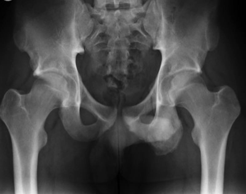
Plain radiograph (figure 1) revealed a grossly expanded ischium and inferior pubic ramus. There was some patchy sclerosis of the left ischium where the hamstrings attach. Subsequent bone scan revealed increased activity at this site. Consideration was given to the possible differential of osteoid osteoma but the increased radioactive uptake was not high enough for this.
Figure 1.

Plain radiograph on presentation demonstrates the asymmetry in ischial tuberosity.
CT scan (figure 2) showed extensive cortical and periosteal thickening along the left ischium, consistent with new bone formation. There was also mild oedema adjacent to the common hamstring origin at the site of the apophyseal avulsion. This was deemed to be consistent with chronic avulsion injury with associated exuberant cortical remodelling. This led to narrowing of the ischiofemoral canal, and the minimum diameter was 8.4 mm at the level of the lesser trochanter indicating ischiofemoral impingement.
Figure 2.
Single-photon emission CT showing chronic expansion of the right ischium with marginally increased tracer uptake.
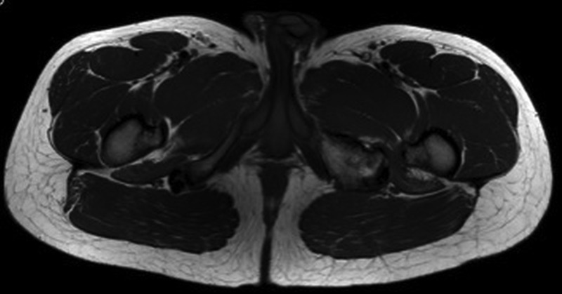
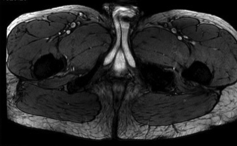
MRI (figures 3 and 4) was consistent with the CT scan findings, supporting the diagnosis of chronic apophyseal avulsion injury of the hamstrings with consequent ischiofemoral impingement, thus explaining his groin pain.
Figure 3.

T1-weighted, transverse MRI showing right-sided narrowing of the ischiofemoral space.
Figure 4.

T2-weighted, transverse MRI demonstrating right-sided ischiofemoral impingement.
Treatment
Non-operative management was undertaken with painkillers as needed, rest, activity modification and physiotherapy exercise regime.
Outcome and follow-up
Over the next 12 months, his symptoms settled and he now reports only a mild, infrequent ache in the groin. He has resumed normal sporting activities without discomfort.
Discussion
Avulsion injuries of the hamstring are common in athletes, particularly football players and sprinters. Ischial tuberosity avulsion fractures arise from forced hyperextension of the hip with the knee in full extension. Ischial apophyseal avulsion is particularly common in the adolescent age group, as in this case. Callus formation as part of the healing process at the site of the chronic avulsion injury is also common and the heterotrophic bone formation can be mistaken for a bone tumour.
In this case, the groin pain is likely to be attributable to the ischiofemoral impingement resulting from the excessive bone formation. The 20 mm distance is usually enough to allow the femur to rotate without making contact with the proximal hamstring tendons or the ischial tuberosity; 8.4 mm is significantly shortened.
With few reports, it is difficult to gauge prevalence. Initially thought to be a potential side effect of surgery, as per Johnson,2 there have been recent studies in which impingement was found in patients without a traumatic incident. The variance in bilateral presentation from 25% to 40%3 5 suggests congenital origin in some cases. There is also a preponderance of cases in women, as seen in all recent reports and studies.3 5 This could, as Tosun et al suggest, indicate a link with female pelvic anatomy where the ischial tuberosities tend to be more prominent and separated.
Presentation is usually with hip/groin pain, as in this case. Some patients present with a snapping or locking hip.3–5 On examination, pain may be elicited by carrying out a combination of hip extension, adduction and external rotation but there is no diagnostic test.
Definitive diagnosis is typically made with MRI, as demonstrated in all reported cases. In contrast to this case, where there is no bony abnormality causing the impingement, plain radiographs are often normal. Quadratus femoris can be visualised on MRI; oedema or fatty change as a result of compression together with reduced space between the ischium and the femur appear to be consistent changes, suggesting the presence of impingement.1 4
In earlier reports,2 resection of the lesser trochanter successfully eradicated the persistent pain. However, as this and other recent cases3 show, a combination of non-steroidal anti-inflammatory drugs, rest and slow reintroduction of sporting activities can be just as successful. Furthermore, excision of the lesser trochanter may cause weakness of hip flexion, a highly undesirable outcome in young, active patients with potentially increased morbidity.1
To conclude, this case report highlights a case of an adolescent boy with acquired unilateral ischiofemoral impingement resulting from excessive bone growth caused by a chronic avulsion fracture of the ischial tuberosity. The diagnosis should be considered alongside more common hip impingement syndromes in presentations of groin pain.
Learning points.
Ischiofemoral impingement must be considered alongside more common hip impingement syndromes, such as femoroacetabular impingement, as a cause of groin pain.
Increasing evidence suggests that it can affect a wider group of patients than initially thought, including younger patients, and those who have not undergone hip arthroplasty procedures.
MRI is diagnostic demonstrating narrowing of the ischiofemoral space, often with associated oedematous changes of the quadratus femoris.
Although surgical treatment can definitively tackle persistent pain, conservative treatment combining activity modification, analgesia and physiotherapy can be successful.
Footnotes
Contributors: SK identified and managed the case. ZH reviewed the literature and drafted the report. SK and RP critically appraised and revised the report for intellectual content.
Competing interests: None.
Patient consent: Obtained.
Provenance and peer review: Not commissioned; externally peer reviewed.
References
- 1.Stafford GH, Villar RN. Ischiofemoral impingement. J Bone Joint Surg Br 2011;93:1300–2 [DOI] [PubMed] [Google Scholar]
- 2.Johnson KA. Impingement of the lesser trochanter on the ischial ramus after total hip arthroplasty. Report of three cases. J Bone Joint Surg Am 1977;59:268–9 [PubMed] [Google Scholar]
- 3.Tosun O, Cay N, Bozkurt M, et al. Ischiofemoral impingement in an 11 year old girl. Diagn Interv Radiol 2012;18:571–3 [DOI] [PubMed] [Google Scholar]
- 4.Patti JW, Ouellette H, Bredella MA, et al. Impingement of lesser trochanter on ischium as a potential cause for hip pain. Skeletal Radiol 2008;37:939–41 [DOI] [PubMed] [Google Scholar]
- 5.Torriani M, Souto SC, Thomas BJ, et al. Ischiofemoral impingement syndrome: an entity with hip pain and abnormalities of the quadratus femoris muscle. AJR Am J Roentgenol 2009;193:186–90 [DOI] [PubMed] [Google Scholar]


