Abstract
Rheumatoid arthritis (RA) is an inflammatory disease associated with intense angiogenesis and vascular expression of integrin αvβ3. Intra-articular administration of a cyclic peptide antagonist of integrin αvβ3 to rabbits with antigen-induced arthritis early in disease resulted in inhibition of synovial angiogenesis and reduced synovial cell infiltrate, pannus formation, and cartilage erosions. These effects were not associated with lymphopenia or impairment of leukocyte function. Furthermore, when administered in chronic, preexisting disease, the αvβ3 antagonist effectively diminished arthritis severity and was associated with a quantitative increase in apoptosis of the angiogenic blood vessels. Therefore, angiogenesis appears to be a central factor in the initiation and persistence of arthritic disease, and antagonists of integrin αvβ3 may represent a novel therapeutic strategy for RA.
Introduction
The rheumatoid arthritic joint is characterized by massive synovial proliferation and changes in synovial architecture resulting in interdigitating folds of tissue, termed pannus. The formation of active inflamed pannus is thought to be central to erosive disease and resulting joint destruction (1). Angiogenesis, the formation of new blood vessels, is one of the earliest histopathologic findings in rheumatoid arthritis (RA) and appears to be required for pannus development (2). This neovascularization is thought not only to maintain the chronic architectural changes via delivery of required blood-borne elements to the pannus but also to play an active role in inflammation as a source of both cytokine and protease activity (3). The expanded vascular-bed volume resulting from angiogenesis may provide increased access for inflammatory cells to infiltrate the synovium (4). Although the factors specifically promoting angiogenesis in RA have not been identified, both synovial tissue and fluid are enriched in angiogenesis-promoting molecules. These include cytokines, such as basic fibroblast growth factor (bFGF) (5), interleukin-8, and vascular endothelial growth factor, and soluble adhesion molecules, such as vascular cell adhesion molecule and E-selectin (6). Interestingly, many of the available treatments for RA have been shown to possess some degree of antiangiogenic activity (7–9). In fact, treatments that suppress the angiogenic process may favorably impact disease course, as suggested by studies in an adjuvant-induced model of arthritis (10).
We have demonstrated previously that integrin αvβ3 is both a marker and crucial effector for blood vessels undergoing angiogenesis (11, 12). Blockade of this integrin by either antibody or peptide antagonists induces apoptosis of angiogenic blood vessels in cytokine and tumor models of angiogenesis (11–13). In agreement with previous immunohistochemical studies (14, 15), we found that synovial blood vessels from RA patients show increased expression of integrin αvβ3. These observations prompted the current study, in which treatment directed against vascular integrin αvβ3 was assessed for its impact on arthritic disease in a rabbit model of RA.
In this report, we demonstrate that intra-articular administration of a cyclic peptide antagonist of αvβ3 in bFGF-augmented antigen-induced arthritis (AIA) was associated with a quantitative increase in vascular apoptosis leading to the inhibition of synovial angiogenesis, as well as a reduction in joint swelling, synovial infiltrate, and pannus formation, in both early and well-established arthritis. Importantly, the αvβ3 antagonist provided significant protection against the development of cartilage erosions. These results substantiate the development of αvβ3 antagonists for future clinical trials in RA.
Methods
Materials.
Ovalbumin (OVA), Freund's complete and incomplete adjuvants, and bovine type II collagen were from Sigma Chemical Co. (St. Louis, Missouri, USA). SDS (Bio-Rad Life Science Research, Hercules, California, USA), sodium chromate (Amersham Life Sciences, Arlington Heights, Illinois, USA), and Percoll (Pharmacia Biotech, Uppsala, Sweden) were purchased from suppliers as indicated. Goat anti–human von Willebrand factor (vWf) affinity-purified antibody (Enzyme Research Laboratories, South Bend, Indiana, USA) was used as a blood vessel marker. Monoclonal antibody (MAB) LM609 directed against integrin αvβ3 (16) was produced and purified from the hybridoma. Fluorescein-conjugated, affinity-purified donkey anti–goat IgG and rhodamine-conjugated, affinity-purified donkey anti–mouse IgG were obtained from Jackson ImmunoResearch Laboratories (West Grove, Pennsylvania, USA). Recombinant bFGF was graciously supplied by J. Abraham (Scios Inc., Mountain View, California, USA). Cyclic peptides (17), cyclic Arg-Gly-Asp-D-Phe-Val (EMD 66203), cyclic Arg-Gly-Asp-D-Phe-[N-methyl]Val (EMD 85189), cyclic Arg–β–Ala-Asp-D-Phe-Val (EMD 69601), and cyclic Arg-Gly-Asp-D-Phe-Lys[fluoresceincarboxylic acid] (EMD 80838) were graciously provided by Merck KGaA (Darmstadt, Germany) and reconstituted in 0.9% saline (2 mg/ml) adjusted to pH 7.4 and stored at −20°C until use.
Establishment of AIA.
All experiments were performed in strict accordance with the guidelines of The Scripps Research Institute Department of Animal Resources. Arthritis was induced as described previously (18). Briefly, male New Zealand White rabbits (2 kg; Western Oregon Rabbit Co., Philomath, Oregon, USA) were immunized in multiple subcutaneous sites with a total of 1 ml OVA (20 mg/ml) in Freund's complete adjuvant and boosted 2 weeks later with 0.6 ml OVA (20 mg/ml) in Freund's incomplete adjuvant. Arthritis was induced 1 week later by bilateral knee intra-articular injection of 0.5 ml OVA/PBS (20 mg/ml) with or without recombinant bFGF (6 μg/ml). Animals were immobilized by predosing with 2 mg acepromazine, followed by inhalation of 2.5% halothane/1.0 l min−1 nitrous oxide/1.0 l min−1 oxygen anesthesia. Analgesia was provided with buprenorphine hydrochloride (Reckitt and Colman Pharmaceuticals Inc., Richmond, Virginia, USA), 0.2 ml twice daily.
Treatment protocol.
OVA-immunized rabbits were randomized, based on ELISA-determined OVA-antibody titers (see ref. 22), into groups treated with the αvβ3 antagonist peptide (EMD 66203) or control peptide (EMD 69601) before arthritis induction with OVA/bFGF. Beginning 24 h after arthritis induction, and again on days 7, 14, and 21, animals were anesthetized and peptides administered by intra-articular injection (0.5 ml/knee, 2 mg/ml). Rabbits were harvested on day 28. To assess the effect of αvβ3 antagonist treatment on chronic disease, rabbits with AIA were randomized (based on day 1 and day 7 knee diameter) into a control group (no treatment) or treatment with the αvβ3 antagonist (0.5 ml/knee, 2 mg/ml administered on days 14, 16, 18, 21, and 25).
Assessment of arthritis.
Joint swelling, defined as increase in extended knee diameter from normal, was measured with dial calipers (0.254 mm; Fisher Scientific, Tustin, California, USA). All assessments were performed by a blinded observer. Immunofluorescence was performed as described previously (12) on cryosections of the infrapatellar fat-pad (5 μm), using anti-vWf as a marker of blood vessels and MAB LM609 as a marker for integrin αvβ3. Rhodamine-conjugated donkey anti–goat and fluorescein-conjugated donkey anti–mouse antibody provided secondary detection. Synovial vascularity was quantified by fluorescent pixel area of synovial sections stained with anti-vWf, with computer analysis (IP Lab Spectrum software; Signal Analytics Corp., Fairfax, Virginia, USA) of three 200× digital images per knee (CCD 1317 camera; Princeton Instruments Inc., Trenton, New Jersey, USA, and Axiovert 100 microscope; Carl Zeiss Inc., Thornwood, New York, USA. Angiogenic index was defined as [(test vWf + area per field) − (nonarthritic vWf + area per field)] / (nonarthritic vWf + area per field). Synovial cell counts were obtained by automated computer counting of nuclei of digital images (three per knee, 400×) from hematoxylin and eosin (H&E)–stained cryosections (5 μm) of the infrapatellar fat-pad. Pannus formation was graded 0–5 as described previously (19) (0 = normal to 5 = pannus accompanied by cartilage erosion). Erosions were assessed as present or absent by visual examination of femoral condyles. Apoptosis was detected using immunofluorescence detection of terminal deoxynucleotidyl transferase–mediated dUTP nick end-labeling (TUNEL) (ApopTag-Red; Oncor Inc., Gaithersburg, Maryland, USA), per supplier's protocol, on infrapatellar cryosections obtained from the right knee of each rabbit. Apoptotic vessels were scored as a percentage of total vessels observed (20 fields [400×] per specimen) by costaining for vWf. Hematological parameters were assessed by Biomedical Testing Services (San Diego, California, USA). Results of early treatment are pooled data from separate experiments expressed as mean ± SE. Statistical evaluation was performed with ANOVA or Student's t test (JMP IN software; Duxbury Press, San Francisco, California, USA), with P < 0.05 considered significant.
Characterization of antagonist activity.
Migration toward RA synovial fluid (1:10 in DMEM) in the presence or absence of peptide antagonists (1 mg/ml) was assessed in transwell assays, using purified radiolabeled AIA rabbit leukocytes as reported previously (20). The relative affinity of the individual antagonists for isolated integrins was determined via soluble ligand-binding studies, as published (21).
Results
The angiogenic growth factor bFGF enhances arthritic disease.
To assess the effect of increased angiogenesis on arthritis development, the proangiogenic cytokine bFGF was added to antigen during arthritis induction. Recent evidence has shown bFGF to be upregulated and localized to the pannus–cartilage interface in RA tissues (5). The inclusion of bFGF in this model resulted in more consistent and accelerated arthritis than with antigen alone, characterized by greater joint swelling (Fig. 1a), increased pannus formation (Fig. 1b), and more frequent erosive disease (Fig. 1c). These results demonstrate that bFGF, an angiogenic growth factor, can exacerbate arthritis.
Figure 1.
The proangiogenic cytokine bFGF intensifies arthritis. (a) The addition of bFGF during induction of AIA enhanced arthritis severity compared with OVA alone, with increased joint swelling, greater pannus formation (b), and earlier and more frequent erosive disease (c). Pannus development was graded on a relative scale 0–5 (0 = normal to 5 = macroscopic cartilage erosion) (ref. 23). All data are expressed as mean ± SE (n = 8). AIA, antigen-induced arthritis; bFGF, basic fibroblast growth factor; OVA, ovalbumin.
Integrin αvβ3 is selectively expressed on synovial blood vessels in AIA and in human RA.
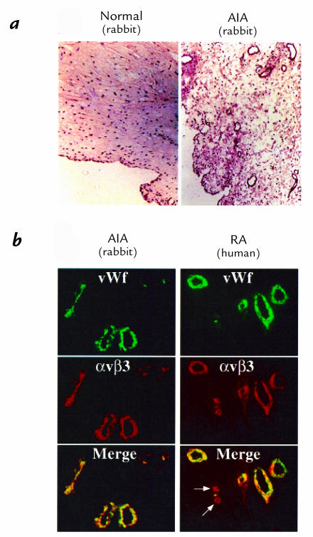
The rabbit AIA model resembles human RA in several important aspects, including histopathology and its response to therapeutic agents (18, 22). Rabbit AIA displays marked neovascularization accompanied by synovial growth and subsynovial inflammatory infiltrate (Fig. 2a). Notably, αvβ3 staining in AIA is restricted to synovial blood vessels (Fig. 2b), as in human RA, which is indicative of ongoing angiogenesis; blood vessels in sections from normal, nonarthritic synovium express little or no αvβ3 (data not shown). The earliest invasive blood vessels, although positive for αvβ3, do not stain for vWf (Fig 2b, arrows), reflecting the fact that vWf is a marker of differentiated endothelium (23). As vessels mature, αvβ3 is lost (24). The hyperproliferative synovial lining and subsynovial tissue also contain numerous fibroblasts and macrophages (Fig. 2a) (1). In contrast to cutaneous wound fibroblasts, which express integrin αvβ3 (25), little or no expression of this integrin was found on synovial fibroblasts (Fig. 2b), in agreement with previous studies (14, 15, 26, 27). With respect to synovial macrophages, αvβ3 expression was found to be restricted to osteoclasts and was not present in other monocyte/macrophage–derived cells (28).
Figure 2.
AIA and human RA exhibit αvβ3 expression on angiogenic blood vessels. (a) Cryosections of infrapatellar tissue in AIA stained with H&E demonstrate marked neovascularization, synovial hypertrophy, and dense synovial inflammatory infiltrate relative to synovium from nonarthritic tissue (×100). (b) Immunofluorescent detection of blood vessels by vWf-specific antibody (green) and MAB LM609 directed to integrin αvβ3 (red) demonstrates colocalization of αvβ3 on the synovial endothelium (yellow merge) in rabbit AIA, as in human RA (×400). Note that microvascular angiogenic sprouts express only αvβ3 (arrows). H&E, hematoxylin and eosin; MAB, monoclonal antibody; RA, rheumatoid arthritis; vWf, von Willebrand factor.
Synovial neovascularization is inhibited by an αvβ3 antagonist that targets angiogenic blood vessels.
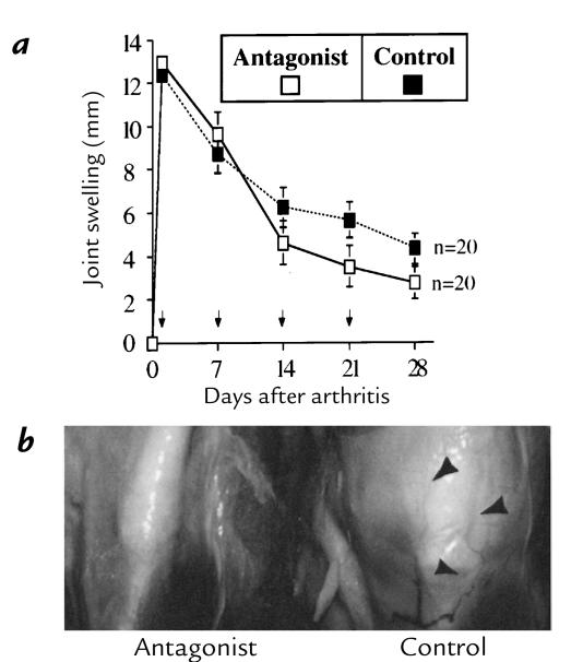
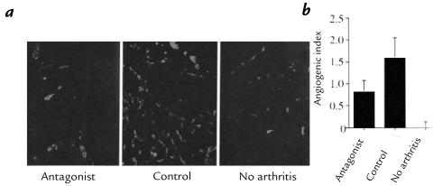
Blockade of integrin αvβ3 by either antibody or peptide antagonists successfully inhibits neovascularization in cytokine and tumor models of angiogenesis in avian, murine, rabbit, and human tissues (11–13). To evaluate whether blockade of integrin αvβ3 could impact arthritis-associated angiogenesis, rabbits with AIA were treated intra-articularly 24 hours after arthritis onset and weekly thereafter for four weeks with the αvβ3 antagonist EMD 66203. This treatment resulted in a modest yet significant (P < 0.05) decrease in joint swelling compared with that in animals treated with the control peptide, EMD 69601 (Fig. 3a). Initial examination of knee joints revealed obvious periarticular vascularization in the control-treated animals (Fig. 3b). Cryosections of the infrapatellar fat-pad were examined with anti-vWf as a blood vessel marker. Tissues from rabbits treated with the αvβ3 integrin antagonist displayed considerably fewer blood vessels than tissues from control-treated rabbits (Fig. 4a). Blinded computer analysis of digital images revealed a 48% decrease in synovial neovascularization in αvβ3 antagonist–treated animals (Fig. 4b) (P < 0.01).
Figure 3.
Joint swelling is reduced after αvβ3 antagonist administration in AIA. (a) Rabbits with AIA were treated with bilateral intra-articular αvβ3 antagonist (EMD 66203; 0.5 ml, 2 mg/ml) or control peptide (EMD 69601) beginning 24 h after arthritis onset, and weekly thereafter for 4 weeks (arrows). Joint swelling, defined as the increase in knee diameter from normal (nonarthritic), was significantly reduced by the αvβ3 antagonist (days 14–28, P < 0.05, ANOVA) compared with control peptide. (b) Periarticular vascularization (arrowheads) was prominent in control-treated AIA compared with antagonist-treated groups.
Figure 4.
Synovial vascularity is decreased after αvβ3 antagonist treatment. (a) Cryosections of the infrapatellar tissue obtained 28 days after arthritis onset in animals treated with the αvβ3 antagonist or control peptide (days 1, 7, 14, and 21) were stained for vWf as a marker of blood vessels, detected with FITC-labeled secondary antibody, and were digitally imaged (×200). (b) The relative increase in area of fluorescent pixels per field vs. normal was computed to determine the angiogenic index as described in Methods. Synovial neovascularization was significantly inhibited by the αvβ3 antagonist (P < 0.050, Student's t test). Data are expressed as mean ± SE (n = 20).
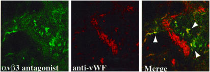
To determine whether the αvβ3 antagonist was targeted to the vasculature in the arthritic synovium, we evaluated the distribution of a fluorescent-labeled form of this peptide, EMD 80838. Twenty-four hours after intra-articular injection, the fluorescent-conjugated αvβ3-binding peptide selectively localized to microvessels within the arthritic synovium, as determined by confocal microscopy (Fig. 5). These studies demonstrate that the αvβ3 antagonist targets synovial blood vessels in this model and inhibits angiogenesis in the arthritic synovium.
Figure 5.
Fluorescent integrin antagonist colocalizes with synovial angiogenic microvessels. An FITC-conjugated αvβ3 antagonist peptide, EMD 80838 (cyclic-Arg-Gly-Asp-D-Phe-Lys-[fluoresceincarboxylic acid]; 0.5 mg/500 μl), was injected intra-articularly in rabbits with AIA. After 24 h, cryostat sections (5 μm) of synovial tissue were examined by confocal microscopy for the presence of peptide (green). Blood vessels were identified by antisera to vWF as detected with TRITC-labeled secondary antibody (red). Colocalization of αvβ3 antagonist is observed on angiogenic microvessels after signal merge (yellow), but not with mature vWF+ vessels (×630). TRITC, tetrarhodamine isothiocyanate.
Animals treated with the αvβ3 antagonist show reduced synovial infiltrate.
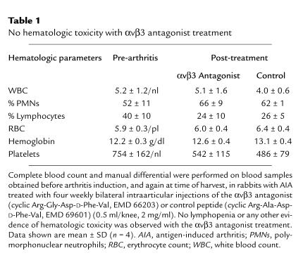
To evaluate whether the observed angiogenesis inhibition in αvβ3 antagonist–treated animals was associated with a change in arthritis, several disease parameters were examined. Angiogenesis inhibition has been postulated to limit accessibility of the tissue to leukocytes (3); therefore, we first examined whether there was any decrease in the degree of synovial inflammatory infiltrate in animals treated with the antiangiogenic αvβ3 antagonist. Histological examination of the infrapatellar fat-pad revealed that treatment with the αvβ3 antagonist resulted in significantly fewer cells infiltrating the synovium (Fig. 6a). Computerized cell counting of digital images of these sections demonstrated a 39% reduction in synovial cellular infiltrate (Fig. 6b) (P < 0.05). Since only a small fraction of peripheral lymphocytes (1%–2%) express integrin αvβ3 (29), it is unlikely that the reduction in cellular infiltrate was due to leukocyte integrin inhibition. In fact, chemotaxis of AIA peripheral blood mononuclear cells (PBMCs) and polymorphonuclear neutrophils (PMNs) toward RA synovial fluid was not influenced by either the αvβ3-directed peptide or the control peptide on collagen (Fig. 6c) or fibronectin (data not shown). No leukopenia or other sign of hematologic or marrow toxicity was observed after treatment in either group (Table 1). Immunohistochemical analysis revealed no difference in the degree of colocalization between apoptotic cells and leukocytes, as determined by TUNEL and KEN-11 (anti–LFA-1) costaining studies between control and αvβ3 antagonist–treated rabbits (data not shown). Thus, the reduced synovial infiltrate observed in the αvβ3 antagonist–treated AIA synovium does not appear to result from direct effects of this compound on leukocyte number or migration.
Figure 6.
Integrin αvβ3 antagonist reduces synovial inflammatory infiltrate in AIA without impairing leukocyte migration. (a) H&E stain of cryosections of the infrapatellar fat-pad 28 days after arthritis onset reveals a marked decrease in synovial cellular infiltrate in animals treated with the αvβ3 antagonist (days 1, 7, 14, and 21) compared with control. (b) Digital assessment of nuclei present in each field (three per joint, ×400) demonstrates that αvβ3 antagonist treatment resulted in a significant reduction in the cellular infiltrate (P < 0.05, Student's t test). Data are expressed as mean ± SE (n = 20). (c) In vitro chemotaxis on type II collagen toward synovial fluid by PBMCs or PMNs isolated from peripheral blood from rabbits with AIA. Data are expressed as mean ± SE of triplicate determinations. No effect of the αvβ3 antagonist on the migratory capacity of leukocytes was observed. PBMCs, peripheral blood mononuclear cells; PMNs, polymorphonuclear neutrophils.
Table 1.
No hematologic toxicity with αvβ3 antagonist treatment
The αvβ3 antagonist protects against erosive disease.
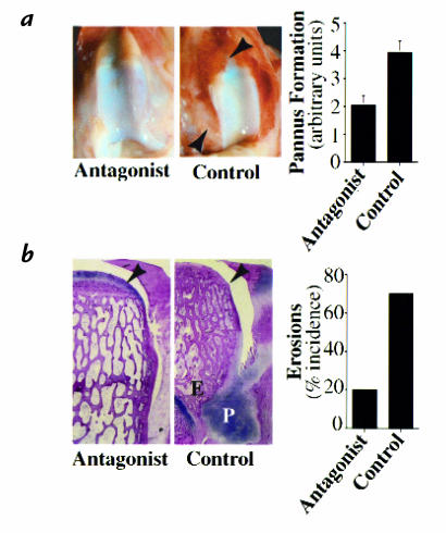
Since pannus formation is thought to initiate progression to erosive disease (1), the effect of the αvβ3 antagonist on the development of pannus was examined. In control-treated animals, extensive pannus formation covering much of the femoral articular cartilage surface was observed, whereas treatment with the αvβ3 antagonist resulted in significant reduction of pannus development (P < 0.05) (Fig. 7a). The αvβ3 antagonist also significantly blocked the development of cartilage erosions. Gross erosions were present in 70% (14 of 20) of femoral condyles in control-treated AIA, yet only 20% (4 of 20) treated with the αvβ3 antagonist exhibited erosions (Fig. 7b) (P < 0.01).
Figure 7.
Blockade of integrin αvβ3 decreases pannus formation and cartilage erosions. (a) Pannus development (arrowheads) was graded on a relative scale 0–5 (0 = normal to 5 = macroscopic erosion). Treatment with the αvβ3 antagonist significantly decreased pannus formation (P < 0.05, Student's t test) relative to control peptide. (b) Frontal sections of decalcified femoral condyle stained by H&E illustrate the protective effect of αvβ3 antagonist treatment on a representative sample of erosive disease (×10). (P indicates pannus, E indicates erosion, and the arrowhead indicates preservation of the articular cartilage in the antagonist-treated group.) Macroscopic cartilage erosions were decreased with αvβ3 antagonist treatment (P < 0.01, Student's t test). All data are expressed as mean ± SE (n = 20).
Vascular integrin αvβ3 is the preferred target in AIA.
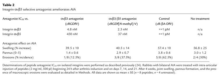
Recent studies suggest that both integrin αvβ3 and integrin αvβ5 can play a role in angiogenesis (30). Therefore, we compared the antiarthritic effects of the cyclic peptide with αvβ3 selectivity, EMD 66203, with that of a peptide with higher αvβ5-binding activity, EMD 85189. Notably, αvβ5 is also highly expressed on synovial fibroblasts and macrophages (14, 27). The peptide antagonist with greatest selectivity for αvβ3 demonstrated strongest inhibition of pannus development and erosive disease (Table 2), suggesting that αvβ3 was the primary therapeutic target in this model.
Table 2.
Integrin αvβ3 selective antagonist ameliorates AIA
Chronic arthritis is ameliorated by the αvβ3 antagonist.
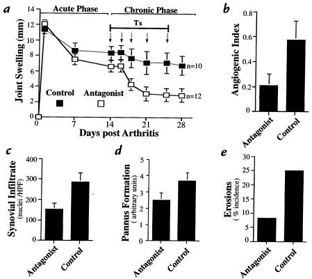
We extended these studies to evaluate the effect of αvβ3 blockade on well-established arthritis. AIA was allowed to progress for two weeks into the chronic phase (18) before the initiation of treatment. Despite the delay, joint swelling was significantly decreased by the αvβ3 antagonist (Fig. 8a). In this chronic model, treatment with the antagonist produced a significant antiangiogenic effect, with a 64% reduction in the angiogenic index compared with untreated AIA (P < 0.05) (Fig. 8b), and blockade of αvβ3 was again associated with a marked reduction in the synovial cell infiltrate (P < 0.05) (Fig. 8c). As observed previously with early administration during acute disease, delayed treatment with the αvβ3 antagonist decreased pannus formation and reduced the incidence of erosive disease compared with controls (Fig. 8, d and e). From these results, we conclude that angiogenesis contributes to disease severity as well as chronicity, and that an antagonist of integrin αvβ3 is an effective antiarthritic agent in well-established disease.
Figure 8.
Chronic arthritis is ameliorated by an αvβ3 antagonist. (a) Administration of the antagonist (arrows) beginning 2 weeks after arthritis onset resulted in decreased joint swelling compared with controls (untreated) (P < 0.05, ANOVA). (b) Angiogenesis, as depicted by the angiogenic index (see Methods), was inhibited by αvβ3 antagonist treatment (P < 0.05, Student's t test). (c) Fewer infiltrating cells were observed in the synovium of αvβ3 antagonist–treated animals (P < 0.05, Student's t test), as assessed by digital computerized counting of nuclei. (d) Pannus was assessed as described in Methods. (e) A decrease in both pannus formation and cartilage erosion was observed in antagonist-treated animals relative to control. Data are expressed as mean ± SE (n = 12, antagonist; n = 10, control). HPF, high-power field; Tx, treatment.
Synovial vascular apoptosis is associated with αvβ3 antagonist treatment.
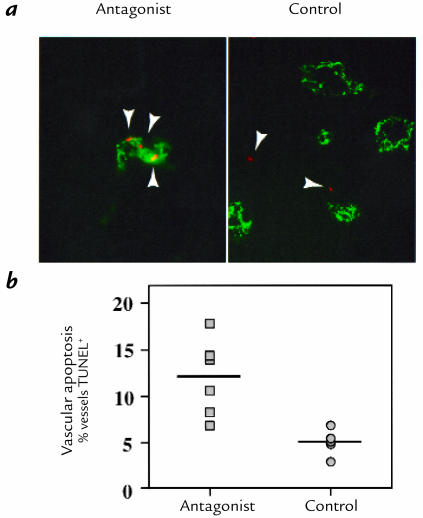
Previous studies have demonstrated that antagonists of integrin αvβ3 induce apoptosis of angiogenic blood vessels (11–13). Therefore, we examined synovial tissues from the chronic arthritis model for the presence of apoptotic cells after treatment with the αvβ3 antagonist. Immunohistochemical detection of apoptosis within AIA synovium demonstrated increased (2.5-fold) TUNEL-positive vascular cells in the αvβ3 antagonist–treated animals (P < 0.01) (Fig. 9). These observations suggest that αvβ3 antagonists may inhibit angiogenesis in the arthritic synovium by inducing apoptosis of angiogenic blood vessels.
Figure 9.
Vascular cell apoptosis is associated with αvβ3 antagonist treatment. (a)Synovium from rabbits in the chronic AIA model were stained with TUNEL immunostaining (red) as an indicator of apoptosis, (arrowheads)and anti-vWF (green) as a marker of blood vessels (×400). (b) Apoptosis associated with blood vessels was detected and quantified blindly as percent blood vessels containing apoptotic cells in 20 fields (×400) per specimen. Apoptosis was increased specifically in the vasculature of arthritic synovium after αvβ3 antagonist treatment (P < 0.01, Student's t test). TUNEL, terminal deoxynucleotidyl transferase–mediated dUTP nick end-labeling.
Discussion
RA has been classified as an angiogenesis-associated disease, based on the finding of extensive neovascularization within the inflamed synovium (3). Clearly, inflammatory cytokines with angiogenic activity can induce or potentiate synovitis (31, 32). However, it has not been determined whether angiogenesis directly contributes to the arthritic process or whether it is merely a consequence of ongoing inflammation. Evidence is provided in this report that angiogenesis contributes to arthritic disease. Addition of the proangiogenic cytokine bFGF to AIA accelerated and exacerbated arthritis. The inclusion of bFGF in this model was based on the facts that bFGF does not possess the inflammatory properties associated with other angiogenic factors (e.g., interleukin-8 [31] and tumor necrosis factor-α [32]), bFGF is upregulated and localized to the pannus–cartilage interface in RA tissues (5), and synovial levels of bFGF in human RA correlate with disease severity (33). In fact, the addition of bFGF to AIA produced an accelerated and more consistent arthritis compared with arthritis induced with antigen alone (Fig. 1), without altering the existing expression and selective distribution of integrin αvβ3, as assessed by staining with LM609 (data not shown).
Not only did bFGF accelerate arthritis in this model, but a peptide antagonist selective for integrin αvβ3 inhibited angiogenesis and reduced the severity of arthritis. Joint swelling, synovial infiltrate, pannus formation, and the incidence of cartilage erosions were all significantly reduced in animals treated early with this integrin antagonist. Inhibition of neovascularization, as demonstrated in this model, may influence many parameters of disease, including inflammation, protease production, and tissue metabolism (3). No evidence for a direct role of the αvβ3 antagonist in impairing peripheral leukocyte migration was detected, nor was acute inflammation affected in antagonist-treated animals upon rechallenge with antigen (data not shown).
Integrin αvβ3 was selectively localized to synovial blood vessels in human RA and rabbit AIA, as determined by costaining with LM609 and anti-vWf (Fig. 2b). The appearance of αvβ3-positive/vWf-negative vessels reflects the fact that vWf is a marker of more mature vessels (23), whereas αvβ3 is present on very early neovessels (34). In addition, the αvβ3 antagonist localized to microvessels 24 hours after its intra-articular administration in the arthritic synovium, supporting previous findings that this integrin is found primarily on angiogenic blood vessels (11, 34).
During maturation and differentiation, mono-cytes/macrophages may express αv integrins (28, 35, 36), and in fact, antagonists of αvβ3 can influence macro-phage-derived osteoclast function (37), which may contribute to the observed decrease in bone erosions after αvβ3 antagonist treatment. However, functionally mature (αvβ3+) osteoclasts are rarely found in the synovium (38), and it is uncertain that the short-term, intermittent dosing regimen of the αvβ3 antagonist used in this study would be sufficient to impact ongoing osteoclast function. Although the αvβ3-selective cyclic peptide antagonist can impact other αv integrins, it is ineffective against β1 integrin–mediated adhesion (data not shown). Antagonism of integrin β1 with small Arg-Gly-Asp –containing linear peptide fragments of fibronectin induces collagenase expression in synovial fibroblasts, and administration of these peptides may thereby actually increase disease (39). Although αvβ3 is present in certain fibroblast populations (25), it was not detected in fibroblasts from synovial tissues in patients with RA (14, 26, 27).
The integrin antagonist used, although selective for integrin αvβ3, exhibits some activity against the closely related integrin αvβ5 (Table 2). Antagonism of integrin αvβ5 is sufficient to block angiogenesis induced by some cytokines (30) and thus may have contributed to the reduced synovial vascularization observed. However, administration of an antagonist with a higher affinity for integrin αvβ5, which is also expressed on synovial fibroblasts and macrophages (14, 27), resulted in only minor improvements in arthritis severity (Table 2), suggesting that vascular integrin αvβ3 is the preferred target in inflammatory arthritis.
The results presented in this report provide evidence for a central pathogenic contribution of angiogenic blood vessels to both the induction and maintenance of arthritic disease. Increased neovascularization was clearly and consistently associated with more severe arthritis. Selective αvβ3 blockade effectively inhibited synovial angiogenesis and provided significant protection against the development of cellular infiltrate, pannus formation, and cartilage erosions. In view of the rather selective distribution of integrin αvβ3 on blood vessels in the arthritic synovium (Fig. 2b) (14, 15, 26–28), the quantitative increase in endothelial apoptosis after αvβ3 antagonist treatment, and the decreased vascularity observed, angiogenesis inhibition is a likely mechanism by which αvβ3 antagonists ameliorate arthritis. The fact that an αvβ3 antagonist was effective when administered in the chronic AIA model is relevant to future clinical applications. The expression of integrin αvβ3 on synovial blood vessels in human RA presents an opportunity to pursue αvβ3 integrin–directed antiangiogenic therapy for the treatment of RA.
Acknowledgments
The authors thank Melinda Storgard for excellent technical assistance. This work was supported in part by grants from the Department of Academic Affairs of Scripps Clinic and the Jeanette Hennings Foundation. C.M. Storgard is a recipient of an Arthritis Foundation Postdoctoral Fellowship. D.G. Stupack is a recipient of a Canadian Arthritis Society Fellowship and a Joseph Drown Foundation Fellowship. D.A. Cheresh was supported by National Institutes of Health grants CA-50286, CA-45726, and HL-54444. This is publication no. 11520–IMM from the Scripps Research Institute.
References
- 1.Zvaifler NJ, Firestein GS. Pannus and pannocytes: alternative models of joint destruction in rheumatoid arthritis. Arthritis Rheum. 1994;37:783–789. doi: 10.1002/art.1780370601. [DOI] [PubMed] [Google Scholar]
- 2.Kimball ES, Gross JL. Angiogenesis in pannus formation. Agents Actions. 1991;34:329–331. doi: 10.1007/BF01988724. [DOI] [PubMed] [Google Scholar]
- 3.Colville-Nash PR, Scott DL. Angiogenesis and rheumatoid arthritis: pathogenic and therapeutic implications. Ann Rheum Dis. 1992;51:919–925. doi: 10.1136/ard.51.7.919. [DOI] [PMC free article] [PubMed] [Google Scholar]
- 4.Paleolog E. Target effector role of vascular endothelium in the inflammatory response: insights from the clinical trial of anti-TNF alpha antibody in rheumatoid arthritis. Mol Pathol. 1997;50:225–233. doi: 10.1136/mp.50.5.225. [DOI] [PMC free article] [PubMed] [Google Scholar]
- 5.Qu Z, et al. Expression of basic fibroblast growth factor in synovial tissue from patients with rheumatoid arthritis and degenerative joint disease. Lab Invest. 1995;73:339–346. [PubMed] [Google Scholar]
- 6.Koch AE. Review: angiogenesis: implications for rheumatoid arthritis. Arthritis Rheum. 1998;4:951–962. doi: 10.1002/1529-0131(199806)41:6<951::AID-ART2>3.0.CO;2-D. [DOI] [PubMed] [Google Scholar]
- 7.Matsubara T, Ziff M. Inhibition of endothelial cell proliferation by gold compounds. J Clin Invest. 1987;79:1440–1446. doi: 10.1172/JCI112972. [DOI] [PMC free article] [PubMed] [Google Scholar]
- 8.Potvin F, Petitclerc E, Marceau F, Poubelle PE. Mechanisms of action of antimalarials in inflammation: induction of apoptosis in human endothelial cells. J Immunol. 1997;158:1872–1879. [PubMed] [Google Scholar]
- 9.Hirata S, Matsubara T, Saura R, Tateishi H, Hirohata K. Inhibition of in vitro vascular endothelial cell proliferation and in vivo neovascularization by low dose methotrexate. Arthritis Rheum. 1989;32:1065–1073. doi: 10.1002/anr.1780320903. [DOI] [PubMed] [Google Scholar]
- 10.Peacock DJ, Banquerigo ML, Brahn E. A novel angiogenesis inhibitor suppresses rat adjuvant arthritis. Cell Immunol. 1995;160:178–184. doi: 10.1016/0008-8749(95)80025-e. [DOI] [PubMed] [Google Scholar]
- 11.Brooks PC, Clark RA, Cheresh DA. Requirement of vascular integrin αvβ3 for angiogenesis. Science. 1994;264:569–571. doi: 10.1126/science.7512751. [DOI] [PubMed] [Google Scholar]
- 12.Brooks PC, et al. Anti-integrin αvβ3 blocks human breast cancer growth and angiogenesis in human skin. J Clin Invest. 1995;96:1815–1822. doi: 10.1172/JCI118227. [DOI] [PMC free article] [PubMed] [Google Scholar]
- 13.Brooks PC, et al. Integrin αvβ3 antagonists promote tumor regression by inducing apoptosis of angiogenic blood vessels. Cell. 1994;79:1157–1164. doi: 10.1016/0092-8674(94)90007-8. [DOI] [PubMed] [Google Scholar]
- 14.Johnson BA, Haines GK, Harlow LA, Koch AE. Adhesion molecule expression in human synovial tissue. Arthritis Rheum. 1993;36:137–146. doi: 10.1002/art.1780360203. [DOI] [PubMed] [Google Scholar]
- 15.Walsh DA, Wade M, Mapp PI, Blake DR. Focally regulated endothelial proliferation and cell death in human synovium. Am J Pathol. 1998;152:691–702. [PMC free article] [PubMed] [Google Scholar]
- 16.Cheresh DA, Spiro RC. Biosynthetic and functional properties of Arg-Gly-Asp-directed adhesion receptor involved in human melanoma cell attachment to vitronectin, fibrinogen, and von Willebrand factor. J Biol Chem. 1987;262:17703–17711. [PubMed] [Google Scholar]
- 17.Aumailley M, et al. Arg-Gly-Asp constrained within cyclic pentapeptides: strong and selective inhibitors of cell adhesion to vitronectin and laminin fragment P1. FEBS Lett. 1991;291:50–54. doi: 10.1016/0014-5793(91)81101-d. [DOI] [PubMed] [Google Scholar]
- 18.Jason HE. Chronic arthritis in rabbits. Methods Enzymol. 1988;162:379–385. doi: 10.1016/0076-6879(88)62093-3. [DOI] [PubMed] [Google Scholar]
- 19.Maksymowych WP, et al. Amelioration of antigen induced arthritis in rabbits treated with a secreted viral serine proteinase inhibitor. J Rheumatol. 1996;23:878–882. [PubMed] [Google Scholar]
- 20.Taub RN. Effects of concanavalin A on the migration of radioactively labeled lymphoid cells. Cell Immunol. 1974;12:263–270. doi: 10.1016/0008-8749(74)90078-1. [DOI] [PubMed] [Google Scholar]
- 21.Haubner R, et al. Cyclic RGD peptides containing β-turn mimetics. J Am Chem Soc. 1996;118:7881–7891. [Google Scholar]
- 22.Hunneyball IM. Use of experimental arthritis in the rabbit for the development of antiarthritic drugs. Adv Inflamm Res. 1984;7:249–262. [Google Scholar]
- 23.Han RN, Tanswell AK, Post M. Ontogeny of reactivity to endothelial cell markers during development of the embryonic and fetal rat lung. Histol Histopathol. 1992;7:591–597. [PubMed] [Google Scholar]
- 24.Clark RA, Tonnesen MG, Gailit J, Cheresh DA. Transient functional expression of αvβ3 on vascular cells during wound repair. Am J Pathol. 1996;148:1407–1421. [PMC free article] [PubMed] [Google Scholar]
- 25.Gailit J, Clark RA. Studies in vitro on the role of alpha v and beta 1 integrins in the adhesion of human dermal fibroblasts to provisional matrix proteins fibronectin, vitronectin, and fibrinogen. J Invest Dermatol. 1996;106:102–108. doi: 10.1111/1523-1747.ep12328177. [DOI] [PubMed] [Google Scholar]
- 26.Rinaldi N, et al. Differential expression and functional behavior of the alpha v and beta 3 integrin subunits in cytokine stimulated fibroblast-like cells derived from synovial tissue of rheumatoid arthritis and osteoarthritis in vitro. Ann Rheum Dis. 1997;56:729–736. doi: 10.1136/ard.56.12.729. [DOI] [PMC free article] [PubMed] [Google Scholar]
- 27.Nikkari L, et al. Localization of the alpha v subfamily of integrins and their putative ligands in synovial lining cell layer. J Rheumatol. 1995;22:16–23. [PubMed] [Google Scholar]
- 28.Zambonin ZA, Teti A, Gaboli M, Marichisio PC. Beta 3 subunit of vitronectin receptor is present in osteoclast adhesion structures and not in other monocyte-macrophage derived cells. Connect Tissue Res. 1989;20:143–149. doi: 10.3109/03008208909023882. [DOI] [PubMed] [Google Scholar]
- 29.Hemler ME. Adhesive protein receptors on hematopoietic cells. Immunol Today. 1988;9:109–113. doi: 10.1016/0167-5699(88)91280-7. [DOI] [PubMed] [Google Scholar]
- 30.Friedlander M, et al. Two pathways of angiogenesis defined by distinct αv integrins. Science. 1995;250:1500–1502. doi: 10.1126/science.270.5241.1500. [DOI] [PubMed] [Google Scholar]
- 31.Endo H, et al. Experimental arthritis induced by continuous infusion of IL-8 into rabbit knee joints. Clin Exp Immunol. 1994;96:31–35. doi: 10.1111/j.1365-2249.1994.tb06225.x. [DOI] [PMC free article] [PubMed] [Google Scholar]
- 32.Lewthwaite J, et al. Role of TNF alpha in the induction of antigen induced arthritis in the rabbit and the anti-arthritic effect of species specific TNF alpha neutralizing monoclonal antibodies. Ann Rheum Dis. 1995;54:366–374. doi: 10.1136/ard.54.5.366. [DOI] [PMC free article] [PubMed] [Google Scholar]
- 33.Nakashima M, et al. Expression of basic fibroblast growth factor in synovial tissues from patients with rheumatoid arthritis: detection by immunohistological staining and in situ hybridisation. Ann Rheum Dis. 1994;53:45–50. doi: 10.1136/ard.53.1.45. [DOI] [PMC free article] [PubMed] [Google Scholar]
- 34.Drake CJ, Cheresh DA, Little CD. An antagonist of integrin αvβ3 prevents maturation of blood vessels during embryonic neovascularization. J Cell Sci. 1995;108:2655–2661. doi: 10.1242/jcs.108.7.2655. [DOI] [PubMed] [Google Scholar]
- 35.De Nichilo MO, Yamada KM. Integrin αvβ5-dependent serine phosphorylation of paxillin in cultured human macrophages adherent to vitronectin. J Biol Chem. 1996;271:11016–11022. doi: 10.1074/jbc.271.18.11016. [DOI] [PubMed] [Google Scholar]
- 36.Andreesen R, et al. Surface phenotype analysis of human monocyte to macrophage maturation. J Leukoc Biol. 1990;47:490–497. doi: 10.1002/jlb.47.6.490. [DOI] [PubMed] [Google Scholar]
- 37.Engleman VW, et al. A peptidomimetic antagonist of the αvβ3 integrin inhibits bone resorption in vitro and prevents osteoporosis in vivo. J Clin Invest. 1997;99:2284–2292. doi: 10.1172/JCI119404. [DOI] [PMC free article] [PubMed] [Google Scholar]
- 38.Ashton BA, Ashton IK, Marshall MJ, Butler RC. Localization of vitronectin receptor immunoreactivity and tartrate resistant acid phosphatase activity in synovium from patients with inflammatory or degenerative arthritis. Ann Rheum Dis. 1993;52:133–137. doi: 10.1136/ard.52.2.133. [DOI] [PMC free article] [PubMed] [Google Scholar]
- 39.Homandber GA, Meyers R, Williams JM. Intraarticular injection of fibronectin fragments causes severe depletion of cartilage proteoglycans in vivo. J Rheumatol. 1993;20:1378–1382. [PubMed] [Google Scholar]


