Abstract
Allergic asthma, which is present in as many as 10% of individuals in industrialized nations, is characterized by chronic airway inflammation and hyperreactivity induced by allergen-specific Th2 cells secreting interleukin-4 (IL-4) and IL-5. Because Th1 cells antagonize Th2 cell functions, it has been proposed that immune deviation toward Th1 can protect against asthma and allergies. Using an adoptive transfer system, we assessed the roles of Th1, Th2, and Th0 cells in a mouse model of asthma and examined the capacity of Th1 cells to counterbalance the proasthmatic effects of Th2 cells. Th1, Th2, and Th0 lines were generated from ovalbumin (OVA)-specific T-cell receptor (TCR) transgenic mice and transferred into lymphocyte-deficient, OVA-treated severe combined immunodeficiency (SCID) mice. OVA-specific Th2 and Th0 cells induced significant airway hyperreactivity and inflammation. Surprisingly, Th1 cells did not attenuate Th2 cell–induced airway hyperreactivity and inflammation in either SCID mice or in OVA-immunized immunocompetent BALB/c mice, but rather caused severe airway inflammation. These results indicate that antigen-specific Th1 cells may not protect or prevent Th2-mediated allergic disease, but rather may cause acute lung pathology. These findings have significant implications with regard to current therapeutic goals in asthma and allergy and suggest that conversion of Th2-dominated allergic inflammatory responses into Th1-dominated responses may lead to further problems.
Introduction
Chronic inflammation of the respiratory mucosa plays a fundamental role in the pathogenesis of asthma, which affects as many as 10% of individuals in industrialized nations (1). The inflammatory response in asthma is tightly associated with airway hyperresponsiveness, a hallmark of asthma, and involves a number of different cell types including eosinophils, basophils, mast cells, and, most importantly, CD4+ Th2 lymphocytes, which can be isolated from the lungs of patients with asthma (2, 3). Th2 cells secreting interleukin-4 (IL)-4 and IL-5 play a central role in initiating and sustaining an asthmatic response by regulating the production of IgE and the growth, differentiation, and recruitment of mast cells, basophils, and eosinophils (4). Persistent wheezing in children is predicted by development of IgE-mediated hypersensitivity to inhaled allergens (5), indicating that the development of an inappropriate adaptive Th2-polarized immune response to environmental antigens plays a critical role in the development of asthma.
While Th2 cells promote airway inflammation in asthma, it has been proposed that Th1 cells, which secrete interferon (IFN)-γ, protect against allergic disease by dampening the activity of Th2 effector cells. The evidence, however, demonstrating that Th1 cells actually render salutary effects in allergic disease and asthma, is indirect. Th1 cells inhibit the proliferation, and therefore the development, of Th2 cells (6), and IFN-γ inhibits IgE synthesis in some instances (7). Allergen-specific Th1 clones can be generated from nonallergic individuals (8), but the frequency of such cells in asymptomatic individuals is not clear, particularly given that such cells are not found in the nasal or pulmonary mucosa of nonallergic individuals. Conventional immunotherapy for allergic diseases and asthma reduces IL-4 production in allergen-specific Th2 cells (9) and increases IFN-γ production by allergen-specific Th1 cells (10, 11). Individuals predisposed toward the production of Th1 cytokines (e.g., patients with multiple sclerosis) have a reduced likelihood of developing allergic disease (12). Finally, infection with Mycobacteria tuberculosis, which requires a Th1-dominated immune response for eradication, reduces the likelihood of developing atopy (13).
These studies together suggest that immunotherapies for asthma should promote the replacement of allergen-specific Th2 cells with Th1 cells, which counterbalance the effects of Th2 cells. No data are available, however, that directly show that Th1 cells actively suppress Th2-dominated allergic responses. Recent studies with Th1 and Th2 cells in autoimmune encephalomyelitis and diabetes mellitus indicate that Th2 cells may not always antagonize Th1 cells and that polarized Th2 cells that are generally assumed to be protective in autoimmunity may in some instances be unexpectedly harmful (14–16). These studies indicate that the Th1/Th2 paradigm may be much more complex than initially appreciated.
The purpose of this study was to examine directly the capacity of Th1 CD4+ cells to counterbalance the allergic effects of Th2 cells in a murine model of asthma. Th1, Th2, and Th0 CD4+ lines expressing identical ovalbumin (OVA)-specific T-cell receptors (TCRs) were generated from OVA-specific TCR transgenic mice, and their effects after transfer into lymphocyte-deficient severe combined immunodeficiency (SCID) and immunocompetent BALB/c mice on allergen-induced airway inflammation and airway hyperreactivity were investigated. Our results demonstrated that OVA-specific Th1 cells caused severe airway inflammation and did not reduce the inflammatory effects of OVA-specific Th2 cells. OVA-specific Th1 cells did not induce airway hyperreactivity but were unable to reduce Th2 cell–induced airway hyperreactivity, either in lymphocyte-deficient SCID mice or in immunocompetent recipients. These results indicate that antigen-specific CD4+ Th1 cells may not be protective in Th2-mediated allergic diseases and asthma, but instead cause significant pulmonary disease. These findings are of particular concern with regard to current therapeutic goals in asthma and allergies, and suggest that conversion of Th2-dominated allergic inflammatory responses into Th1-dominated responses may be problematic.
Methods
Animals.
BALB/cBy mice were obtained from The Jackson Laboratory (Bar Harbor, Maine, USA), and histocompatible C.B-17Icrscid/scid mice (17) were obtained from the Stanford Medical Center Division of Laboratory Animal Medicine (Stanford, California, USA). DO11.10 mice, which are transgenic for TCR recognizing OVA peptide 323–339 (pOVA323–339) and backcrossed to BALB/c (18), were kindly provided by D. Loh and were bred in our facilities. Animals were used between 6 and 10 weeks of age and were age and sex matched within each experiment. All animal protocols were approved by the Stanford University Committee on Animal Welfare.
Monoclonal antibodies and reagents.
Anti–IFN-γ monoclonal antibody (MAB) R46A2 (HB170, ATCC) and anti–IL-4 MAB (11B11) (both generously provided by J. Ohara and B. Paul, National Institutes of Health, Bethesda, Maryland, USA) were prepared from serum-free culture supernatants by ammonium sulfate precipitation. Anti–IL-2 MAB S4B6, anti–IFN-γ antibody XMG1.2, and anti–IL-5 MABs TRFK-4 and TRFK-5 were obtained from T. Mosmann (University of Alberta, Edmonton, Alberta, Canada). Anti–IL-4 MABs BVD4-1D11 and BVD6-24G2 were obtained from M. Howard (DNAX Research Institute, Palo Alto, California, USA). Each of these antibodies was purified from ascites by ammonium sulfate precipitation and ion-exchange chromatography. Neutralizing anti–IL-12 MAB C17.8, a generous gift of G. Trinchieri (The Wistar Institute, Philadelphia, Pennsylvania, USA), was purified from ascites by affinity chromatography. Hybridoma cells producing anticlonotype antibody KJ1-26.1 were generously provided by P. Marrack (National Jewish Medical Center, Denver Colorado, USA)(19). M. Howard (Anergen Inc., Redwood City, California, USA) generously provided murine rIL-4, and the Genetics Institute (Boston, Massachusetts, USA) generously provided rIL-12.
Preparation of OVA-specific Th1 and Th2 cells.
OVA-specific Th1, Th2, and Th0 lines were generated from spleen and lymph node cells from DO11.10 (OVA TCR Tg) mice. T cells bearing the DO11.10 TCR were purified from spleen by negative selection on anti-Ig–coated plates, followed by positive selection using the anticlonotype antibody KJ1-26.1 and MACS anti–mouse IgG2a plus IgG2b microbeads. T cells were cultured at 5 × 10⁵ per well with 4 × 10⁶ irradiated spleen cells as Antigen-presenting celts (APC) and OVA (ICN Biomedicals, Costa Mesa, California, USA) (50 μg/ml). To generate Th2 lines, rIL-4 (40 U/ml) and anti–IL-12 MAB (10 μg/ml) were added to the cultures. Th0 lines were generated with the addition of rIL-4 (20 U/ml) and rIFN-γ (5 ng/ml), but not anti–IL-12, to the cultures. Th1 lines were generated by culturing T cells purified from lymph nodes of the DO11.10 mice in the presence of anti–IL-4 MAB and rIL-12 (0.1 ng/ml). After 7 days, cells were washed, counted, and restimulated with APC and antigen under the same conditions used for the initial stimulation. After the T cell lines achieved a stable committed phenotype (by the third cycle of stimulation), the T cell lines were maintained by weekly stimulation with APC and OVA without addition of anti-cytokine antibodies. Th1 lines were maintained with periodic addition of rIL-2. The cytokine profiles of the Th1, Th0, and Th2 lines were confirmed, by direct examination of supernatants generated from these lines, using ELISA for IL-4, IL-5, and IFN-γ, assays all well- established in our laboratory.
Transfer of cells.
Th cell lines were transferred into histocompatible SCID mice. To facilitate homing of the T cell lines to the lungs, SCID mice were given 50 μg OVA in 50 μl NaCl 0.9% intranasally 1 day before intravenous transfer of cells. To facilitate pulmonary aspiration, mice were lightly anesthetized intraperitoneally with 0.25 ml of ketamine (0.44 mg/ml)/xylazine (6.3 mg/ml) in normal saline. Seventy-five percent of the intranasally administered antigen can be subsequently detected in the lungs (20).
Cultured Th1 or Th2 cells were harvested and washed with PBS. The results of pilot experiments suggested that transfer of 2.5 × 10⁶ Th cells into syngeneic SCID recipients produced results similar to those observed when 5 × 10⁶ or 7.5 × 10⁶ Th cells were transferred. Therefore, 2.5 × 10⁶ cells were adoptively transferred intravenously in most experiments. Some mice received a mixture of Th1 and Th2 cells (2.5 × 10⁶ cells each or 5 × 10⁶ Th1 plus 2.5 × 10⁶ Th2 cells). Control mice received either OVA intranasally but NaCl 0.9% instead of T cells intravenously, or they received T cells intravenously but NaCl 0.9% instead of OVA intranasally. One and 2 days after the adoptive transfer of cells, OVA was again administered intranasally. Airway hyperreactivity was determined 24 h after the last intranasal dose of antigen was administered. Bronchoalveolar lavage (BAL) and lung fixation were performed the following day.
Immunization protocol of normal BALB/c mice to induce airway hyperreactivity.
Th1 cell lines were also transferred into OVA-immunized BALB/c mice. BALB/c mice were immunized with OVA intraperitoneally (50 μg) complexed with aluminum potassium sulfate (alum) on days 1 and 14, and intranasally (50 μg OVA in 50 μl of PBS) on days 14, 25, 26, and 27. Control mice received intraperitoneal injections of alum alone and intranasal PBS. Some mice received Th1 cells intravenously (2.5 × 10⁶ cells per mouse) on days 14 and 25. Airway hyperreactivity to inhaled methacholine was measured 24 h after the last intranasal dose of OVA (day 28). BAL and lung fixation were performed the following day (day 29).
Measurement of airway responsiveness.
Airway responsiveness was assessed by methacholine-induced airflow obstruction from conscious mice placed in a whole-body plethysmograph (model PLY 3211; Buxco Electronics Inc., Troy, New York, USA). Pulmonary airflow obstruction was measured by Penh (see FDR RID="E1">). Penh = enhanced pause (dimensionless), Te = expiratory time, RT = relaxation time, PEF = peak expiratory flow (ml/s), and PIF = peak inspiratory flow (ml/s) (21). Enhanced pause (Penh), minute volume, tidal volume, and breathing frequency were obtained from chamber pressure, measured with a transducer (model TRD5100; Buxco Electronics) connected to preamplifier modules (model MAX2270; Buxco Electronics), and analyzed by system XA software (model SFT 1810; Buxco Electronics). Measurements of methacholine responsiveness were obtained by exposing mice for 2 min to NaCl 0.9% (Portable Ultrasonic 5500D; DeVilbiss Health Care Inc., Sommerset, Pennsylvania, USA), followed by incremental doses (2.5–40 mg/ml) of aerosolized methacholine and monitoring Penh. Results were expressed for each methacholine concentration as the percentage of baseline Penh values after NaCl 0.9% exposure.
(Equation 1)
 |
1 |
Collection of BAL fluid.
Animals were injected intraperitoneally with a lethal dose of phenobarbital (450 mg/kg). After tying off the left lung at the mainstem bronchus, the trachea was cannulated; the right lung was then lavaged with 0.4 ml of PBS three times and the fluid pooled. Cells in the lavage fluid were counted using a hemocytometer, and BAL cell differentials were determined on slide preparations stained with Hansel Stain (Lide Laboratories, Florissant, Montana, USA). At least 200 cells were differentiated by light microscopy based on conventional morphologic criteria.
Lung histology.
After washing with PBS, one part of the left lung was fixed in 10% formalin and stained with hematoxylin and eosin. The other part of the lung was embedded in OCT compound (Tissue Tec; Miles Inc., Elkhart, Indiana, USA), snap-frozen in liquid nitrogen, and stored at –80°C until sectioning. Then, 6-μm sections were fixed in acetone and stained with the biotinylated anticlonotypic antibody KJ1-26.1 and Streptavidin-FITC (PharMingen, San Diego, California, USA). A biotinylated anti–human IL-10 antibody (JES-312G8-biotinylated) was used as control antibody.
Cytokine ELISA.
ELISAs were performed as described previously (22). The antibody pairs used were as follows, listed by capture/biotinylated detection: IFN-γ, R4-6A2/XMG1.2; IL-4, 11B11/BVD6-24G2; IL-5, TRFK-5/TRFK-4. The standards were recombinant cytokine curves generated in 1:2 dilutions from 20 to 0.156 ng/ml for IFN-γ, 500 to 7.5 pg/ml for IL-4, and 5,000 to 40 pg/ml for IL-5.
Results
Generation and characterization of OVA-specific Th1, Th0, and Th2 cells.
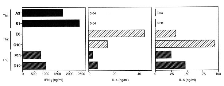
Th1, Th0, and Th2 cell lines were generated from OVA-specific T cells purified from the lymph nodes or spleen of OVA-specific TCR DO11.10 transgenic mice. CD4+ T cells were stimulated by OVA in the presence of rIL-4 and anti–IL-12 MAB to generate Th2 cell lines, whereas Th0 lines were generated by addition of rIL-4 and IFN-γ, but not anti–IL-12 MAB, to the culture. Th1 lines were generated by culturing OVA-specific T cells in the presence of anti–IL-4 MAB and rIL-12. They were maintained by weekly stimulation with irradiated APC and OVA, and Th1 lines required periodic addition of rIL-2. Figure 1 shows the cytokine content of supernatants from each Th cell line, as analyzed by ELISA for IL-4, IL-5, and IFN-γ. The Th1 cell lines produced high levels of IFN-γ but no detectable IL-4. In contrast, the Th2 cell lines produced high levels of IL-4 but no detectable IFN-γ. The Th0 cell lines produced both IFN-γ and IL-4. The quantity of IL-4 produced by the Th0 lines was significant, although less than that of Th2 lines. The Th1, Th2, and Th0 lines were used after at least three cycles of stimulation, after the T cell lines achieved a stable committed phenotype (23).
Figure 1.
Cytokine profiles of OVA-specific Th cell lines used in these studies. Th1, Th2, and Th0 cell lines (106 cells/ml) were stimulated with ConA (1 μg/ml) for 18 h. Supernatants were collected and analyzed by ELISA for IFN-γ, IL-4, and IL-5. IFN, interferon; IL, interleukin; OVA, ovalbumin.
In vivo transfer and detection of OVA-specific T cells to the lung.
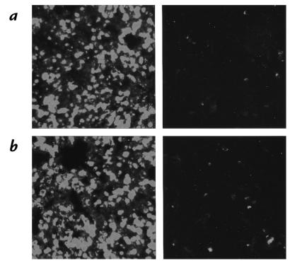
To determine the effects of Th cell lines on the development of airway hyperreactivity and airway inflammation, we adoptively transferred each cell line into histocompatible SCID mice. In these experiments, we chose SCID mice to eliminate the contribution of endogenous lymphocytes, which might inhibit the function of the transferred Th cells, and which could also contribute to airway hyperreactivity, as the recipients were exposed to OVA administered intranasally. To enhance the localization of the transferred T cell lines into the lungs of mice, we administered OVA intranasally one day before adoptive transfer of the T cell lines. One and two days later, OVA was again administered intranasally. Control mice received NaCl 0.9% instead of OVA intranasally. Four days after cell transfer, the lungs were stained with anti-TCR clonotypic antibody KJ1-26.1. Figure 2 shows that lungs from mice that received Th1 or Th2 cells and intranasal OVA stained brightly with the anti-TCR antibody, indicating that the OVA-specific T cell lines migrated to the lungs of these mice. In contrast, lungs from mice that received Th1 or Th2 cells, but no intranasal OVA, had no staining (data not shown), indicating that migration of the OVA-specific T cell lines was antigen dependent.
Figure 2.
OVA-TCR transgenic Th cells migrate to the lungs of recipient mice. Four days after adoptive transfer of OVA-specific Th cell lines (2.5 × 10⁶ cells per mouse), mice were sacrificed and lung tissue was embedded in OCT compound. Frozen sections were obtained and stained with biotinylated anticlonotype antibody, KJ1-26.1 or biotinylated control antibody JES-312G8 and Streptavidin-FITC. (a) Lung tissue from recipient of Th1 cells. Left: Lung section stained with KJ1-26.1-biotin plus Streptavidin-FITC; Right: Lung section stained with biotinylated control antibody plus Streptavidin-FITC. (b) Lung tissue from recipient of Th2 cells. Left: Lung section stained with KJ1-26.1-biotin plus Streptavidin-FITC; Right: Lung section stained with biotinylated control antibody plus Streptavidin-FITC. TCR, T-cell receptor.
Airway histology in recipients of Th1, Th2, and Th0 cell lines.
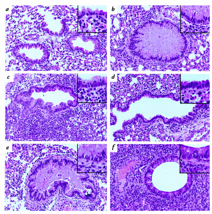
Mice were sacrificed four days after T-cell transfer. Lungs were removed and fixed, and lung sections were stained with hematoxylin and eosin to reveal lung histology. SCID mice that received OVA but no T cells had normal lung histology (Fig. 3a). Adoptive transfer of OVA-specific Th2 cells induced significant bronchiolar mucus production and induced peribronchiolar and perivascular infiltrates, consisting of lymphocytes, eosinophils, and some neutrophils (Fig. 3b). These results were similar to those reported in previous studies with Th2 cells (24). Transfer of OVA-specific Th1 cells also resulted in significant airway inflammation, with peribronchiolar and perivascular infiltrates (Fig. 3c). Th1 cells induced much less mucus production than did Th2 cells, and far more lymphocytes were present in the lungs when Th1 cells rather than Th2 cells were transferred. The inflammatory reaction with Th1 cells resembled that of moderate to severe lung allograft rejection, with dense perivascular and interstitial accumulations of small lymphocytes and immunoblasts with neutrophils and occasional eosinophils (25, 26). The inflammatory response with Th1 cells depended on the presence of OVA, as mice that received Th1 cells, but not OVA, had minimal lung disease (Fig. 3d). Furthermore, this inflammatory response with Th1 cells plus intranasal OVA was confined only to the lungs and was not observed in any other organ system.
Figure 3.
Histologic examination of lungs from SCID mice receiving Th1, Th2, and Th0 cells. (a) Lung tissue from control SCID mouse that received intranasal OVA but no Th cells. H&E, ×250. Inset: High-power magnification of normal bronchiolar epithelium. H&E, ×400. (b) Lung tissue from SCID mouse that received Th2 cells and intranasal OVA. Peribronchiolar mononuclear cell infiltrates are noted. The airway lumen is filled and expanded by thick mucus. H&E, ×250. Inset: High-power magnification of the airway epithelium showing tall columnar cells exhibiting abundant cytoplasmic mucin and a collarette of inflammatory cells. H&E, ×400. (c) Lung tissue from SCID mouse that received Th1 cells and intranasal OVA. Dense peribronchiolar inflammatory infiltrates are seen. The airway lumen does not contain mucus plugs. H&E, ×250. Inset: Lymphocytes are penetrating the airway epithelium and surrounding tissue spaces. H&E, ×400. (d) Lung tissue from control SCID mouse that received Th1 cells but not intranasal OVA. The bronchiole is normal with rare mononuclear cells in the peribronchiolar tissue; H&E ×250. Inset: The airway epithelium is normal. H&E, ×400. (e) Lung tissue from SCID mouse that received Th0 cells and intranasal OVA. Peribronchiolar infiltrates are noted, and the lumen is filled with mucus; scattered inflammatory cells are noted. H&E, ×250. Inset: The airway epithelium resembles that of mice that received OVA-specific Th2 cells, with the presence of tall columnar cells with abundant cytoplasmic mucin (b). (f) Lung tissue from SCID mouse that received both Th1 and Th2 cells and intranasal OVA. Significant airway inflammation is noted, without airway mucus. H&E, ×250. Inset: The epithelium displays reactive-appearing columnar cells, with inflammatory cells at the bases. H&E, ×400. H&E, hematoxylin and eosin; SCID, severe combined immunodeficiency.
Adoptive transfer of Th0 cell lines produced results similar to that observed with Th2 cell lines and induced significant airway inflammation (Fig. 3e). The production of IFN-γ by Th0 cells did not limit the inflammatory response, as large numbers of lymphocytes, neutrophils, and some eosinophils were present in the perivascular and peribronchiolar areas of Th0 cell recipients. Transfer of both Th1 and Th2 cells together did not result in reduction of airway inflammation, although the amount of bronchiolar mucus was reduced (Fig. 3f). This indicated that OVA-specific Th1 cells were not effective in counterbalancing Th2-induced airway inflammation.
Th2 cell–induced eosinophilia is significantly reduced by Th1 cells.
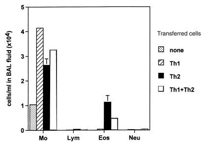
The histopathologic analysis was extended by examination of the cell types and numbers in BAL fluid, which was harvested four days after adoptive T-cell transfer into SCID mice. The total number of cells recovered in the BAL for Th1-, Th2-, or Th1 plus Th2–treated mice was 4.2, 3.8, and 3.8 × 10⁴/ml, respectively (Fig. 4). Macrophages were the predominant cell type in all groups of mice. Transfer of Th2 cells, however, significantly increased the proportion of eosinophils to 29.4% of total BAL cells (1.13 × 10⁴ cells/ml), compared with 0.94% of total BAL cells (0.04 × 10⁴ cells/ml) with Th1 cells. Transfer of a mixture of Th1 and Th2 cells in a ratio of 1:1 significantly decreased the number of eosinophils seen after transfer of Th2 cells from 29% to 12% of total BAL cells (0.47 × 10⁴ cells/ml), suggesting that Th1 cells were able to reduce Th2 cell–induced airway eosinophilia. All other cell types together did not reach more than 2% in any group, and the proportions of these cell types did not differ significantly between the groups.
Figure 4.
OVA-specific Th1 cells significantly reduce the number of eosinophils induced by OVA-specific Th2 cells in BAL fluid of OVA-treated SCID mice. Four days after transfer of Th1 (2.5 × 10⁶ cells per mouse), Th2 (2.5 × 10⁶ cells per mouse), or Th1 plus Th2 (2.5 × 10⁶ Th1 + 2.5 × 10⁶ Th2 cells per mouse) cells in OVA-treated SCID mice, BAL was performed with three aliquots of 0.4 ml PBS per mouse (n = 6 for each group). The relative number of different types of leukocytes (lung cell differentials) was determined from Hansel Stain slide preparations of BAL fluid. The data are expressed as mean ± SEM of the percentage of each cell type derived from differentials based on 200 cells. Results are given as cells per milliliter in BAL fluid. BAL, bronchoalveolar lavage; Eos, eosinophils; Lym, lymphocytes; Mo, macrophages; Neu, neutrophils.
Th1 cells do not induce airway hyperreactivity.
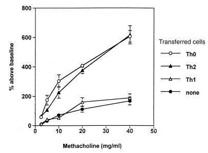
Three days after intravenous transfer of different T cell lines, we examined the recipients of Th1, Th2, and Th0 lines for airway hyperreactivity by challenging OVA-treated SCID mice with increasing concentrations of methacholine in a whole-body plethysmograph. Figure 5 shows that, as expected, SCID mice that had received Th2 lines developed significant airway hyperreactivity. Control SCID mice that received NaCl 0.9% rather than T cells intravenously and were challenged with intranasal OVA, or SCID mice that received Th2 cells intravenously but not intranasal OVA, did not develop airway hyperreactivity, indicating that this response depended on the presence of Th2 cells and on the presence of airway antigen. Recipients of Th0 cell lines developed significant airway hyperreactivity that was not statistically different from that induced with Th2 cells, suggesting that the production of IFN-γ by the T cells was not effective in reducing airway hyperreactivity. Transfer of Th1 lines, although inducing significant airway inflammation, did not induce airway hyperreactivity to methacholine. This indicated that airway hyperreactivity depends on factors produced by Th2-polarized cells and cannot be induced with inflammation-causing Th1-polarized cells.
Figure 5.
Th2 and Th0 cells, but not Th1 cells, increase airway hyperreactivity. SCID mice received Th1, Th2, or Th0 cells (2.5 × 10⁶ cells per mouse) intravenously plus intranasal OVA. Control mice received either OVA only or cells only. Three days after adoptive cell transfer, airway hyperreactivity in response to increasing concentrations of inhaled methacholine was measured in a whole-body plethysmograph. Data are expressed as percent above baseline (mean ± SEM); n ≥ 7 for each data point. Cell transfer without intranasal administration of OVA had no effect on airway hyperreactivity (data not shown).
Th1 cells do not counterbalance airway hyperreactivity induced by Th2 cells.
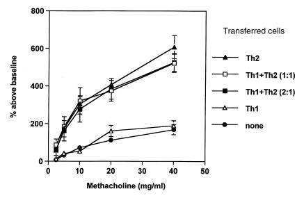
We next directly examined the capacity of Th1 cells to counterbalance airway hyperreactivity induced by Th2 cells by transferring a mixture of Th1 cells and Th2 cells into SCID mice. Figure 6 shows that although recipients of Th2 cells (2.5 × 10⁶ cells per mouse) expressed significant airway hyperreactivity, recipients of Th1 cells plus Th2 cells in a ratio of 1:1 (2.5 × 10⁶ cells each, Th1 + Th2 [1:1]) expressed equally strong airway hyperreactivity on methacholine challenge. Even on transfer of twice as many Th1 as Th2 cells (5 × 10⁶ Th1 cells + 2.5 × 10⁶ Th2 cells, Th1 + Th2 [2:1]), Th1 cells did not limit Th2 cell–induced airway hyperreactivity. These results indicate that antigen-specific Th1 cells do not inhibit airway hyperreactivity induced by Th2 cells even when given in a 2:1 excess.
Figure 6.
Th1 cells do not counterbalance airway hyperreactivity induced by Th2 cells. SCID mice received Th1 (2.5 × 10⁶ cells per mouse) or Th2 (2.5 × 10⁶ cells per mouse) cells intravenously plus intranasal OVA. Other SCID mice received a mixture of Th1 and Th2 cells in a ratio of 1:1 (2.5 × 10⁶ cells each) or 2:1 (5 × 10⁶ Th1 cells plus 2.5 × 10⁶ Th2 cells) (n ≥ 5 for each data point). Airway hyperreactivity in response to inhaled methacholine was measured in a whole-body plethysmograph. Results are demonstrated as percent above baseline (mean ± SEM).
Th1 cells do not reduce airway hyperreactivity in OVA-immunized BALB/c mice.
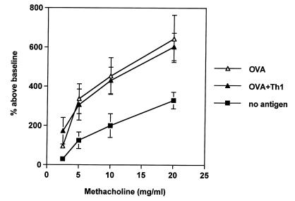
To determine whether Th1 cells could inhibit the development of Th2 cells in normal mice rather than reversing the function of established Th2 effector cells, we examined the capacity of adoptively transferred Th1 cell lines to prevent the development of allergen-induced airway hyperreactivity in normal BALB/c mice. Figure 7 shows that immunization of control BALB/c mice with OVA intraperitoneally and intranasally resulted in the development of significant airway hyperreactivity. Adoptive transfer of Th1 cells during the four-week sensitization phase on days 14 and 25 did not reduce this airway hyperreactivity, although Th1 cells migrated to the lungs as proved by fluorescence staining of lung sections with anti-TCR clonotypic antibody KJ1-26.1 one day after airway measurement (data not shown). These results indicated that Th1 cells were not able to inhibit the development of a Th2 response in sensitized immunocompetent BALB/c mice.
Figure 7.
OVA-specific Th1 cells do not reduce airway hyperreactivity in OVA-immunized BALB/c mice. BALB/c mice were immunized with OVA (50 μg) in alum intraperitoneally on days 0 and 14, and intranasally (50 μg OVA in 50 μl PBS) on days 14, 25, 26, and 27 (OVA; n = 6). A second group of mice also received OVA-specific Th1 cells intravenously on days 14 and 25 (2.5 × 10⁶ cells per mouse at each time point) (OVA + Th1; n = 5). Control mice received alum intraperitoneally and PBS intranasally (no antigen; n = 6). Airway hyperreactivity to methacholine was determined as in Figs. 5 and 6. Results are expressed as mean ± SEM. Transfer of Th1 cells without administration of antigen did not have any effect on airway hyperreactivity (data not shown).
Airway eosinophilia in OVA-immunized BALB/c mice is significantly reduced by transfer of Th1 cells.
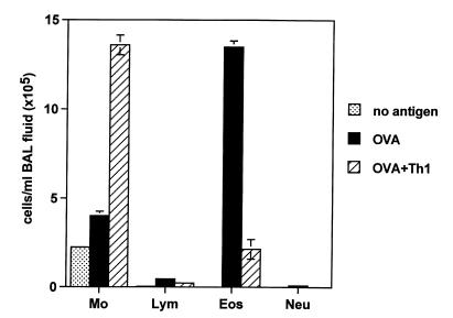
Although Th1 cells were unable to reduce airway hyperreactivity in OVA-immunized mice, Th1 cells did significantly reduce airway eosinophilia (Fig. 8), indicating that the Th1 cells functioned in vivo. Th1 cells transferred during the sensitization phase reduced the number of BAL eosinophils by more than fivefold (from 13.5 × 10⁵ to 2.3 × 10⁵ eosinophils/ml), although the total number of cells in BAL fluid from OVA-sensitized BALB/c mice was not significantly reduced (18.1 × 10⁵ cells/ml in BAL from control mice vs. 15.9 × 10⁵ cells/ml in BAL from Th1-cell recipients). Lymphocytes and neutrophils were relatively rare in BAL fluid and did not differ significantly between the groups. However, histologic analysis revealed that the inflammatory response was not reduced by transfer of Th1 cells. Hematoxylin and eosin–stained lung sections of BALB/c mice after OVA immunization and Th1 cell transfer showed dense perivascular and peribronchiolar infiltrates containing numerous lymphocytes and eosinophils (data not shown). This pattern was comparable to the histology of the OVA-immunized control group.
Figure 8.
OVA-specific Th1 cells significantly reduce the number of eosinophils in OVA-immunized BALB/c mice. BALB/c mice were immunized with OVA (50 μg) in alum intraperitoneally on days 0 and 14 and intranasally (50 μg OVA in 50 μl PBS) on days 14, 25, 26, and 27 (OVA; n = 6). A second group of mice was immunized with OVA and additionally received OVA-specific Th1 cells intravenously on days 14 and 25 (2.5 × 10⁶ cells per mouse at each time point) (OVA + Th1; n = 5). Control mice received alum intraperitoneally and PBS intranasally (no antigen; n = 6). BAL was performed on day 29 with three aliquots of 0.4 ml PBS per mouse, and the relative number of different types of leukocytes (lung cell differentials) was determined. The data are expressed as mean ± SEM of the percentage of each cell type derived from differentials based on 200 cells.
Discussion
The major goal of this study was to examine directly the capacity of Th1 cells to counterbalance the proasthmatic effects of Th2 cells in a murine model of asthma. The Th1/Th2 paradigm suggests that Th1 and Th2 cells counterbalance each other and that Th1 cells protect or prevent Th2-mediated allergic disease and asthma (27). However, using well-defined, phenotypically committed OVA-specific Th1 and Th2 cells expressing identical TCRs and adoptively transferred into either SCID mice or into OVA-immunized BALB/c mice, we found that OVA-specific Th1 cells failed to reverse Th2-mediated airway inflammation and airway hyperreactivity, even when given in twofold excess. These studies indicate that the Th1/Th2 paradigm, which predicts that Th1 cells downregulate allergic disease and asthma, may be more complex than initially appreciated and that suppression of allergic inflammation and Th2 activity in vivo may depend on cells other than Th1 lymphocytes.
Inasmuch as asthma is associated with the presence of lymphocytes producing Th2 cytokines (28), it is not surprising that OVA-specific Th2 cells induced severe allergic inflammatory responses and airway hyperreactivity when adoptively transferred into normal (24, 29) or SCID recipients (Figs. 3 and 5). We also demonstrated, however, that antigen-specific Th0 cells caused severe airway inflammation and hyperreactivity equal to those observed with Th2 cells. This indicated that production of IFN-γ, a cytokine thought to inhibit the development of allergy and asthma (30, 31), by Th0 cells was insufficient to neutralize the effects of IL-4 and IL-5. Our studies differed from previous studies with Th2 cells (24, 29) in that we used SCID mice as recipients to eliminate the effects of host cells that could inhibit the function of the transferred Th cells or that could contribute to airway hyperreactivity on immunization with antigen. These results with SCID mice demonstrated that airway hyperreactivity can be induced by Th2 or Th0 cells in the absence of other T cells, B cells, IgE, or γδ T cells. In contrast, previous studies with B cell–deficient mice (32) and γδ T cell–deficient mice (33) suggested that B cells, IgE, and γδ T cells were required for the induction of airway hyperreactivity or allergic airway inflammation. It is possible, however, that B cells or γδ T cells are required for the development of Th2 effector cells from naive precursor cells but that once Th2 cells have become activated, such “accessory cells” are not essential for expression of airway inflammation and hyperreactivity.
Our results demonstrating the inability of Th1 cells to neutralize the effects of Th2 cells are surprising in view of numerous studies that suggested that allergen-specific Th1 cells might suppress allergic inflammation. For example, we and others have shown that conventional allergen immunotherapy, which improves symptoms in allergic and asthmatic patients, reduces IL-4 production (9) and increases IFN-γ production in an allergen-specific fashion (10, 11). Immunization with IL-12–modified allergen (34), with heat-killed Listeria monocytogenes as adjuvant (35), with intratracheal IL-12, or with naked DNA plasmids containing cDNA for allergens (36, 37) also resulted in a switch in cytokine production in allergen-specific CD4+ T cells and caused a reduction in allergen-induced airway hyperreactivity (38). These observations suggest that allergen-specific Th1-polarized responses suppress allergic inflammation, although no previous studies have examined the capacity of Th1 cells to inhibit Th2-induced airway hyperreactivity directly.
On the other hand, there is increasing evidence that Th1 and Th2 cells may not always antagonize each other. First, IFN-γ has been identified in BAL fluid and serum of asthmatic patients, suggesting that Th1-like cells may in fact contribute to, rather than inhibit, the pathology in asthma (39, 40). In addition, Th1 cells are not found in large numbers in the lungs or mucus membranes of nonallergic or nonasthmatic individuals after allergen exposure, as would be predicted if Th1 cells reduced airway inflammation. Moreover, in a murine model of autoimmunity, myelin basic protein–specific Th2 cells, when transferred into immunodeficient mice, exacerbated rather than prevented experimental autoimmune encephalomyelitis (14). Similarly, attempts to use autoantigen-specific Th2 cells to confer protection against Th1-induced diabetes mellitus unexpectedly produced intense and generalized pancreatitis and subsequent diabetes mellitus (15). These studies together indicate that Th1 and Th2 cells may not always balance each other's function in a dichotomous paradigm, but rather may in some instances be unexpectedly harmful, not only in autoimmune disease but also in allergic disease and asthma.
We found that antigen-specific Th1 cells when adoptively transferred to SCID mice induced considerable airway inflammation, resembling severe delayed-type hypersensitivity reactions, lung allograft rejection (25), or hypersensitivity pneumonitis (41). The inflammatory response caused by Th1 cells depended on the presence of antigen and occurred only in the lungs of the recipient mice. Surprisingly, even though Th1 cells induced severe airway inflammation, they did not cause airway hyperreactivity. These studies indicate that inflammation in and of itself is not important for the development of airway hyperreactivity, although CD4+ T cells appear to be essential for the development of airway hyperreactivity (42). The precise factors produced by CD4+ T cells that are important for the development of airway hyperreactivity, however, have not yet been identified, but they appear not to be IL-4 and IL-5 (42) nor those produced by Th1 cells (Fig. 5). Further studies are necessary to determine whether other factors produced by Th2 cells, such as IL-9 (43) or IL-13 (44), are essential for the induction of airway hyperreactivity.
Although we found that Th1 cells could not reverse Th2-induced airway hyperreactivity in SCID or in OVA-immunized BALB/c mice, Th1 cells were able to reduce the number of airway eosinophils and reduce intrabronchiolar mucus production induced by Th2 cells. Similarly, we reported previously (45) that adoptively transferred Th1 clones could inhibit the capacity of Th2 clones to induce antigen-specific IgE synthesis. This indicated not only that the adoptively transferred Th1 cells functioned in vivo after transfer and could modify some, although not all, functions of Th2 effector cells, but also that the presence of eosinophils may not be essential for the development of airway hyperreactivity, as has been suggested previously (46). The resistance of Th2 cell function to modification by Th1 cells may be due in part to the fact that Th2 effector cells are terminally differentiated with fixed cytokine profiles (23) and cease to express IL-12 and IL-18 receptors (47, 48).
It is possible that Th1 cells may in fact be beneficial in asthma if they are present early on in high numbers before activated effector Th2 cells are present. In such instances, while being ineffective in reversing the function of effector Th2 cells, Th1 cells may be capable of blocking the development of Th2 cells from naive T cells and of preventing the induction of airway hyperreactivity. Alternatively, other cell types — e.g., transforming growth factor-β–producing CD4+ antigen-specific Th3 cells (49), Tr1 cells (50), γδ cells (51), or CD8+ cells (52, 53) — may be more important than Th1 cells in downregulating allergic inflammation and hyperreactivity. Further studies are required to determine how Th1 cells or other cell types regulate allergic inflammation and airway hyperreactivity. Such studies are particularly important as prerequisites for the development of therapies focused on immunomodulation of deleterious Th2-polarized responses.
In summary, using well-defined polarized Th cells, we showed that antigen-specific Th1 cells were ineffective in reducing airway hyperreactivity induced by Th2 cells and caused serious airway inflammation but not airway hyperreactivity. In contrast, Th2 and Th0 cells induced considerable airway hyperreactivity, in the absence of B cells, IgE, or other lymphocytes. Our results raise concerns regarding the protective effects of Th1 cells in allergy and asthma and regarding the feasibility of Th1-based therapies for these clinical problems.
Acknowledgments
We thank G. Trinchieri for the anti–IL-12 MAB; J. Ohara and B. Paul for MAB 11B11; M. Howard for MABs BVD4, BVD6, SXC.1, and 2A5; J. Kappler and P. Marrack for anti-clonotype antibody KJ1-26.1; T. Mosmann for MABs S4B6 and XMG1.2; S. Levy (Stanford University) for MAB 4G10; and D.B. Lewis (Stanford University) for assistance in measuring airway hyperreactivity in mice. This work was supported by grants RO1AI24571 and RO1AI26322 from the US Public Health Service/National Institutes of Health. G. Hansen is a Fellow of the Deutsche Forschungsgemeinschaft (Ha2799/1-1).
References
- 1.Gergen PJ, Weiss KB. Changing patterns of asthma hospitalization among children: 1979 to 1987. JAMA. 1990;264:1688–1692. [PubMed] [Google Scholar]
- 2.Kirby JG, Hargreave FE, Gleich GJ, O’Byrne PM. Bronchoalveolar cell profiles of asthmatic and non-asthmatic subjects. Am Rev Respir Dis. 1987;136:379–383. doi: 10.1164/ajrccm/136.2.379. [DOI] [PubMed] [Google Scholar]
- 3.Bradley LB, et al. Eosinophils, T-lymphocytes, mast cells, neutrophils and macrophages in bronchial biopsy specimens from atopic subjects with asthma: comparison with biopsy specimens from atopic subjects without asthma and normal control subjects and relationship to bronchial hyperresponsiveness. J Allergy Clin Immunol. 1991;88:661–674. doi: 10.1016/0091-6749(91)90160-p. [DOI] [PubMed] [Google Scholar]
- 4.Umetsu DT, DeKruyff RH. Th1 and Th2 CD4+ cells in human allergic diseases. J Allergy Clin Immunol. 1997;100:1–6. doi: 10.1016/s0091-6749(97)70186-6. [DOI] [PubMed] [Google Scholar]
- 5.Martinez FD, et al. Asthma and wheezing in the first six years of life. N Engl J Med. 1995;332:133–138. doi: 10.1056/NEJM199501193320301. [DOI] [PubMed] [Google Scholar]
- 6.Abbas AK, Murphy KM, Sher A. Functional diversity of helper T lymphocytes. Nature. 1996;383:787–793. doi: 10.1038/383787a0. [DOI] [PubMed] [Google Scholar]
- 7.Coffman RL, et al. The role of helper T cell products in mouse B cell differentiation and isotype regulation. Immunol Rev. 1988;102:5–28. doi: 10.1111/j.1600-065x.1988.tb00739.x. [DOI] [PubMed] [Google Scholar]
- 8.Wierenga EA, et al. Human atopen-specific types 1 and 2 T helper cell clones. J Immunol. 1991;147:2942–2949. [PubMed] [Google Scholar]
- 9.Secrist H, Chelen CJ, Wen Y, Marshall JD, Umetsu DT. Allergen immunotherapy decreases interleukin 4 production in CD4+ T cells from allergic individuals. J Exp Med. 1993;178:2123–2130. doi: 10.1084/jem.178.6.2123. [DOI] [PMC free article] [PubMed] [Google Scholar]
- 10.Jutel M, et al. Bee venom immunotherapy results in decrease of IL-4 and IL-5 and increase of IFN-gamma secretion in specific allergen-stimulated T cell cultures. J Immunol. 1995;154:4187–4194. [PubMed] [Google Scholar]
- 11.Varney VA, et al. Influence of grass pollen immunotherapy on cellular infiltration and cytokine mRNA expression during allergen-induced late-phase cutaneous responses. J Clin Invest. 1993;92:644–651. doi: 10.1172/JCI116633. [DOI] [PMC free article] [PubMed] [Google Scholar]
- 12.Oro AS, Guarino TJ, Driver R, Steinman L, Umetsu DT. Regulation of disease susceptibility: decreased prevalence of IgE-mediated allergic disease in patients with multiple sclerosis. J Allergy Clin Immunol. 1996;97:1402–1408. doi: 10.1016/s0091-6749(96)70210-5. [DOI] [PubMed] [Google Scholar]
- 13.Shirakawa T, Enomoto T, Shimazu S, Hopkin JM. The inverse association between tuberculin responses and atopic disorder. Science. 1997;275:77–79. doi: 10.1126/science.275.5296.77. [DOI] [PubMed] [Google Scholar]
- 14.Lafaille JJ, et al. Myelin basic protein-specific T helper 2 (Th2) cells cause experimental autoimmune encephalomyelitis in immunodeficient hosts rather than protect them from the disease. J Exp Med. 1997;186:307–312. doi: 10.1084/jem.186.2.307. [DOI] [PMC free article] [PubMed] [Google Scholar]
- 15.Pakala SV, Kurrer MO, Katz JD. T helper 2 cells induce acute pancreatitis and diabetes in immune-compromised nonobese diabetic mice. J Exp Med. 1997;186:299–306. doi: 10.1084/jem.186.2.299. [DOI] [PMC free article] [PubMed] [Google Scholar]
- 16.Genain CP, et al. Late complications of immune deviation therapy in a nonhuman primate. Science. 1996;274:2054–2056. doi: 10.1126/science.274.5295.2054. [DOI] [PubMed] [Google Scholar]
- 17.Bosma GC, Custer RP, Bosma MJ. A severe combined immunodeficiency mutation in the mouse. Nature. 1983;301:527–530. doi: 10.1038/301527a0. [DOI] [PubMed] [Google Scholar]
- 18.Murphy KM, Heimberger AB, Loh DY. Induction by antigen of intrathymic apoptosis of CD4+CD8+TCRlo thymocytes in vivo. Science. 1990;250:1720–1723. doi: 10.1126/science.2125367. [DOI] [PubMed] [Google Scholar]
- 19.Marrack P, Shimonkevitz R, Hannum C, Haskins K, Kappler J. The major histocompatibility complex-restricted antigen receptor on T cells. IV. An antiidiotypic antibody predicts both antigen and I-specificity. J Exp Med. 1983;158:1635–1646. doi: 10.1084/jem.158.5.1635. [DOI] [PMC free article] [PubMed] [Google Scholar]
- 20.Tsuyuki S, Stuyuki J, Einsle K, Kopf M, Coyle AJ. Costimulation through B7-2 (CD86) is required for the induction of a lung mucosal Th2 immune response and altered airway responsiveness. J Exp Med. 1997;185:1671–1679. doi: 10.1084/jem.185.9.1671. [DOI] [PMC free article] [PubMed] [Google Scholar]
- 21.Hamelmann E, et al. Noninvasive measurement of airway responsiveness in allergic mice using barometric plethysmography. Am J Respir Crit Care Med. 1997;156:766–775. doi: 10.1164/ajrccm.156.3.9606031. [DOI] [PubMed] [Google Scholar]
- 22.Macaulay AE, DeKruyff RH, Umetsu DT. Antigen-primed T cells from B cell-deficient JHD mice fail to provide B cell help. J Immunol. 1998;160:1694–1700. [PubMed] [Google Scholar]
- 23.Murphy E, et al. Reversibility of T helper 1 and 2 populations is lost after long-term stimulation. J Exp Med. 1996;183:901–913. doi: 10.1084/jem.183.3.901. [DOI] [PMC free article] [PubMed] [Google Scholar]
- 24.Cohn L, Homer RJ, Marinov A, Rankin J, Bottomly K. Induction of airway mucus production By T helper 2 (Th2) cells: a critical role for interleukin 4 in cell recruitment but not mucus production. J Exp Med. 1997;186:1737–1747. doi: 10.1084/jem.186.10.1737. [DOI] [PMC free article] [PubMed] [Google Scholar]
- 25.Berry, G.J., and Billingham, M.E. 1995. The pathology of combined heart-lung and lung transplantation. In Thoracic transplantation. S.J. Shumway, and N.E. Shumway, editors. Blackwell Science Inc. Cambridge, United Kingdom. 331–347.
- 26.Ohori, N.P., and Yousem, S.A. 1996. Pathology of lung transplantation. In Solid organ transplant rejection: mechanism, pathology and diagnosis. K. Solez, L.C. Racusen, and M.E. Billingham, editors. Marcel Dekker Inc. New York, NY. 161–185
- 27.Romagnani S. Lymphokine production by human T cells in disease states. Ann Rev Immunol. 1994;12:227–257. doi: 10.1146/annurev.iy.12.040194.001303. [DOI] [PubMed] [Google Scholar]
- 28.Robinson DS, et al. Predominant Th2-like bronchoalveolar T-lymphocyte population in atopic asthma. N Engl J Med. 1992;326:298–304. doi: 10.1056/NEJM199201303260504. [DOI] [PubMed] [Google Scholar]
- 29.Li XM, Schofield BH, Wang QF, Kim KH, Huang SK. Induction of pulmonary allergic responses by antigen-specific Th2 cells. J Immunol. 1998;160:1378–1384. [PubMed] [Google Scholar]
- 30.Tang ML, Kemp AS, Thorburn J, Hill DJ. Reduced interferon-gamma secretion in neonates and subsequent atopy. Lancet. 1994;344:983–985. doi: 10.1016/s0140-6736(94)91641-1. [DOI] [PubMed] [Google Scholar]
- 31.Boguniewicz M, et al. The effects of nebulized recombinant interferon-gamma in asthmatic airways. J Allergy Clin Immunol. 1995;95:133–135. doi: 10.1016/s0091-6749(95)70162-1. [DOI] [PubMed] [Google Scholar]
- 32.Hamelmann E, et al. Allergic airway sensitization induces T cell activation but not airway hyperresponsiveness in B cell-deficient mice. Proc Natl Acad Sci USA. 1997;94:1350–1355. doi: 10.1073/pnas.94.4.1350. [DOI] [PMC free article] [PubMed] [Google Scholar]
- 33.Zuany-Amorim C, et al. Requirement for gd T cells in allergic airway inflammation. Science. 1998;280:1265–1267. doi: 10.1126/science.280.5367.1265. [DOI] [PubMed] [Google Scholar]
- 34.Kim TS, et al. An OVA-IL-12 fusion protein is more effective than OVA plus rIL-12 in inducing a Th1-dominated immune response and inhibiting antigen-specific IgE production. J Immunol. 1997;158:4137–4144. [PubMed] [Google Scholar]
- 35.Yeung VP, Gieni RS, Umetsu DT, DeKruyff RH. Heat killed Listeria monocytogenes as an adjuvant converts established Th2-dominated immune responses into Th1-dominated responses. J Immunol. 1998;161:4146–4152. [PubMed] [Google Scholar]
- 36.Hsu CH, et al. Immunoprophylaxis of allergen-induced immunoglobulin E synthesis and airway hyperresponsiveness in vivo by genetic immunization. Nat Med. 1996;2:540–544. doi: 10.1038/nm0596-540. [DOI] [PubMed] [Google Scholar]
- 37.Raz E, et al. Preferential induction of a Th1 immune response and inhibition of specific IgE antibody formation by plasmid DNA immunization. Proc Natl Acad Sci USA. 1996;93:5141–5145. doi: 10.1073/pnas.93.10.5141. [DOI] [PMC free article] [PubMed] [Google Scholar]
- 38.Gavett SH, et al. Interleukin 12 inhibits antigen-induced airway hyperresponsiveness, inflammation, and Th2 cytokine expression in mice. J Exp Med. 1995;182:1527–1536. doi: 10.1084/jem.182.5.1527. [DOI] [PMC free article] [PubMed] [Google Scholar]
- 39.Cembrzynska-Nowak M, Szklarz E, Inglot AD, Teodorczyk-Injeyan JA. Elevated release of tumor necrosis factor-alpha and interferon-gamma by bronchoalveolar leukocytes from patients with bronchial asthma. Am Rev Respir Dis. 1993;147:291–295. doi: 10.1164/ajrccm/147.2.291. [DOI] [PubMed] [Google Scholar]
- 40.Hessel EM, et al. Development of airway hyperresponsiveness is dependent on interferon-gamma and independent of eosinophil infiltration. Am J Respir Cell Mol Biol. 1997;16:325–334. doi: 10.1165/ajrcmb.16.3.9070618. [DOI] [PubMed] [Google Scholar]
- 41.Schuyler M, Gott K, Cherne A, Edwards B. Th1 CD4+ cells adoptively transfer experimental hypersensitivity pneumonitis. Cell Immunol. 1997;169:169–175. doi: 10.1006/cimm.1997.1107. [DOI] [PubMed] [Google Scholar]
- 42.Hogan SP, et al. A novel T cell–regulated mechanism modulating allergen-induced airways hyperreactivity in BALB/c mice independently of IL-4 and IL-5. J Immunol. 1998;161:1501–1509. [PubMed] [Google Scholar]
- 43.Temann U-A, Geba GP, Rankin JA, Flavell RA. Expression of Interleukin 9 in the lungs of transgenic mice causes airway inflammation, mast cell hyperplasia, and bronchial hyperresponsiveness. J Exp Med. 1998;188:1307–1320. doi: 10.1084/jem.188.7.1307. [DOI] [PMC free article] [PubMed] [Google Scholar]
- 44.Urban JF, Jr, et al. IL-13, IL-4Ralpha, and Stat6 are required for the expulsion of the gastrointestinal nematode parasite Nippostrongylus brasiliensis. Immunity. 1998;8:255–264. doi: 10.1016/s1074-7613(00)80477-x. [DOI] [PubMed] [Google Scholar]
- 45.Rizzo LV, DeKruyff RH, Umetsu DT, Capsi RR. Regulation of the interaction between Th1 and Th2 T cell clones to provide help for antibody production in vivo. Eur J Immunol. 1995;25:708–716. doi: 10.1002/eji.1830250312. [DOI] [PubMed] [Google Scholar]
- 46.Corry DB, et al. Interleukin 4, but not interleukin 5 or eosinophils, is required in a murine model of acute airway hyperreactivity. J Exp Med. 1996;183:109–117. doi: 10.1084/jem.183.1.109. [DOI] [PMC free article] [PubMed] [Google Scholar]
- 47.Szabo SJ, Dighe AS, Gubler U, Murphy KM. Regulation of the interleukin (IL)-12R beta 2 subunit expression in developing T helper 1 (Th1) and Th2 cells. J Exp Med. 1997;185:817–824. doi: 10.1084/jem.185.5.817. [DOI] [PMC free article] [PubMed] [Google Scholar]
- 48.Xu D, et al. Selective expression and functions of interleukin 18 receptor on T helper (Th) type 1 but not Th2 cells. J Exp Med. 1998;188:1485–1492. doi: 10.1084/jem.188.8.1485. [DOI] [PMC free article] [PubMed] [Google Scholar]
- 49.Chen Y, Kuchroo VK, Inobe J, Hafler DA, Weiner HL. Regulatory T cell clones induced by oral tolerance: suppression of autoimmune encephalomyelitis. Science. 1994;265:1237–1240. doi: 10.1126/science.7520605. [DOI] [PubMed] [Google Scholar]
- 50.Groux H, et al. A CD4+ T-cell subset inhibits antigen-specific T-cell–responses and prevents colitis. Nature. 1997;389:737–742. doi: 10.1038/39614. [DOI] [PubMed] [Google Scholar]
- 51.Ferrick DA, et al. Differential production of interferon-gamma and interleukin-4 in response to Th1- and Th2-stimulating pathogens by gamma delta T cells in vivo. Nature. 1995;373:255–257. doi: 10.1038/373255a0. [DOI] [PubMed] [Google Scholar]
- 52.McMenamin C, Holt PG. The natural immune response to inhaled soluble protein antigens involves major histocompatibility complex (MHC) class I-restricted CD8+ T cell-mediated but MHC class II-restricted CD4+ T cell-dependent immune deviation resulting in selective suppression of immunoglobulin E production. J Exp Med. 1993;178:889–899. doi: 10.1084/jem.178.3.889. [DOI] [PMC free article] [PubMed] [Google Scholar]
- 53.Rocklin RE, Sheffer A, Greineder DK, Melmon KL. Generation of antigen-specific suppressor cells during allergy desensitization. N Engl J Med. 1980;302:1213–1219. doi: 10.1056/NEJM198005293022201. [DOI] [PubMed] [Google Scholar]


