Abstract
Nosema spp. fungal gut parasites are among myriad possible explanations for contemporary increased mortality of western honey bees (Apis mellifera, hereafter honey bee) in many regions of the world. Invasive Nosema ceranae is particularly worrisome because some evidence suggests it has greater virulence than its congener N. apis. N. ceranae appears to have recently switched hosts from Asian honey bees (Apis cerana) and now has a nearly global distribution in honey bees, apparently displacing N. apis. We examined parasite reproduction and effects of N. apis, N. ceranae, and mixed Nosema infections on honey bee hosts in laboratory experiments. Both infection intensity and honey bee mortality were significantly greater for N. ceranae than for N. apis or mixed infections; mixed infection resulted in mortality similar to N. apis parasitism and reduced spore intensity, possibly due to inter-specific competition. This is the first long-term laboratory study to demonstrate lethal consequences of N. apis and N. ceranae and mixed Nosema parasitism in honey bees, and suggests that differences in reproduction and intra-host competition may explain apparent heterogeneous exclusion of the historic parasite by the invasive species.
Introduction
Western honey bees (Apis mellifera, hereafter honey bees) are among the most vital and versatile pollinators, contributing to production of 39 of the world's 57 most important crops [1]. Unfortunately, today's beekeepers face significant hurdles to maintain healthy colonies that are capable of crop pollination because of dramatic honey bee colony mortalities in many regions of the world. A great deal of attention has focussed on these mortalities because humanity's reliance on pollinator-dependent crops has increased significantly in the last half century [2]. Honey bee mortality is believed to result from multiple stressors acting alone or in combination, including nutritional deficiencies, management issues, agro-chemicals, and especially introduced parasites [3]–[5].
Significant interest has recently focussed on the newly detected microsporidian gut parasite Nosema ceranae because unusually high honey bee colony mortality coincided with its apparent host-switch from Asian honey bees (Apis cerana) to honey bees [6], [7], as well as its subsequent widespread dispersal [8]–[12]. N. ceranae can cause tissue damage [13]–[15], nutritional stress [16]–[18], and suppression of host immunity [19]. In Spain, N. ceranae is typically associated with reduced colony survivorship [20], whereas in other parts of Europe [21] and in North America [22]–[25], its virulence is debated. Possible explanations for this variation include parasite or host genetics [15], [26]–[28], climate [29], [30], nutrition [18], or interactions with other stressors such as environmental contaminants or other parasites [31]–[35]. Although biological mechanisms underlying relationships among stressors of honey bees are not well understood, it is likely that exploitative competition for limited resources, as well as host stress resulting from tissue pathology and immune suppression, play important roles [14], [31], [33], and could lead to numerical (i.e., intensity) or functional (i.e., realised niche) responses by parasites that are either symmetrical (both species experience equal responses) or asymmetrical [36].
It is rare for multiple microsporidian species to be parasitic within sympatric individuals of the same insect species [37]. Nonetheless, sympatric honey bee populations, and even individuals, can be co-parasitized by both N. ceranae and Nosema apis [38], [39], the latter being the historical microsporidian species of honey bees [12], [24], [40]. Similar to N. ceranae, N. apis can cause significant tissue damage in the gut that ultimately results in increased winter colony mortality or poor build-up of surviving colonies in spring [40]. Within the last decade, N. ceranae has been detected on all continents where honey bees are maintained, while the occurrence of N. apis has diminished [10], [12], [24], [41]–[43], suggesting a numerical response by N. apis to co-infection that has resulted in decreased prevalence and distribution of the parasite. This apparent exclusion appears to be geographically heterogeneous, and is likely governed by previously discussed genetic and environmental factors influencing dispersal and competition for limited resources during density-dependent parasite regulation [15], [18], [26]–[30], [44].
Few studies have investigated host honey bee responses to both Nosema parasites simultaneously or parasite reproduction under experimental conditions. Paxton et al. [45] observed higher mortality in N. ceranae-infected worker honey bees compared to those parasitized by N. apis, and no difference in spore intensity (number of vegetative parasite cells per host) between the two species. Forsgren and Fries [46] similarly found no difference in spore intensity between N. ceranae and N. apis, but they did not observea difference in mortality between workers infected by either N. apis or N. ceranae. Furthermore, using molecular techniques they did not detect a competitive advantage during co-infection by either parasite congener. Lastly, Martín-Hernández et al. [47] reported higher mortality and increased nutritional demand by workers infected with N. ceranae compared to N. apis, whereas Huang and Solter [48] reported consistently higher spore production by N. ceranae.
Because of the conflicting results regarding differences in host mortality caused by N. ceranae and N. apis, and because the former has only recently spread from Asia to become a global concern, comparative studies focusing on these congeneric parasites are of significant interest. Here we present an experiment that compared host mortality and nutritional demand, as well as parasite reproduction (quantified by spore intensity and DNA amount) and interspecific interactions, using honey bees artificially infected by N. apis, N. ceranae, or both. Uniquely, experimental Nosema and honey bees were obtained from outside of Europe, and experimental hosts were observed for over four weeks, the typical length of time that worker honey bees spend performing intra-hive duties [49]. Previous work used European-collected parasites and hosts, and terminated experiments between days 7 and 15 post inoculation. Based on laboratory and field investigations previously discussed, we hypothesised that Nosema-infected honey bees, in particular those parasitized by N. ceranae, would exhibit greater mortality than controls. We also predicted greater N. ceranae reproduction compared to N. apis. This could help to explain apparent exclusion of N. apis by N. ceranae in many regions of the world [41].
Materials and Methods
Experimental design
Laboratory experiments consisted of four treatment groups (1. control, 2. N. apis, 3. N. ceranae, and 4. N. apis/N. ceranae (hereafter, mixed)) housed at Acadia University in Wolfville, Nova Scotia, Canada. Each treatment group had 60 Buckfast honey bee workers housed in hoarding cages (wooden frame with hardware cloth and plexiglass sides; volume = 2,652 cm3; 20 workers per cage) in a growth chamber maintained at 33°C, ∼45% relative humidity, and in complete darkness [50].
Combs of similarly aged pupae obtained from two Buckfast colonies were used to collect workers for the experiment. In the laboratory, emerging individuals were randomly assigned to one of four treatment groups, and orally inoculated with 5 µl 75% (weight/volume) sucrose solution within 48 h of emergence. Inoculum for each worker belonging to the Nosema treatments contained a total of 35,000 freshly obtained local spores of the respective parasite species, enough to ensure 100% infection [46]; the mixed inoculum contained equal parts N. apis and N. ceranae. Nosema species confirmation was performed molecularly as described below and in Burgher-MacLellan et al. [38]. Post-inoculation, workers were group fed 50% (w/v) sucrose solution ad libitum for the duration of the experiment using a 10-ml syringe with the adaptor removed. The experiment was terminated at 30 d when no living workers remained for one of the treatment groups because they had either died in the cage or had been removed to quantify Nosema infection.
Host mortality and food consumption
Mortality was recorded daily; dead individuals were removed from cages and stored at −80°C for later Nosema sp. quantification (see below). Food consumption was also measured daily to quantify nutritional demand [16] by visually recording quantities of sucrose solution depleted from syringes; per worker daily consumption was calculated by using the number of living workers at the end of each 24-h interval. Food was replaced every week to limit microbial growth and to ensure sucrose solution was provided ad libitum [51]. Comparison of food consumption among groups continued only until 25 d post inoculation, when one cage contained a single living worker.
Parasite reproduction
Nosema spores (spores per bee) and DNA were quantified on all workers that died between 28 and 30 d (n = 2, 8, 7, and 9 for control, N. apis, N. ceranae, and mixed treatments, respectively) immediately prior to experiment termination. Spores were further quantified at 7, 14, and 21 d post inoculation using three randomly chosen living workers per treatment (one per cage). All workers were stored immediately at −80°C after collection from cages until laboratory analyses.
Nosema quantification – microscopy
For each individual honey bee, suspensions were created by crushing its abdomen with a pellet pestle in 1 ml distilled water. Nosema spores were counted in these suspensions using a haemocytometer and light microscopy (Thermo Fisher Scientific, Waltham, Massachusetts, USA) [52], [53].
Nosema quantification - simplex real-time PCR
Nosema DNA (ng) was quantified using methods outlined by Burgher-MacLellan et al. [38]. This included use of primer pairs 218MITOC (N. ceranae) and 321APIS (N. apis) that were originally optimized by Martin-Hernandez et al. [54], as well as qPCR methods developed by Forsgren and Fries [46] that applied external DNA standards of serial diluted PCR amplicons. Briefly, genomic DNA was isolated from each honey bee by pre-treating a 250-µl aliquot of a crushed abdomen suspension (described in the previous section) with 10 µl proteinase K (20 mg/ml) (Sigma-Aldrich Canada, Oakville, Ontario, Canada) for 20 min at 37°C. DNA was then purified using a modified protocol (steps 1–3 omitted) from the Ultra Clean Tissue DNA Extraction Kit (Mo Bio Laboratories, Carlsbad, California, USA). DNA was quantified using a Nanodrop 1000 spectrophotometer (Fisher Scientific, Ottawa, Ontario, Canada), and samples stored at −20°C until real-time PCR was performed.
Simplex quantitative real-time PCR (qPCR) was performed using an Mx4000 thermocycler (Stratagene, La Jolla, California, USA). Each separate qPCR reaction consisted of 12.5 µl Maxima SYBR Green/Rox qPCR master mix (Thermo Scientific, Rockford, Illinois, USA), 0.2 µl N. apis or N. ceranae primer sets [54], 1 µl template (100 ng genomicDNA) and nuclease-free water to a final volume of 25 µl. For each primer pair, the PCR reactions were performed in triplicate on the same plate and contained negative and positive controls (no template DNA and DNA isolated from N. apis or N. ceranae spores). Triplicate means were reported. PCR amplification parameters included an initial 10-min denaturing period at 95°C followed by 40 cycles of 30-s denaturing at 95°C, 30-s annealing at 60°C, and 30-s extension at 72°C, and a final 5-min extension period at 72°C. Amplified products were confirmed using melting curve analysis plots where temperature profiles were 1 min at 95°C, 30 s at 55°C, followed by forty 30-s increases of 1°C, and a final holding temperature at 4°C. Each simplex qPCR run included the appropriate quantification standard curve (i.e. R2>0.98 and primer efficiency >94%) prepared using serial dilutions (1.0−1 to 1.0−7) ng of purified PCR products (N. apis and N. ceranae) for target DNA. Bee DNA samples were quantified for Nosema DNA amount by plotting cycle threshold (Ct) values against nanograms of target DNA.
Statistical analyses
All statistical analyses were performed using R 2.15.2 (R Development Core Team; Vienna, Austria), except for the survival analysis which was performed using Minitab 16 (Minitab Inc., State College, Pennsylvania, USA). Cumulative mortality was analysed using the Kaplan-Meier Log-Rank survival analysis for ‘censored’ data because time of death for some workers was not known (i.e., some living workers were killed periodically to quantify spore intensity during the experiment, and some were still living when the experiment was terminated) [55]. Food consumption and Nosema intensities were evaluated using ANOVAs or Repeated Measures ANOVAs; Tukey's HSD post hoc tests were used for multiple comparisons among treatments. Where appropriate, data were square-root transformed to improve fit to normality.
Results
Host mortality and food consumption
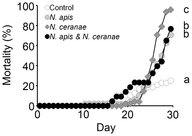
Mortality at 30 d post-inoculation was 25.0, 70.0, 95.0, and 76.7% for control, N. apis, N. ceranae, and mixed treatments, respectively (Fig. 1). Workers in the N. ceranae treatment had significantly increased mortality compared to workers from the other treatments (Kaplan-Meier Log-Rank Test, all Ps<0.002), whereas controls had significantly lower mortality compared to all other treatments (Kaplan-Meier Log-Rank, all Ps<0.001). Mortality did not differ significantly between workers in the N. apis and mixed treatments (Kaplan-Meier Log-Rank, P = 0.67) (Fig. 1).
Figure 1. Effect of Nosema infection on mortality of adult worker western honey bees (Apis mellifera).
Mortality is shown as the cumulative percentage of dead individuals from control, Nosema apis, Nosema ceranae and mixed N. apis/N. ceranae treatments each day. The experiment was terminated at 30 d post inoculation. Treatments with different letters had significant differences in mortality.
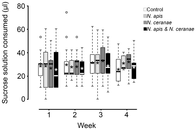
No difference was observed among treatments in food consumption (Repeated Measures ANOVA, F 3,8 = 0.4, P = 0.79) (Fig. 2).
Figure 2. Effect of Nosema infection on adult worker western honey bee (Apis mellifera) nutritional demand.
Consumption is shown as volume of 50% (weight/volume) sucrose-water mixture per bee per week post inoculation for control, Nosema apis, Nosema ceranae, and mixed N. apis/N. ceranae treatments (Week 4 included only consumption from 22–25 d post inoculation). Boxplots show interquartile range (box), median (black or white line within interquartile range), data range (dashed vertical lines), and outliers (open dots); asterisks (black or white) represent means. No significant differences were observed among treatments for daily consumption per worker.
Parasite reproduction
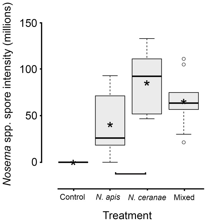
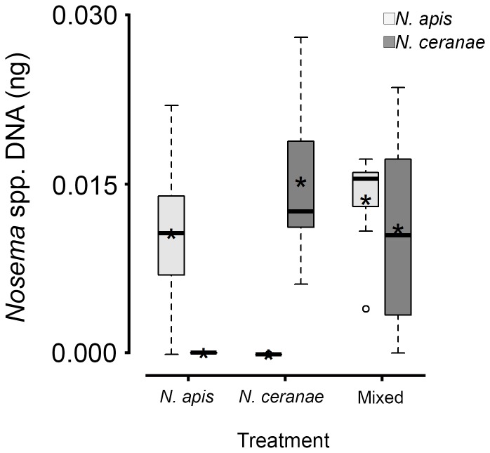
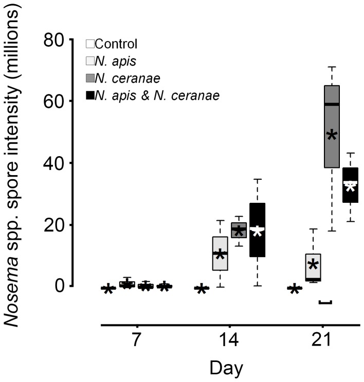
In workers that died between 28 and 30 d post inoculation, Nosema spore intensities were significantly different among groups (Fig. 3). Spore intensity in N. ceranae workers was greater than in N. apis workers (Tukey's HSD, adjusted P = 0.03), but not compared to workers from the mixed group (Tukey's HSD, adjusted P = 0.60). No difference in spore intensity was observed between workers from the N. apis and mixed treatments (Tukey's HSD, adjusted P = 0.16) (Fig. 3). Additionally, no difference in the quantity of N. apis DNA was observed between N. apis and mixed treatments (all Tukey's HSD, adjusted P≥0.50), or of N. ceranae DNA quantity between N. ceranae and mixed treatments (all Tukey's HSD, adjusted P≥0.62) (Fig. 4). Despite greater spore intensities for N. apis and N. ceranae treatments at 7 and 14 d in live sampled workers, respectively, no statistical differences were observed (both ANOVAs, F 2,6≤0.5, Ps≥0.62; Fig. 5). At 21 d, however, spore intensity was significantly greater in the N. ceranae than in the N. apis treatment (Tukey's HSD, P = 0.05).
Figure 3. Level of Nosema infection in dead adult worker western honey bees (Apis mellifera) 28–30 d post oral inoculation for control, Nosema apis, Nosema ceranae and mixed N. apis/N. ceranae treatments.
Boxplots show interquartile range (box), median (black line within interquartile range), data range (dashed vertical lines), and outliers (open dots); asterisks (black) represent means. Horizontal square parenthesis under boxplots indicates a significant difference; controls were excluded from analyses because no infections were observed.
Figure 4. Levels of Nosema apis and Nosema ceranae DNA (square root-transformed) in adult worker western honey bees (Apis mellifera) that died between 28 and 30 d post inoculation in N. apis, N. ceranae or mixed N. apis/N. ceranae treatments (same workers shown in Fig. 3 ).
Boxplots show interquartile range (box), median (black or white line within interquartile range), data range (dashed vertical lines), and outliers (open dots); asterisks (black or white) represent means. No significant differences were observed in quantities among the four instances where we expected to find DNA (i.e., the boxes with means above 0).
Figure 5. Nosema infection intensities in live-sampled adult worker western honey bees (Apis mellifera) at 7, 14, and 21 d post oral inoculation in control, Nosema apis, Nosema ceranae, and mixed N. apis/N. ceranae treatments.
Boxplots show interquartile range (box), median (black or white line within interquartile range), data range (dashed vertical lines), and outliers (open dots); asterisks (black or white) represent means. Horizontal square parenthesis under boxplots indicates a significant difference; controls were excluded from analyses because no infections were observed.
Discussion
Our experiment demonstrated that Nosema infection significantly increased honey bee worker mortality but had no influence on food consumption. Spore intensity and mortality was significantly greater for N. ceranae-infected individuals compared to those infected by N. apis. This supports claims that N. ceranae could be one stressor responsible for elevated colony losses that have been observed recently [3], [4], [20], [23], and suggests that high spore production could be a mechanism by which apparent rapid range extension of this horizontally-introduced species occurred.
To our knowledge this is the first laboratory study to follow simultaneously both N. apis and N. ceranae intensity beyond two weeks to measure effects on hosts using species from outside of Europe [45]–[47]. Length of observation is particularly important because mean honey bee worker longevity during the foraging season (when this study was performed) is between 15 and 60 d [49]. Nosema spore intensities in workers that died between 28 and 30 d post-inoculation were consistent with spore intensity data collected from live workers at 21 d post inoculation, wherein N. ceranae reproduction was significantly greater than that of N. apis. Conversely, quantity of Nosema DNA did not differ between congeners. It is likely that Nosema DNA that we detected represented immature stages within host cells rather than mature spores due to a dense wall surrounding each spore [40], [56]. Spore dimorphism (thin-walled spores germinate within hosts whereas thick-walled spores are released into the environment) are known from the family Nosematidae, including N. apis [40]. It is possible that higher spore intensity of N. ceranae compared to N. apis is the result of a faster multiplication rate and greater investment in environmentally resistant spores that do not reinfect gut epithelial cells, but rather reside in the rectum until they are released into the environment via contaminated faeces [48]. Unfortunately, little is known about the biology, including life cycle and spore production, of N. ceranae in honey bees. Greater potential for faecal-oral horizontal transmission resulting from high levels of N. ceranae spores in the environment could explain why the distribution of N. ceranae has increased rapidly in recent years, and why the parasite can be found in contaminated materials in the hive or on forage [57], [58].
Based on spore intensity, it appears that carrying capacity within honey bees, or at least maximum population size, can be much greater for N. ceranae than for N. apis. Despite our extended observation of workers, neither our data nor those of previous studies that observed spore intensities regularly for shorter time periods obtained asymptotic N. ceranae intensities [15], [45]. It is possible that smaller spore size [59], broader tissue tropism [56], and limited time for co-evolution [36], at least compared to N. apis, could help to explain this.
Results from mixed infections suggested competition between N. apis and N. ceranae. If full infection occurs regardless of initial spore inocula [60], [61], we would expect parasite intensities from the mixed treatment to be the sum of both single Nosema infections; this was clearly not observed because spore intensity was of intermediate intensity. Unfortunately, similar size and shape of N. apis and N. ceranae spores did not make it possible to accurately distinguish species [59] using light microscopy; therefore, we could not determine if symmetrical or asymmetrical competition occurred for a particular species. Recently, Martin-Hernandez et al. [62] demonstrated that N. ceranae does not replace N. apis, at least in Spain. This suggests that both parasites in our study could have responded equally to competition rather than asymmetrically wherein one out-competes the other. Conversely, DNA quantities in single and mixed infections did not suggest competition because no difference in parasite intensity was observed, regardless of treatment. Forsgren and Fries [46] similarly did not observe competition between Nosema species based on molecular methods; they did not investigate spore levels using light microscopy. This could suggest a functional response by one or both parasites, whereby host cells can be parasitized by Nosema equally but reproductive output (i.e., number of environmental spores) is unaffected.
We did not observe differences in energetic demand, as measured by sucrose consumption, among treatment groups. This was unexpected because parasites usually, but not always [63], compete with their hosts for nutrients [64] to increase nutritional demand. In previous studies, Nosema-infected workers had significantly increased demand for energy, which was also measured by carbohydrate sucrose consumption [16], [31], [47], as well as increased sugar metabolism [14]. However, not all studies have observed this phenomenon [34]. Possibly, experimental methods (e.g., spore inoculation dose, observation period, testing arena) explain these differences.
Controversy remains over the role of Nosema gut parasites in the recent high honey bee colony mortalities observed in many parts of North America and Europe [20]–[22], [24], [25], [32]. This could be due to both genetic and environmental (or methodological) factors. As suspected for Nosema bombi microsporidians in bumble bees [65], genetic variants of Nosema species infecting honey bees may differ in virulence [26], [66], but likewise host genetics could also affect susceptibility [15], [23]. Additionally, some commonly used agro-chemicals may interact with N. ceranae [31], [34], [61], and Deformed wing and Black queen cell viruses were negatively and positively correlated with N. ceranae and N. apis, respectively [33], [67]. Unfortunately, broad-scale screening for these extrinsic factors in experimental workers, as well as their source colonies, is costly and not regularly performed during standard laboratory assays. Furthermore, variation in laboratory methods employed by researchers could further contribute to our foggy understanding of these host-parasite systems as recently highlighted by Fries et al. [53] and Williams et al. [51].
Here, in a long-term laboratory cage study using parasites and hosts residing outside of Europe, we demonstrated that parasitism by Nosema, in particular by the invasive N. ceranae compared to the historic N. apis, increased honey bee worker mortality. We also observed higher spore intensity in honey bees parasitized by N. ceranae compared to N. apis, and a numerical response in spore production during co-infection; this is likely important to inter-host horizontal parasite transmission that relies on ingestion of spores, and that should be further investigated to better understand epidemiology of these important honey bee parasites.
Acknowledgments
We thank G. Williams' doctoral committee (S. Walde, S. Adamo, T. Romanuk) for fruitful scientific discussions and Acadia University's KCIC Irving Environmental Centre for providing use of a growth chamber. We also thank L. Charbonneau and M. Colwell for laboratory assistance, and K. Spicer and D. Amirault for allowing us access to their bees.
Funding Statement
This research was supported by a Natural Sciences and Engineering Research Council of Canada (NSERC) postgraduate scholarship to G.R.W. and a NSERC Discovery Grant to D.S. Additional financial assistance was provided by Bayer CropScience for consumable expenses. The funders had no role in study design, data collection and analysis, decision to publish, or preparation of the manuscript.
References
- 1. Klein AM, Vaissiere BE, Cane JH, Steffan-Dewenter I, Cunningham SA, et al. (2007) Importance of pollinators in changing landscapes for world crops. Proceedings of the Royal Society B-Biological Sciences 274: 303–313. [DOI] [PMC free article] [PubMed] [Google Scholar]
- 2. Aizen MA, Harder LD (2009) The global stock of domesticated honey bees is growing slower than agricultural demand for pollination. Current Biology 19: 915–918. [DOI] [PubMed] [Google Scholar]
- 3. Neumann P, Carreck NL (2010) Honey bee colony losses. Journal of Apicultural Research 49: 1–6. [Google Scholar]
- 4. vanEngelsdorp D, Meixner MD (2010) A historical review of managed honey bee populations in Europe and the United States and the factors that may affect them. Journal of Invertebrate Pathology 103: S80–S95. [DOI] [PubMed] [Google Scholar]
- 5. Williams GR, Tarpy DR, Vanengelsdorp D, Chauzat MP, Cox-Foster DL, et al. (2010) Colony Collapse Disorder in context. Bioessays 32: 845–846. [DOI] [PMC free article] [PubMed] [Google Scholar]
- 6. Fries I, Feng F, da Silva A, Slemenda SB, Pieniazek NJ (1996) Nosema ceranae n. sp. (Microspora, Nosematidae), morphological and molecular characterization of a microsporidian parasite of the Asian honey bee Apis cerana (Hymenoptera, Apidae). European Journal of Protistology 32: 356–365. [Google Scholar]
- 7. Botias C, Anderson DL, Meana A, Garrido-Bailon E, Martin-Hernandez R, et al. (2012) Further evidence of an oriental origin for Nosema ceranae (Microsporidia: Nosematidae). Journal of Invertebrate Pathology 110: 108–113. [DOI] [PubMed] [Google Scholar]
- 8. Higes M, Martín R, Meana A (2006) Nosema ceranae, a new microsporidian parasite in honeybees in Europe. Journal of Invertebrate Pathology 92: 93–95. [DOI] [PubMed] [Google Scholar]
- 9. Klee J, Tay WT, Paxton RJ (2006) Specific and sensitive detection of Nosema bombi (Microsporidia : Nosematidae) in bumble bees (Bombus spp.; Hymenoptera : Apidae) by PCR of partial rRNA gene sequences. Journal of Invertebrate Pathology 91: 98–104. [DOI] [PubMed] [Google Scholar]
- 10. Chen Y, Evans JD, Smith IB, Pettis JS (2008) Nosema ceranae is a long-present and wide-spread microsporidian infection of the European honey bee (Apis mellifera) in the United States. Journal of Invertebrate Pathology 97: 186–188. [DOI] [PubMed] [Google Scholar]
- 11. Huang W-F, Jiang J-H, Wang C-H (2007) A Nosema ceranae isolate from the honeybee Apis mellifera . Apidologie 38: 30–37. [Google Scholar]
- 12. Williams GR, Sampson MA, Shutler D, Rogers REL (2008) Does fumagillin control the recently detected invasive parasite Nosema ceranae in western honey bees (Apis mellifera)? Journal of Invertebrate Pathology 99: 342–344. [DOI] [PubMed] [Google Scholar]
- 13. Higes M, García-Palencia P, Martín-Hernández R, Meana A (2007) Experimental infection of Apis mellifera honeybees with Nosema ceranae (Microsporidia). Journal of Invertebrate Pathology 94: 211–217. [DOI] [PubMed] [Google Scholar]
- 14. Dussaubat C, Brunet JL, Higes M, Colbourne JK, Lopez J, et al. (2012) Gut pathology and responses to the microsporidium Nosema ceranae in the honey bee Apis mellifera . Plos One 7: e37017. [DOI] [PMC free article] [PubMed] [Google Scholar]
- 15. Dussaubat C, Sagastume S, Gomez-Moracho T, Botias C, Garcia-Palencia P, et al. (2013) Comparative study of Nosema ceranae (Microsporidia) isolates from two different geographic origins. Veterinary Microbiology 162: 670–678. [DOI] [PubMed] [Google Scholar]
- 16. Mayack C, Naug D (2009) Energetic stress in the honeybee Apis mellifera from Nosema ceranae infection. Journal of Invertebrate Pathology 100: 185–188. [DOI] [PubMed] [Google Scholar]
- 17. Mayack C, Naug D (2010) Parasitic infection leads to decline in hemolymph sugar levels in honeybee foragers. Journal of Insect Physiology 56: 1572–1575. [DOI] [PubMed] [Google Scholar]
- 18. Alaux C, Ducloz F, Crauser D, Le Conte Y (2010) Diet effects on honeybee immunocompetence. Biology Letters 6: 562–565. [DOI] [PMC free article] [PubMed] [Google Scholar]
- 19. Chaimanee V, Chantawannakul P, Chen YP, Evans JD, Pettis JS (2012) Differential expression of immune genes of adult honey bee (Apis mellifera) after inoculated by Nosema ceranae . Journal of Insect Physiology 58: 1090–1095. [DOI] [PubMed] [Google Scholar]
- 20. Higes M, Martin-Hernandez R, Botias C, Bailon EG, Gonzalez-Porto AV, et al. (2008) How natural infection by Nosema ceranae causes honeybee colony collapse. Environmental Microbiology 10: 2659–2669. [DOI] [PubMed] [Google Scholar]
- 21. Genersch E, von der Ohe W, Kaatz H, Schroeder A, Otten C, et al. (2010) The German bee monitoring project: a long term study to understand periodically high winter losses of honey bee colonies. Apidologie 41: 332–352. [Google Scholar]
- 22. Guzman-Novoa E, Eccles L, Calvete Y, McGowan J, Kelly PG, et al. (2010) Varroa destructor is the main culprit for the death and reduced populations of overwintered honey bee (Apis mellifera) colonies in Ontario, Canada. Apidologie 41: 443–450. [Google Scholar]
- 23. Villa JD, Bourgeois AL, Danka RG (2013) Negative evidence for effects of genetic origin of bees on Nosema ceranae, positive evidence for effects of Nosema ceranae on bees. Apidologie 44: 511–518. [Google Scholar]
- 24. Williams GR, Shutler D, Rogers REL (2010) Effects at Nearctic north-temperate latitudes of indoor versus outdoor overwintering on the microsporidium Nosema ceranae and western honey bees (Apis mellifera). Journal of Invertebrate Pathology 104: 4–7. [DOI] [PubMed] [Google Scholar]
- 25. Williams GR, Shutler D, Little CM, Burgher-MacLellan KL, Rogers REL (2011) The microsporidian Nosema ceranae, the antibiotic Fumagilin-B (R), and western honey bee (Apis mellifera) colony strength. Apidologie 42: 15–22. [Google Scholar]
- 26. Williams GR, Shafer ABA, Rogers REL, Shutler D, Stewart DT (2008) First detection of Nosema ceranae, a microsporidian parasite of European honey bees (Apis mellifera), in Canada and central USA. Journal of Invertebrate Pathology 97: 189–192. [DOI] [PubMed] [Google Scholar]
- 27. Chaimanee V, Warrit N, Chantawannakul P (2010) Infections of Nosema ceranae in four different honeybee species. Journal of Invertebrate Pathology 105: 207–210. [DOI] [PubMed] [Google Scholar]
- 28. Medici SK, Sarlo EG, Porrini MP, Braunstein M, Eguaras MJ (2012) Genetic variation and widespread dispersal of Nosema ceranae in Apis mellifera apiaries from Argentina. Parasitology Research 110: 859–864. [DOI] [PubMed] [Google Scholar]
- 29. Gisder S, Hedtke K, Mockel N, Frielitz MC, Linde A, et al. (2010) Five-year cohort study of Nosema spp. in Germany: does climate shape virulence and assertiveness of Nosema ceranae? Applied and Environmental Microbiology 76: 3032–3038. [DOI] [PMC free article] [PubMed] [Google Scholar]
- 30. Chen YW, Chung WP, Wang CH, Softer LF, Huang WF (2012) Nosema ceranae infection intensity highly correlates with temperature. Journal of Invertebrate Pathology 111: 264–267. [DOI] [PubMed] [Google Scholar]
- 31. Alaux C, Brunet JL, Dussaubat C, Mondet F, Tchamitchan S, et al. (2010) Interactions between Nosema microspores and a neonicotinoid weaken honeybees (Apis mellifera). Environmental Microbiology 12: 774–782. [DOI] [PMC free article] [PubMed] [Google Scholar]
- 32. Bromenshenk JJ, Henderson CB, Wick CH, Stanford MF, Zulich AW, et al. (2010) Iridovirus and microsporidian linked to honey bee colony decline. Plos One 5: e13181. [DOI] [PMC free article] [PubMed] [Google Scholar]
- 33. Costa C, Tanner G, Lodesani M, Maistrello L, Neumann P (2011) Negative correlation between Nosema ceranae spore loads and deformed wing virus infection levels in adult honey bee workers. Journal of Invertebrate Pathology 108: 224–225. [DOI] [PubMed] [Google Scholar]
- 34. Aufauvre J, Biron DG, Vidau C, Fontbonne R, Roudel M, et al. (2012) Parasite-insecticide interactions: a case study of Nosema ceranae and fipronil synergy on honeybee. Scientific Reports 2: 326. [DOI] [PMC free article] [PubMed] [Google Scholar]
- 35. Pettis JS, Lichtenberg EM, Andree M, Stitzinger J, Rose R, et al. (2013) Crop pollination exposes honey bees to pesticides which alters their susceptibility to the gut pathogen Nosema ceranae . Plos One 8: e70182. [DOI] [PMC free article] [PubMed] [Google Scholar]
- 36.Poulin R (2006) Evolutionary Ecology of Parasites. Princeton: Princeton University Press. 342 p. [Google Scholar]
- 37. Solter LF, Siegel JP, Pilarska DK, Higgs MC (2002) The impact of mixed infection of three species of microsporidia isolated from the gypsy moth, Lymantria dispar L. (Lepidoptera : Lymantriidae). Journal of Invertebrate Pathology 81: 103–113. [DOI] [PubMed] [Google Scholar]
- 38. Burgher-MacLellan KL, Williams GR, Shutler D, MacKenzie K, Rogers REL (2010) Optimization of duplex real-time PCR with melting-curve analysis for detecting the microsporidian parasites Nosema apis and Nosema ceranae in Apis mellifera . Canadian Entomologist 142: 271–283. [Google Scholar]
- 39. Traver BE, Fell RD (2011) Prevalence and infection intensity of Nosema in honey bee (Apis mellifera L.) colonies in Virginia. Journal of Invertebrate Pathology 107: 43–49. [DOI] [PubMed] [Google Scholar]
- 40. Fries I (1993) Nosema apis - a parasite in the honey bee colony. Bee World 74: 5–19. [Google Scholar]
- 41. Klee J, Besana AM, Genersch E, Gisder S, Nanetti A, et al. (2007) Widespread dispersal of the microsporidian Nosema ceranae, an emergent pathogen of the western honey bee, Apis mellifera . Journal of Invertebrate Pathology 96: 1–10. [DOI] [PubMed] [Google Scholar]
- 42. Invernizzi C, Abud C, Tomasco IH, Harriet J, Ramallo G, et al. (2009) Presence of Nosema ceranae in honeybees (Apis mellifera) in Uruguay. Journal of Invertebrate Pathology 101: 150–153. [DOI] [PubMed] [Google Scholar]
- 43. Higes M, Martin-Hernandez R, Garrido-Bailon E, Botias C, Meana A (2009) The presence of Nosema ceranae (Microsporidia) in North African honey bees (Apis mellifera intermissa). Journal of Apicultural Research 48: 217–219. [Google Scholar]
- 44. Williams GR, Head K, Burgher-MacLellan KL, Rogers REL, Shutler D (2010) Parasitic mites and microsporidians in managed western honey bee colonies on the island of Newfoundland, Canada. Canadian Entomologist 142: 584–588. [Google Scholar]
- 45. Paxton RJ, Klee J, Korpela S, Fries I (2007) Nosema ceranae has infected Apis mellifera in Europe since at least 1998 and may be more virulent than Nosema apis . Apidologie 38: 558–565. [Google Scholar]
- 46. Forsgren E, Fries I (2010) Comparative virulence of Nosema ceranae and Nosema apis in individual European honey bees. Veterinary Parasitology 170: 212–217. [DOI] [PubMed] [Google Scholar]
- 47. Martin-Hernandez R, Botias C, Barrios L, Martinez-Salvador A, Meana A, et al. (2011) Comparison of the energetic stress associated with experimental Nosema ceranae and Nosema apis infection of honeybees (Apis mellifera). Parasitology Research 109: 605–612. [DOI] [PubMed] [Google Scholar]
- 48. Huang WF, Solter LF (2013) Comparative development and tissue tropism of Nosema apis and Nosema ceranae . Journal of Invertebrate Pathology 113: 35–41. [DOI] [PubMed] [Google Scholar]
- 49.Winston ML (1987) The biology of the honey bee. Cambridge: Harvard University Press. 294 p. [Google Scholar]
- 50. Kraus B, Velthuis HHW (1997) High humidity in the honey bee (Apis mellifera L) brood nest limits reproduction of the parasitic mite Varroa jacobsoni Oud. Naturwissenschaften 84: 217–218. [Google Scholar]
- 51. Williams GR, Alaux C, Costa C, Csaki T, Doublet V, et al. (2013) Standard methods for maintaining adult Apis mellifera in cages under in vitro laboratory conditions. Journal of Apicultural Research 52: 52.51.04. [Google Scholar]
- 52. Cantwell GE (1970) Standard methods for counting Nosema spores. American Bee Journal 110: 222–223. [Google Scholar]
- 53. Fries I, Chauzat MP, Chen YP, Doublet V, Genersch E, et al. (2013) Standard methods for Nosema research. Journal of Apicultural Research 52: 52.51.14. [Google Scholar]
- 54. Martin-Hernandez R, Meana A, Prieto L, Salvador AM, Garrido-Bailon E, et al. (2007) Outcome of colonization of Apis mellifera by Nosema ceranae . Applied and Environmental Microbiology 73: 6331–6338. [DOI] [PMC free article] [PubMed] [Google Scholar]
- 55.Crawley MJ (2009) Statistics: an introduction using R Ascot: John Wiley & Sons, Ltd. 327 p. [Google Scholar]
- 56. Chen YP, Evans JD, Murphy C, Gutell R, Zuker M, et al. (2009) Morphological, molecular, and phylogenetic characterization of Nosema ceranae, a microsporidian parasite isolated from the European honey bee, Apis mellifera . Journal of Eukaryotic Microbiology 56: 142–147. [DOI] [PMC free article] [PubMed] [Google Scholar]
- 57. Higes M, Martin-Hernandez R, Garrido-Bailon E, Garcia-Palencia P, Meana A (2008) Detection of infective Nosema ceranae (Microsporidia) spores in corbicular pollen of forager honeybees. Journal of Invertebrate Pathology 97: 76–78. [DOI] [PubMed] [Google Scholar]
- 58. Copley TR, Giovenazzo P, Jabaji SH (2012) Detection of Nosema apis and N. ceranae in honeybee bottom scraps and frass in naturally infected hives. Apidologie 43: 753–760. [Google Scholar]
- 59. Fries I, Meana A, García-Palencia P, Martín R, Higes M (2006) Natural infections of Nosema ceranae in European honey bees. Journal of Apicultural Research 45: 230–233. [Google Scholar]
- 60. Fries I (1988) Infectivity and multiplication of Nosema apis Z. in the ventriculus of the honey bee. Apidologie 19: 319–328. [Google Scholar]
- 61. Pettis JS, vanEngelsdorp D, Johnson J, Dively G (2012) Pesticide exposure in honey bees results in increased levels of the gut pathogen Nosema . Naturwissenschaften 99: 153–158. [DOI] [PMC free article] [PubMed] [Google Scholar]
- 62. Martin-Hernandez R, Botias C, Bailon EG, Martinez-Salvador A, Prieto L, et al. (2012) Microsporidia infecting Apis mellifera: coexistence or competition. Is Nosema ceranae replacing Nosema apis? Environmental Microbiology 14: 2127–2138. [DOI] [PubMed] [Google Scholar]
- 63. Kyriazakis I, Tolkamp BJ, Hutchings MR (1998) Towards a functional explanation for the occurrence of anorexia during parasitic infections. Animal Behaviour 56: 265–274. [DOI] [PubMed] [Google Scholar]
- 64. Schmid-Hempel P (2005) Evolutionary ecology of insect immune defenses. Annual Review of Entomology 50: 529–551. [DOI] [PubMed] [Google Scholar]
- 65. Tay WT, O'Mahony EM, Paxton RJ (2005) Complete rRNA gene sequences reveal that the microsporidium Nosema bombi infects diverse bumblebee (Bombus spp.) hosts and contains multiple polymorphic sites. Journal of Eukaryotic Microbiology 52: 505–513. [DOI] [PubMed] [Google Scholar]
- 66. Chen YP, Evans JD, Zhou L, Boncristiani H, Kimura K, et al. (2009) Asymmetrical coexistence of Nosema ceranae and Nosema apis in honey bees. Journal of Invertebrate Pathology 101: 204–209. [DOI] [PubMed] [Google Scholar]
- 67. Bailey L, Ball BV, Perry JN (1983) Association of viruses with two protozoal pathogens of the honey bee. Annals of Applied Biology 103: 13–20. [Google Scholar]


