Summary
Background
Measuring coronary artery calcium score (CACS) using a dual-source CT scanner is recognized as a major indicator for assessing coronary artery disease. The present study aimed to validate the clinical significance of CACS in predicting coronary artery stenosis and its severity.
Material/Methods
This prospective study was conducted on 202 consecutive patients who underwent both conventional coronary angiography and dual-source (256-slice) computed tomography coronary angiography (CTA) for any reason in our cardiac imaging center from March to September 2013. CACS was measured by Agatston algorithm on non-enhanced CT. The severity of coronary artery disease was assessed by Gensini score on conventional angiography.
Results
There was a significant relationship between the number of diseased coronary vessels and mean calcium score, i.e. the mean calcium score was 202.25±450.06 in normal coronary status, 427.50±607.24 in single-vessel disease, 590.03±511.34 in two-vessel disease, and 953.35±1023.45 in three-vessel disease (p<0.001). There was a positive association between calcium score and Gensini score (r=0.636, p<0.001). In a linear regression model, calcium score was a strong determinant of the severity of coronary artery disease. Calcium scoring had an acceptable value for discriminating coronary disease from normal condition with optimal cutoff point of 350, yielding a sensitivity and specificity of 83% and 70%, respectively.
Conclusions
Our study confirmed the strong relationship between the coronary artery calcium score and the presence and severity of stenosis in coronary arteries assessed by both the number of diseased coronary vessels and also by the Gnesini score.
MeSH Keywords: Calcium, Coronary Artery Disease, Coronary Stenosis, Multidetector Computed Tomography, Vascular Calcification
Background
Coronary artery disease (CAD) has been well proven as the most common cause of death worldwide and thus early diagnosis and timely treatment of this disease can lead to significant reductions in its morbidity and mortality in both younger and older people. Over the past years computed tomographic angiography (CTA) of coronary arteries has been shown to be a reliable technique to exclude CAD non-invasively [1–3]. On the other hand, CT is currently the technique of choice to find and quantitatively measure the calcified plaques of coronary arteries, aortic valve and aorta plaques [4–7]. Besides, although invasive coronary angiography remains the standard tool for detecting coronary artery disease, the use of coronary calcification scoring by multi-detector CT angiography has been recently debated as a noninvasive method for assessment of coronary atherosclerosis [8]. Coronary artery calcification is the main part of the process of atherosclerotic degeneration in the arterial walls [9,10]. In 1990, the method of ultrafast CT or electron beam CT was introduced as a new method for quantitative assessment of coronary calcium [11]. Nowadays, measuring calcium score was recognized as a major indicator for assessing extensive coronary artery disease [12]. In this regard, Agatston scoring system as a combination of size and density of calcium deposits in coronary arteries is now employed to assess coronary artery involvement so that Agatston score higher than 400 is indicative of the presence of significant and extensive obstructive coronary artery disease in at least one coronary artery [13]. Coronary artery calcification and severity of stenosis have a non-linear relationship so that some non-calcified plaques can lead to a significant coronary stenosis [14–17]. Thus, it is important to identify all coronary plaques including non-calcified plaques and also to assess the relationship between plaque formation and degree of stenosis. Furthermore, it has been repeatedly observed that calcification score below 100 can be also associated with significant coronary stenosis and it was shown that the absence of coronary artery calcification could not definitely rule out significant stenosis of the coronary arteries [18,19]. Hence, the value of calcium scoring system to discriminate significant coronary stenosis (especially mild to moderate stenosis) from normal condition should be more valued. Therefore, the present study aimed to validate the clinical significance of coronary artery calcium score in predicting coronary artery stenosis.
Material and Methods
Study population
With institutional review board approval, and after obtaining written informed consent from all participants, this prospective study was conducted on 202 consecutive patients (mean age=59.75±11.91 years; 69% male) who underwent both diagnostic coronary angiography and dual-source (256-slice) computed tomography coronary angiography (CTA) for any reason in our cardiac imaging center from March to September 2013. Patients were excluded if they had allergy to contrast medium, impaired renal function, or arrhythmia. Moreover, patients with a history of coronary artery bypass surgery or coronary stenting and those who underwent aortic valve or thoracic aorta surgery were also excluded from the study since the deployed stents or surgical clips made evaluation of their coronary calcium score impossible. All baseline characteristics and clinical data were collected before performing CTA.
Conventional coronary angiography
All patients with suspected coronary artery disease due to the appearance of typical symptoms, presence of coronary risk factors, and experiencing at least one cardiac ischemic event underwent first conventional coronary angiography to assess the number of diseased coronary vessels as well as the severity of coronary disease assessed by the Gensini score. Selective coronary angiography was performed by the Judkins technique in each patient with a minimum of two biplane projections for the left coronary artery system and one biplane projection for the right coronary artery by the use of a HICOR System (Siemens, Germany). Luminal narrowing of 50% or more was defined as significant stenosis. Patients with significant stenoses (>50% diameter reduction) in one or at least two major vessels were defined as having one-vessel or multi-vessel coronary artery disease, respectively. Patients with significant stenoses and those with intermediate (≥30% to 50% luminal diameter) stenoses at more than one site in the coronary system were classified as having moderate to severe atherosclerotic disease [20]. Gensini score was calculated according to the coronary pathological characteristics shown by angiography by summation of stenosis score multiplied by functional significance score. CAD severity was assessed by Gensini score, which is based on the percentage of luminal narrowing (25%: 1 point; 50%: 2 points; 75%: 4 points; 90%: 8 points; 99%: 16 points, and total occlusion: 32 points). Each coronary lesion score was calculated using percentage of luminal narrowing multiplied by coefficient of coronary segment: the left main coronary artery (LMCA) ×5; the proximal segment of the left anterior descending coronary artery (LAD) ×2.5; the proximal segment of the circumflex artery (CX) ×2.5; the mid-segment of the LAD ×1.5; the distal segment of the LAD, all segments of the right coronary artery (RCA) and the obtuse marginal artery ×1; and other segments ×0.5. The Gensini score was calculated by summation of individual coronary segment scores [21].
Coronary CTA and calcium scoring technique
Coronary CTA was performed using a dual-source (256-slice) CT scanner (SOMATOM Definition Flash, Siemens Healthcare, Forchheim, Germany). Before injecting contrast medium, non-contrasted cardiac CT was performed in a longitudinal scan field from tracheal carina down to the diaphragm. The corresponding images for calcium scoring were reconstructed with a slice width of 2.5–3 mm and slice interval of 1.25–1.5 mm and the tube voltage was 120 kVp. Total calcium score was calculated using dedicated Vitrea2 software. Calcium score based on the Agatston method was defined as the presence of a lesion with an area greater than 1 mm2, and peak intensity greater than 130 Hounsfield Units, which was automatically identified and marked with color by the software. All lesions were added to calculate the total calcium score with the Agatston method [22].
Statistical analysis
Results were presented as mean ± standard deviation (SD) for quantitative variables and were summarized by absolute frequencies and percentages for categorical variables. Continuous variables were compared using t-test or non-parametric Mann-Whitney U test or whenever the data did not appear to have normal distribution or when the assumption of equal variances was violated across the groups. Categorical variables were, on the other hand, compared using chi-square test or Fisher’s exact test when more than 20% of cells with expected count of less than 5 were observed. The Pearson’s correlation test was applied to examine the association between study measures. Multivariate linear regression analysis was employed to assess the value of coronary calcium score to predict severity of coronary artery involvement. A receiver operating characteristic (ROC) curve was used to identify the best cutoff point to maximize the sensitivity and specificity of discriminating coronary disease from normal state. The value of calcium score for discriminating coronary disease from normal condition was also assessed by this curve. For the statistical analysis, the statistical software SPSS, version 20.0 for windows (SPSS Inc., Chicago, IL) was used. P values of 0.05 or less were considered statistically significant.
Results
Regarding the number of diseased coronary vessels, 24 patients (11.9%) had three-vessel disease, 39 (19.3%) had two-vessel disease, 69 (34.2%) had single-vessel disease, and others had normal coronary condition. Left main lesions were revealed in 7 patients (3.5%), left anterior descending artery (LAD) was involved in 108 patients (53.5%), left circumflex artery (LCX) in 58 patients (28.7%), and right coronary artery (RCA) in 53 patients (26.2%). As concerns the severity of coronary artery disease, the mean of Gensini score was 20.31±20.18, with the range of 0–82.
As concerns the severity of stenosis (Table 1), moderate and severe stenosis was observed in 21.3% and 25.2% in LAD artery, 13.9% and 11.4% in LCX artery, and 9.4% and 10.4% in RCA artery, respectively. The mean calcium score was 210.93±338.24 in LAD, 80.16±177.39 in LCX, and 139.31±270.66 in RCA artery. The mean total calcium score was 443.30±647.41. Calcium score of >400 was found in 15.4% of involved LAD arteries, 4.5% of involved LCX arteries, and 12.4% of defected RCS arteries. In total, 26.2% of diseased coronary vessels had the calcium score of 101–400 and 35.2% of the involved vessels had the calcium score higher than 400 (Table 2).
Table 1.
Mean calcium score according to the severity of coronary stenosis.
| Degree of stenosis | LAD | LCX | RCA |
|---|---|---|---|
| No | 130.03±262.18 | 43.59±102.91 | 72.74±201.46 |
| Minimal | 191.28±166.19 | 62.00±92.93 | 121.27±130.32 |
| Mild | 229.50±372.33 | 127.12±231.16 | 99.65±150.18 |
| Moderate | 245.90±328.65 | 146.56±228.90 | 263.09±428.09 |
| Severe | 204.70±267.44 | 167.14±243.22 | 254.72±273.31 |
| P-value | 0.016 | 0.006 | <0.001 |
Table 2.
Calcium score classification according to the involved coronary artery.
| 0 | 1–10 | 11–100 | 101–400 | 401–1000 | >1000 | |
|---|---|---|---|---|---|---|
| Left main | 168 (83.2%) | 7 (3.5%) | 19 (9.4%) | 7 (3.5%) | 1 (0.5%) | 0 (0.0%) |
| LAD | 30 (14.9%) | 18 (8.9%) | 49 (24.3%) | 74 (36.6%) | 25 (12.4%) | 6 (3.0%) |
| LCX | 81 (40.1%) | 19 (9.4%) | 59 (29.2%) | 34 (16.8%) | 8 (4.0%) | 1 (0.5%) |
| RCA | 79 (39.1%) | 23 (11.4%) | 40 (19.8%) | 35 (17.3%) | 21 (10.4%) | 4 (2.0%) |
| Total | 26 (12.9%) | 11 (5.4%) | 41 (20.3%) | 53 (26.2%) | 44 (21.8%) | 27 (13.4%) |
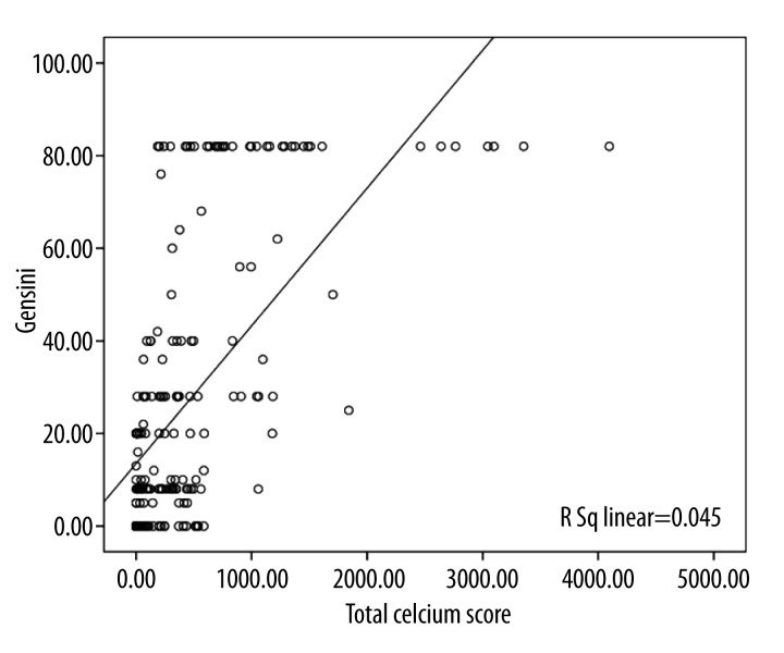
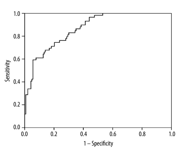
There was a significant correlation between the number of diseased coronary vessels and the mean calcium score, i.e. the mean calcium score was 202.25±450.06 in subjects with normal coronary status, 427.50±607.24 in patients with single-vessel disease, 590.03±511.34 in two-vessel disease, and 953.35 ± 1023.45 in those with three-vessel disease (p<0.001) (Figures 1 and 2). Using the Pearson’s correlation test, a direct association was found between calcium score and Gensini score (r=0.636, p<0.001) (Figure 3), According to the linear regression model and in the presence of baseline parameters including demographics, left ventricular ejection fraction, lipid profile, body mass index, and blood pressure, calcium score measurement was the main determinant of the severity of coronary artery disease indicated by Gensini score (beta=0.025, standard error=0.003, p<0.001). According to the ROC curve analysis (Figure 4), calcium score measurement revealed an acceptable value for discriminating coronary disease from normal condition (AUC=0.867, 95% CI: 0.817–0.917, p<0.001). The optimal cutoff point of calcium score for discriminating these two types of coronary conditions was 350, yielding sensitivity of 83% and specificity of 70%.
Figure 1.
(A) Calcium scoring of a 69-year-old female shows a total calcium score of 985.1. (B) Conventional coronary angiogram shows moderate stenosis of the first diagonal branch (white arrow) and severe stenosis over the second diagonal branch (black arrow) of the left anterior descending artery (LAD). (C). A moderate stenosis of origin (long arrow) and mild stenosis of mid part (short arrow) of the right coronary artery (RCA) are also seen.
Figure 2.
A 71-year-old male with calcium score of 495. Conventional coronary angiogram (A) shows severe stenosis of the left anterior descending artery (LAD) (arrow) extending into diagonal ostium and ectatic proximal part. There is also a significant stenosis in the mid-part of the right coronary artery (arrowhead) with anomalous origin from LAD. These findings are also evident in 3D volume rendering (B). Axial image of coronary CT angiography (C) shows a heavily calcified plaque in the proximal LAD.
Figure 3.
Linear association between total calcium score and Gensini score.
Figure 4.
ROC curve analysis to determine the calcium score for discriminating normal coronary condition from coronary artery disease.
Discussion
Various tools are now being discovered to assess the presence and severity of coronary artery involvement in suspected patients. More importantly, scoring systems that are based on pathophysiological mechanisms of atherosclerotic plaque formation can be more interesting. In this regard, because calcium deposition plays the main role in cardiac events following coronary artery involvement, measuring calcium density in the affected site can have a certain role in predicting coronary events. Based on this hypothesis, the present study goaled to assess the calcium score to discriminate normal coronary condition from coronary disease. Moreover, we identified its value for determining the extension of coronary artery disease in patients who underwent both diagnostic DSA coronary angiography and CTA. In this study, we considered two indicators of CAD severity, including the number of involved coronary vessels and Gensini score that encompasses both the type of diseased artery and also the extension of arterial involvement. The correlation between calcium score and extension and severity of coronary artery disease was proven with these two indicators.
In fact, calcium score has a high value in differentiating normal conditions from affected coronaries, as well as in discriminating intermediate from severe coronary involvement. In a similar study by Budoff et al. [23] the distribution of calcification within the major coronary arteries was used to determine the severity of angiographic disease. Schmermund et al. [24] utilized calcium scoring to distinguish patients with and those without the 3-vessel disease. In another study by de Carvalho et al., in the group of patients with a calcium score of zero, 12.4% had coronary plaques on contrast CT, 10.8% had non-obstructive CAD and only 1.6% had obstructive CAD. In their study, although the absence of coronary artery calcification did not exclude the presence of coronary artery disease, the prevalence of obstructive diseases was considerably low [25]. Alqarqaz et al. [26] also showed that nearly one in five patients with zero calcium score had non-calcified plaques and one in three patients with zero calcium score and intermediate to high FRS had evidence of non-calcified plaques on coronary computed tomography angiography.
While some other studies have found a moderate correlation between coronary artery calcium score and the incidence of atherosclerotic disease based on vessel analysis [24], our study revealed more comprehensive findings. We showed a strong association between the degree of stenosis assessed by the Gensini score and calcium score (p<0.001). Thus, the presence of coronary artery calcium is strongly associated with a greater risk of cardiovascular events and is able to predict their severity. Therefore, our study was in line with previous studies with respect to predictive value of calcium score for presence and severity of coronary artery involvement.
The strength of our study is that it provided the best cutoff value for calcium score to discriminate coronary involvement from normal coronary condition. In this regard, the cutoff was 350 for coronary calcium score, yielding acceptable sensitivity and specificity for predicting coronary disease. Most previous studies included calcium score categorizations for determining coronary involvement. A systematic review of 49 studies revealed that coronary artery calcium score had a high negative predictive value (99%) for ruling out acute coronary syndrome [27]. Similarly, another series found that coronary artery disease was present in 7% of patients with zero calcium score and in 17% of those with calcium score ranging from 1 to 100 [28]. In our study, on a per-patient basis, 12.1% of patients with zero coronary score had coronary artery disease, while 26.2% of patients with score from 101 to 400 and 21.8% of those with score from 401 to 1000 suffered from coronary disease.
Conclusions
Our study confirmed the strong relationship between the coronary artery calcium score and the presence and extension of coronary artery disease assessed by both the number of diseased coronary vessels and also by the Gnesini score. In this regard, patients with a more severe coronary disease revealed a higher coronary calcium score. According to ROC analysis, the best cut point of calcium score for predicting coronary artery involvement was 350 yielding acceptable diagnostic value. Our study emphasized calcium screening as an additional filter before coronary angiography in suspected cases, especially those with mild to moderate risk for having CAD.
References
- 1.Leber AW, Knez A, von Ziegler F, et al. Quantification of obstructive and nonobstructive coronary lesions by 64-slice computed tomography. J Am Coll Cardiol. 2005;46:147–54. doi: 10.1016/j.jacc.2005.03.071. [DOI] [PubMed] [Google Scholar]
- 2.Shabestari AA, Abdi S, Akhlaghpoor S, et al. Diagnostic performance of 64-channel multislice computed tomography in assessment of significant coronary artery disease in symptomatic subjects. Am J Cardiol. 2007;99:1656–61. doi: 10.1016/j.amjcard.2007.01.040. [DOI] [PubMed] [Google Scholar]
- 3.Ropers D, Rixe J, Anders K, et al. Usefulness of multidetector row spiral computed tomography with 64×0.6-mm collimation and 330-ms rotation for the noninvasive detection of significant coronary artery stenoses. Am J Cardiol. 2006;97:343–48. doi: 10.1016/j.amjcard.2005.08.050. [DOI] [PubMed] [Google Scholar]
- 4.Groen JM, Greuter MJ, Vliegenthart R, et al. Calcium scoring using 64-slice MDCT, dual source CT and EBT: a comparative phantom study. Int J Cardiovasc Imaging. 2008;2:547–56. doi: 10.1007/s10554-007-9282-0. [DOI] [PMC free article] [PubMed] [Google Scholar]
- 5.Shabestari AA, Akhlaghpoor S, Shadmani M, et al. Agreement Determination between Coronary Calcium-Scoring and Coronary Stenosis Significance on CT-Angiography. Iran J Radiol. 2006;3:85–90. [Google Scholar]
- 6.Grayburn PA. Interpreting the coronary-artery calcium score. N Engl J Med. 2012;366:294–96. doi: 10.1056/NEJMp1110647. [DOI] [PubMed] [Google Scholar]
- 7.Shabestari AA, Pourghorban R, Tehrai M, et al. Comparison of aortic root dimension changes during cardiac cycle between the patients with and without aortic valve calcification using ECG-gated 64-slice and dual-source 256-slice computed tomography scanners: results of a multicenter study. Int J Cardiovasc Imaging. 2013;29:1391–400. doi: 10.1007/s10554-013-0217-7. [DOI] [PubMed] [Google Scholar]
- 8.Otsuka F, Sakakura K, Yahagi K, et al. Has Our Understanding of Calcification in Human Coronary Atherosclerosis Progressed? Arterioscler Thromb Vasc Biol. 2014;34:724–36. doi: 10.1161/ATVBAHA.113.302642. [DOI] [PMC free article] [PubMed] [Google Scholar]
- 9.Noll D, Kruk M, Pręgowski J, et al. Lumen and calcium characteristics within calcified coronary lesions. Postepy Kardiol Interwencyjnej. 2013;9:1–8. doi: 10.5114/pwki.2013.34022. [DOI] [PMC free article] [PubMed] [Google Scholar]
- 10.Gussenhoven EJ, Essed CE, Lancée CT, et al. Arterial wall characteristics determined by intravascular ultrasound imaging: an in vitro study. J Am Coll Cardiol. 1989;14:947–52. doi: 10.1016/0735-1097(89)90471-3. [DOI] [PubMed] [Google Scholar]
- 11.Hulten E, Bittencourt MS, Ghoshhajra B, et al. Incremental prognostic value of coronary artery calcium score versus CT angiography among symptomatic patients without known coronary artery disease. Atherosclerosis. 2014;233:190–95. doi: 10.1016/j.atherosclerosis.2013.12.029. [DOI] [PMC free article] [PubMed] [Google Scholar]
- 12.Yerramasu A, Lahiri A, Venuraju S, et al. Diagnostic role of coronary calcium scoring in the rapid access chest pain clinic: prospective evaluation of NICE guidance. Eur Heart J Cardiovasc Imaging. 2014 doi: 10.1093/ehjci/jeu011. [Epub ahead of print] [DOI] [PubMed] [Google Scholar]
- 13.Agatston AS, Janowitz WR, Hildner FJ, et al. Quantification of coronary artery calcium using ultrafast computed tomography. J Am Coll Cardiol. 1990;15:827–32. doi: 10.1016/0735-1097(90)90282-t. [DOI] [PubMed] [Google Scholar]
- 14.Schmermund A, Bailey KR, Rumberger JA, et al. An algorithm for noninvasive identification of angiographic three-vessel and/or left main coronary artery disease in symptomatic patients on the basis of cardiac risk and electron-beam computed tomographic calcium scores. J Am Coll Cardiol. 1999;33:444–52. doi: 10.1016/s0735-1097(98)00565-8. [DOI] [PubMed] [Google Scholar]
- 15.Gussenhoven EJ, Essed CE, Lancée CT, et al. Arterial wall characteristics determined by intravascular ultrasound imaging: an in vitro study. J Am Coll Cardiol. 1989;14:947–52. doi: 10.1016/0735-1097(89)90471-3. [DOI] [PubMed] [Google Scholar]
- 16.Schmermund A, Bailey KR, Rumberger JA, et al. An algorithm for noninvasive identification of angiographic three-vessel and/or left main coronary artery disease in symptomatic patients on the basis of cardiac risk and electron-beam computed tomographic calcium scores. J Am Coll Cardiol. 1999;33:444–52. doi: 10.1016/s0735-1097(98)00565-8. [DOI] [PubMed] [Google Scholar]
- 17.Rumberger JA, Brundage BH, Rader DJ, et al. Electron beam computed tomographic coronary calcium scanning: a review and guidelines for use in asymptomatic persons. Mayo Clin Proc. 1999;74:243–52. doi: 10.4065/74.3.243. [DOI] [PubMed] [Google Scholar]
- 18.Palumbo AA, Maffei E, Martini C, et al. Coronary calcium score as gatekeeper for 64-slice computed tomography coronary angiography in patients with chest pain: per-segment and per-patient analysis. Eur Radiol. 2009;19:2127–35. doi: 10.1007/s00330-009-1398-2. [DOI] [PubMed] [Google Scholar]
- 19.Park MJ, Jung JI, Choi YS, et al. Coronary CT angiography in patients with high calcium score: evaluation of plaque characteristics and diagnostic accuracy. Int J Cardiovasc Imaging. 2011;27(Suppl 1):43–51. doi: 10.1007/s10554-011-9970-7. [DOI] [PubMed] [Google Scholar]
- 20.Schmermund A, Baumgart D, Görge G, et al. Coronary artery calcium in acute coronary syndromes: a comparative study of electron-beam computed tomography, coronary angiography, and intracoronary ultrasound in survivors of acute myocardial infarction and unstable angina. Circulation. 1997;96:1461–69. doi: 10.1161/01.cir.96.5.1461. [DOI] [PubMed] [Google Scholar]
- 21.Budoff S, Achenbach R, Blumenthal S, et al. Assessment of coronary artery disease by cardiac computed tomography: a scientific statement from the American Heart Association Committee on Cardiovascular Imaging and Intervention, Council on Cardiovascular Radiology and Intervention, and Committee on Cardiac Imaging, Council on Clinical Cardiology. Circulation. 2006;114:1761–91. doi: 10.1161/CIRCULATIONAHA.106.178458. [DOI] [PubMed] [Google Scholar]
- 22.Gökdeniz T, Kalaycıoğlu E, Aykan AC, et al. Value of coronary artery calcium score to predict severity or complexity of coronary artery disease. Arq Bras Cardiol. 2014;102(2):120–27. doi: 10.5935/abc.20130241. [DOI] [PMC free article] [PubMed] [Google Scholar]
- 23.Budoff MJ, Diamond GA, Raggi P, et al. Continuous probabilistic prediction of angiographically significant coronary artery disease using electron beam tomography. Circulation. 2002;105:1791–96. doi: 10.1161/01.cir.0000014483.43921.8c. [DOI] [PubMed] [Google Scholar]
- 24.Ma ES, Yang ZG, Li Y, et al. Correlation of calcium measurement with low dose 64- slice CT and angiographic stenosis in patients with suspected coronary artery disease. Int J Cardiol. 2010;4:249–52. doi: 10.1016/j.ijcard.2008.11.043. [DOI] [PubMed] [Google Scholar]
- 25.de Carvalho MS, de Araújo Gonçalves P, Garcia-Garcia HM, et al. Prevalence and predictors of coronary artery disease in patients with a calcium score of zero. Int J Cardiovasc Imaging. 2013;29(8):1839–46. doi: 10.1007/s10554-013-0267-x. [DOI] [PubMed] [Google Scholar]
- 26.Alqarqaz M1, Zaidan M, Al-Mallah MH. Prevalence and predictors of atherosclerosis in symptomatic patients with zero calcium score. Acad Radiol. 2011;18(11):1437–41. doi: 10.1016/j.acra.2011.07.012. [DOI] [PubMed] [Google Scholar]
- 27.Rubinshtein R, Gaspar T, Halon DA, et al. Prevalence and extent of obstructive coronary artery disease in patients with zero or low calcium score undergoing 64-slice cardiac multidetector computed tomography for evaluation of a chest pain syndrome. Am J Cardiol. 2007;99:472–75. doi: 10.1016/j.amjcard.2006.08.060. [DOI] [PubMed] [Google Scholar]
- 28.Sekiya M, Mukai M, Suzuki M, et al. Clinical significance of the calcification of coronary arteries in patients with angiographically normal coronary arteries. Angiology. 1992;43:401–7. doi: 10.1177/000331979204300505. [DOI] [PubMed] [Google Scholar]


