Abstract
Background
Pathology in the long head of the biceps tendon often occurs in patients with rotator cuff tears. Arthroscopic tenotomy is the most common treatment. However, the role of the long head of the biceps at the shoulder and the consequences of surgical detachment on the remaining shoulder structures remain unknown.
Questions/purposes
We hypothesized that detachment of the long head of the biceps, in the presence of supraspinatus and infraspinatus tears, would decrease shoulder function and decrease mechanical and histologic properties of both the subscapularis tendon and the glenoid articular cartilage.
Methods
We detached the supraspinatus and infraspinatus or the supraspinatus, infraspinatus, and long head of the biceps after 4 weeks of overuse in a rat model. Animals were gradually returned to overuse activity after detachment. At 8 weeks, the subscapularis and glenoid cartilage biomechanical and histologic properties were evaluated and compared.
Results
The group with the supraspinatus, infraspinatus, and long head of the biceps detached had greater medial force and decreased change in propulsion, braking, and vertical force. This group also had an increased upper and lower subscapularis modulus but without any differences in glenoid cartilage modulus. Finally, this group had a significantly lower cell density in both the upper and lower subscapularis tendons, although cartilage histology was not different.
Conclusions
Detachment of the long head of the biceps tendon in the presence of a posterior-superior cuff tear resulted in improved shoulder function and less joint damage in this animal model.
Clinical Relevance
This study provides evidence in an animal model that supports the use of tenotomy for the management of long head of the biceps pathology in the presence of a two-tendon cuff tear. However, long-term clinical trials are required.
Introduction
The complex anatomy of the shoulder includes both static and dynamic structures to provide stability. The static restraints at the shoulder (ie, ligaments, joint capsule, and bony anatomy) provide only some stability, which can place the joint at risk for mechanical instability. Therefore, to maintain joint congruency and stability during functional motions, the rotator cuff muscles must work together to dynamically stabilize the joint by balancing the muscle forces in all directions. The anterior-posterior force balance of the shoulder, which is primarily composed of the subscapularis anteriorly and the infraspinatus and teres minor posteriorly, has been described previously and is a major component to the dynamic stability [1]. Disruption of this force balance (from rotator cuff tears) is thought to lead to increased translation of the humeral head and may contribute to significant pain and dysfunction. The long head of the biceps tendon crosses the shoulder and may serve as an important joint stabilizer, particularly in the presence of cuff tears; however, its role in joint stability remains controversial. Specifically, some believe that it provides minimal stability at the shoulder [11, 27], while others believe it serves mainly as a humeral head depressor, assisting the supraspinatus [7, 8].
Rotator cuff tendon tears are common injuries, occurring in 20% of the general population; the occurrence rate increases with age [28]. Large rotator cuff tears involving both the supraspinatus and infraspinatus are more common in patients who remain active in repetitive overhead activities, such as manual labor and recreation. Initially, patients often present with shoulder pain and dysfunction, including the inability to perform certain activities of daily living. Patients with rotator cuff tears frequently develop long head of the biceps tendon pathology [2], which may lead to increasing pain and loss of function. Previous studies have demonstrated structural long head of the biceps tendon damage in the presence of cuff tears both clinically [13] and in a rat rotator cuff tear model [14]. Commonly, these long head of the biceps tendon symptoms persist and surgeons often will recommend arthroscopic tenodesis or tenotomy. These surgical techniques can reduce pain and improve function [9, 24]. However, the functional and mechanical consequences on the remaining (intact) joint structures (glenoid cartilage and subscapularis) after detachment of the long head of the biceps tendon in the presence of a combined supraspinatus and infraspinatus rotator cuff tear remain unknown.
We therefore hypothesized the additional detachment of the long head of the biceps tendon would decrease (1) shoulder function, (2) mechanical and histologic properties of the subscapularis tendon, and (3) mechanical and histologic properties of the glenoid articular cartilage.
Materials and Methods
Study Design
After a 2-week training period, 36 adult male Sprague-Dawley rats (400–450 g) underwent 4 weeks of overuse (downhill, 10° treadmill running at 17 m/minute for 1 hour/day, 5 days/week) [22] to create a tendinopathic condition in the supraspinatus tendon. Subsequently, animals were randomized into two surgical groups: unilateral detachment of the supraspinatus and infraspinatus tendons or the supraspinatus, infraspinatus, and long head of the biceps tendons, as previously described [26]. This model has been approved by our Institutional Animal Care and Use Committee.
After tendon detachment, animals were returned to overuse activity. Postsurgery, animals were subjected to 1 week of cage activity before gradually returning to the overuse protocol over 2 weeks. Subsequently, all animals completed 5 weeks of overuse activity. All animals were then sacrificed 8 weeks after surgical tendon detachment. For histology (n = 4), tissues were immediately fixed in formalin. The remaining animals (n = 11) designated for mechanical testing were stored intact at −20 °C.
Quantitative Ambulatory Assessment
To assess shoulder joint function [20], forelimb gait and ground reaction forces were recorded using an instrumented walkway 1 day before detachment surgery (baseline) and at 3, 7, 14, 28, 42, and 56 days after tendon detachment. Ground reaction force data, including medial/lateral, braking, propulsion, and vertical forces, were determined for each walk. Parameters were averaged across walks on a given day for each animal and normalized to body weight, and the change in force from baseline was calculated.
Tendon Mechanical Testing
The animals were thawed, and the scapula and humerus were dissected with the subscapularis tendons intact. Tendon testing was performed as described [25]. Briefly, stain lines for local optical strain measurement (at insertion and midsubstance) were placed on the upper and lower bands of the subscapularis tendon. Cross-sectional area was measured using a custom laser device. The humerus was embedded in a holding fixture using polymethylmethacrylate, gripped with cyanoacrylate annealed sand paper, and immersed in phosphate-buffered saline (PBS) at 37 °C. Tensile testing was performed as follows: preload to 0.08 N, preconditioning (10 cycles of 0.1–0.5 N at a rate of 1% strain/second), stress relaxation to 5% strain at a rate of 5% strain/second for 600 seconds, and ramp to failure at 0.3% strain/second. Stress was calculated as force divided by initial area, and two-dimensional Lagrangian optical strain was determined from stain line displacements using texture tracking software.
Cartilage Mechanical Testing
The glenoid was prepared for cartilage mechanical testing by sharply detaching the long head of the biceps at its insertion on the superior rim of the glenoid in the supraspinatus and infraspinatus group. The glenoid was then preserved by wrapping in soft tissue and frozen (−20 °C).
For cartilage thickness measurement [16], each scapula was thawed and immersed in PBS containing protease inhibitors (5 mM benzamidine hydrochloride, 1 mM phenylmethylsulfonyl fluoride, 1 M N-ethylmaleimide) at room temperature. Specimens were scanned at 0.25-mm increments using a 55-MHz ultrasound probe (VisualSonics, Inc, Toronto, Ontario, Canada) in plane with the scapula. Captured B-mode images of each scan were segmented by selecting the cartilage and bony surfaces of the glenoid. The three-dimensional positions of these surfaces were reconstructed and used to determine cartilage thickness maps. Each map was divided into six regions (center, posterior-superior, posterior-inferior, anterior-superior, anterior-inferior, and superior) and a mean thickness was computed for each region. After ultrasound scanning, specimens were wrapped in soft tissue and frozen (−20 °C) until mechanical testing.
For cartilage mechanical testing [16], each scapula was thawed and immersed in PBS containing the protease inhibitor cocktail at room temperature. Utilizing a 0.5-mm-diameter, nonporous spherical indenter, cartilage indentation testing was performed. Briefly, a preload (0.005 N) was followed by eight stepwise stress relaxation tests (8-μm ramp at 2 μm/seconds followed by a 300-second hold). The scapula was repositioned for each localized region such that the indenter tip was perpendicular to the cartilage surface in each region. Cartilage thickness for indentation testing was determined by identifying the indentation location on each thickness map. Equilibrium elastic modulus was calculated, as described [5], at 20% indentation and assuming Poisson’s ratio (ν = 0.30).
Histology
For histology, rotator cuff samples were left intact as bone-tendon-muscle units. For the glenoid cartilage, the glenoid was detached from the rest of the scapula at the glenoid neck. All samples were processed, longitudinal sections (7 μm) were collected, and tendon samples were stained with hematoxylin and eosin, while cartilage samples were stained with safranin O, fast green, and iron hematoxylin. Hematoxylin and eosin-stained tendon sections were imaged at the insertion site and midsubstance of each tendon at ×200 magnification. Cell density (number of cells/mm2) and cell shape (aspect ratio; 0–1, with 1 being a circle) were quantified using a bioquantification software system (Bioquant Osteo II; BIOQUANT Image Analysis Corp, Nashville, TN, USA). Cartilage sections were imaged at ×100 magnification in five regions (center, posterior-superior, posterior-inferior, anterior-superior, anterior-inferior, and superior) corresponding to the indentation locations and graded using a modified Mankin score (including scores for cellularity, structure, and matrix staining). Due to the presence of an intact tidemark in all images, scoring for this category was removed. Three blinded investigators (RV, GF, AD) performed the cartilage histology scoring. Cartilage cell density was also quantified using the bioquantification software system.
Statistics
For the ambulatory assessment, significance was assessed using a two-way ANOVA with repeated measures on time with followup t-tests between groups at each time point. In this data set, points were occasionally absent (~18%) for a specific animal on a specific day [19]. Therefore, multiple imputations were conducted using the Markov chain Monte Carlo method on the ambulation data to allow for a repeated-measures analysis. The mean of five imputations was used for the final analysis. Tissue mechanics and tendon histology were assessed using one-tailed and two-tailed t-tests, respectively. For cartilage thickness and histology scoring, median grades were compared between groups using a Mann-Whitney test. Significance was set at p values of less than 0.05. We used the statistical program SPSS® for Windows® (Version 20.0; IBM Corp, Armonk, NY, USA) for data analysis.
Results
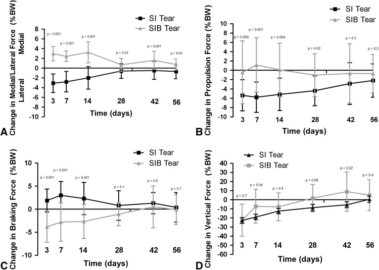
The additional detachment of the long head of the biceps in the presence of a supraspinatus and infraspinatus detachment demonstrated a larger change in ground reaction forces over time compared to the biceps being left intact (Fig. 1). Specifically, rats in which a biceps tenotomy (95% CI = 4.24–2.24) was performed directed the change in joint force more medially (p < 0.001, effect size [ES] = 2.38) than those with an intact biceps (95% CI = −0.9 to −3.1) at all time points. The rats with a biceps tenotomy (95% CI = 2.75 to −2.57) had a decreased change in propulsion force (p = 0.004, ES = 1.14) at 3, 7, 14, and 28 days compared to those with an intact biceps (95% CI = −3.54 to −6.76). The rats with a biceps tenotomy (95% CI = −0.96 to −4.36) had a decreased change in braking force (p = 0.001, ES = 1.38) at 3, 7, and 14 days compared to those with an intact biceps (95% CI = 3.9–0.7). The rats with a biceps tenotomy (95% CI = −0.66 to −14.88) had a decreased change in vertical force (p = 0.4, ES = 0.325) at 7 and 42 days compared to those with an intact biceps (95% CI = −6.25 to −18.67). This suggests that rats with a biceps tenotomy had less detrimental changes in shoulder function over time compared to those that did not receive a biceps tenotomy.
Fig. 1A–D.
Compared to the group without biceps tenotomy, the group with biceps tenotomy had (A) a change to a more medial force at all time points, (B) a decreased change in propulsion force at 3, 7, 14, and 28 days after detachment, (C) a decreased change in braking force at 3, 7, and 14 days after detachment, and (D) a decreased change in vertical force at 7 and 42 days after detachment. Data are shown as mean with SD. SI = supraspinatus and infraspinatus; SIB = supraspinatus, infraspinatus, and long head of the biceps; BW = body weight.
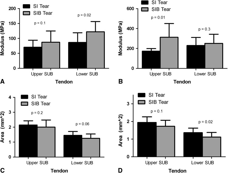
Tendon elastic modulus was greater in the upper subscapularis midsubstance (95% CI: 188.1–158.3 versus 414.6–200.4; p = 0.01, ES = 1.3) and lower subscapularis insertion site (95% CI: 294.5–165.9 versus 381.1–219.5; p = 0.02, ES = 1.19) for the group with biceps tenotomy than for the group without biceps tenotomy (Fig. 2). Additionally, the lower subscapularis tendon cross-sectional area (95% CI: 1.5–1.2 versus 1.3–0.9; p = 0.02, ES = 0.929) was less at the midsubstance region for the group with biceps tenotomy. No other group differences were observed for cross-sectional area for the upper or lower subscapularis. Histology results demonstrated a lower cell density for the group with biceps tenotomy at both the insertion (Figs. 3, 4) and midsubstance of both the upper and lower bands of the subscapularis tendon (Table 1). Additionally, a more rounded cell shape was observed in this group at the insertion sites of both the upper and lower bands of the subscapularis and at the midsubstance of the lower band of the subscapularis.
Fig. 2A–D.
Compared to the group without biceps tenotomy, the group with biceps tenotomy had (A) an increased elastic modulus for the lower subscapularis insertion site, (B) an increased elastic modulus for the upper subscapularis midsubstance, (C) a similar cross-sectional area at the insertion site of either tendon, and (D) a decreased cross-sectional area for the lower subscapularis midsubstance. Data are shown as mean with SD. SI = supraspinatus and infraspinatus; SIB = supraspinatus, infraspinatus, and long head of the biceps; SUB = subscapularis.
Fig. 3A–B.
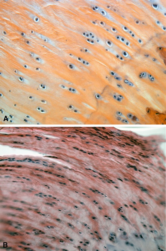
Representative images for the upper subscapularis insertion are displayed (stain, hematoxylin and eosin; original magnification, ×200). Cell density was significantly decreased in (A) the group with biceps tenotomy compared to (B) the group with biceps tenotomy. Note: quantification was performed on original images (Fig. 4). For publication, filters were applied to the images: autotone, autocontrast, autocolor (all three to each individually).
Fig. 4A–B.
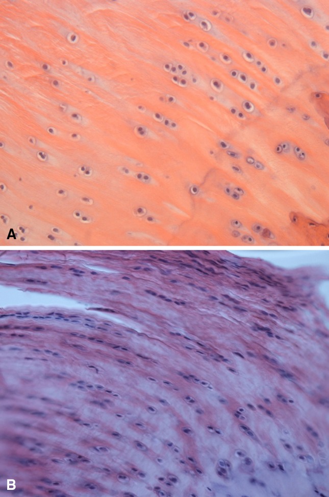
Original, unfiltered tendon histology versions of the images shown in Figure 3 show upper subscapularis insertion in (A) the group with biceps tenotomy and (B) the group without biceps tenotomy (both images: stain, hematoxylin and eosin; original magnification, ×200).
Table 1.
Tendon histology data
| Tendon | Group | Region | Cell density (cells/mm2)* | p value | Effect size | 95% CI | Cell shape (aspect ratio)* | p value | Effect size | 95% CI |
|---|---|---|---|---|---|---|---|---|---|---|
| Upper subscapularis | SI | Insertion | 1736 ± 288 | 0.001 | 5.38 | 2018–1454 | 0.52 ± 0.05 | 0.008 | 3.5 | 0.57–0.47 |
| SIB | 415 ± 203 | 613.9–216.1 | 0.66 ± 0.03 | 0.69–0.63 | ||||||
| SI | Midsubstance | 1859 ± 352 | 0.002 | 5.07 | 2204–1514 | 0.44 ± 0.04 | 0.1 | 2.0 | 0.48–0.4 | |
| SIB | 421 ± 215 | 631.7–210.3 | 0.38 ± 0.02 | 0.4–0.36 | ||||||
| Lower subscapularis | SI | Insertion | 2883 ± 1591 | 0.05 | 2.88 | 4474–1324 | 0.51 ± 0.03 | 0.002 | 6.33 | 0.54–0.48 |
| SIB | 302 ± 200 | 498–106 | 0.70 ± 0.03 | 0.7 –0.67 | ||||||
| SI | Midsubstance | 2772 ± 928 | 0.009 | 5.33 | 368–1863 | 0.35 ± 0.04 | 0.04 | 3.2 | 0.39–0.31 | |
| SIB | 253 ± 18 | 270.6–235.4 | 0.43 ± 0.01 | 0.44–0.42 |
* Values are expressed as mean ± SD; SI = unilateral detachment of the supraspinatus and infraspinatus; SIB = unilateral detachment of the supraspinatus, infraspinatus, and long head of the biceps.
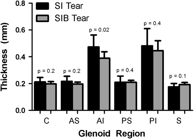
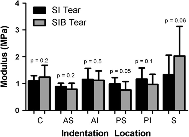
Cartilage thickness was decreased (95% CI: 0.523–0.417 versus 0.418–0.362; p = 0.02, ES = 1.14) at the anterior-inferior region in the group with biceps tenotomy (Fig. 5). However, no differences were observed between groups for cartilage equilibrium elastic modulus in any region (Fig. 6). Additionally, cartilage histology results did not demonstrate any differences for modified Mankin score grading or quantitative cell density between groups in any region (Table 2).
Fig. 5.
Compared to the group without biceps tenotomy, the group with biceps tenotomy had a decrease in cartilage thickness at the anterior-inferior region. Data are shown as mean with SD. SI = supraspinatus and infraspinatus; SIB = supraspinatus, infraspinatus, and long head of the biceps; C = center; AS = anterior-superior; AI = anterior-inferior; PS = posterior-superior; PI = posterior-inferior; S = superior.
Fig. 6.
There were no between-group differences observed for cartilage equilibrium elastic modulus. Data are shown as mean with SD. SI = supraspinatus and infraspinatus; SIB = supraspinatus, infraspinatus, and long head of the biceps; C = center; AS = anterior-superior; AI = anterior-inferior; PS = posterior-superior; PI = posterior-inferior; S = superior.
Table 2.
Cartilage histology data
| Region | Group | Cell density (cells/mm2)* | p value | Mankin score† | p value |
|---|---|---|---|---|---|
| Center | SI | 573 ± 141 | 0.33 | 5.0 (3.5–5.0) | 1.0 |
| SIB | 522 ± 122 | 2.0 (2.0–5.0) | |||
| Anterior-superior | SI | 528 ± 128 | 0.09 | 6.0 (4.0–6.0) | 0.64 |
| SIB | 364 ± 116 | 6.0 (5.5–7.0) | |||
| Anterior-inferior | SI | 495 ± 57 | 0.09 | 6.0 (5.0–6.5) | 0.81 |
| SIB | 375 ± 116 | 6.0 (5.0–6.0) | |||
| Posterior-superior | SI | 667 ± 79 | 0.14 | 2.0 (2.0–4.0) | 0.38 |
| SIB | 520 ± 187 | 4.0 (3.5–5.5) | |||
| Posterior-inferior | SI | 580 ± 151 | 0.17 | 6.0 (3.5–6.5) | 0.70 |
| SIB | 440 ± 171 | 7.0 (4.5–7.0) |
* Values are expressed as mean ± SD; †values are expressed as mean, with range in parentheses; SI = unilateral detachment of the supraspinatus and infraspinatus; SIB = unilateral detachment of the supraspinatus, infraspinatus, and long head of the biceps.
Discussion
Patients with tears of the supraspinatus and infraspinatus tendons commonly present with long head of the biceps tendon pain [2, 10, 24]. This pain is often persistent and prevents patients from performing activities of daily living. Currently, this is commonly treated using surgical tenodesis or tenotomy of the long head of the biceps tendon [9, 24, 29]. However, the effect on the remaining shoulder structures (subscapularis and glenoid cartilage) after detachment of the long head of the biceps tendon in the presence of a posterior-superior rotator cuff tear is unknown. Therefore, we hypothesized that the additional detachment of the long head of the biceps tendon would decrease (1) shoulder function, (2) mechanical and histologic properties of the subscapularis tendon, and (3) mechanical and histologic properties of the glenoid cartilage.
There are some limitations to the study that should be acknowledged. A well-established rat rotator cuff model was used in this study, and although the rat shoulder has similar anatomy, the use of a quadruped animal does not exactly replicate the human shoulder. However, the rat shoulder produces large amounts of glenohumeral forward flexion, which places the rotator cuff and long head of the biceps tendons at risk for subacromial impingement and therefore replicates repetitive overhead activity in the human [21]. In addition, we acknowledge limitations in our surgical procedure. Rotator cuff tears typically develop gradually over time in chronic, degenerative tendons; however, in this study, we performed an acute surgical detachment of the rotator cuff tendons. Although this does not mimic the chronic condition observed clinically, the animals did receive an overuse treadmill protocol for 4 weeks before detachment, which has been shown to create a tendinopathic condition of the supraspinatus tendon [22], and thus, this model represents an acute-on-chronic condition quite well. In addition, the long head of the biceps tendon was detached at the same time as the rotator cuff tear was created. Clinically, the long head of the biceps is typically detached several months or even years after the initial rotator cuff tear. Although there is commonly a delay between the initial rotator cuff tear and surgical detachment of the long head of the biceps tendon, our study design allows for a well-controlled comparison to investigate the role of the long head of the biceps tendon in the development of joint damage. Despite these limitations, our results clearly demonstrate that the additional detachment of the long head of the biceps tendon improves shoulder function and subscapularis mechanical and histologic properties and does not affect the mechanical or histologic properties of the glenoid cartilage.
We demonstrated that shoulder function, as measured by a change in ground reaction forces from baseline, was different between groups. Specifically, the group without biceps tenotomy had a more laterally directed force, a decreased propulsion force, and an increased braking force. These changes have also been shown to occur after shoulder injury in this animal model system [17, 18] and may be indicative of an altered loading environment that could be present in the human condition. However, this finding disagrees with previous studies. Specifically, a cadaveric study examined the role of the long head of the biceps in the presence of supraspinatus and infraspinatus tears [23] and found that loading of the long head of the biceps tendon decreased humeral head translations. Results from a clinical EMG study suggested that the long head of the biceps tendon assists the supraspinatus with humeral head depression during shoulder elevation [8]. However, due to confounding variables in clinical and cadaver studies, it is difficult to determine true cause and effect relationships. Therefore, the true functional role of the biceps at the shoulder has yet to be determined. As discussed previously, shoulder function is thought to decrease in the presence of tears involving the supraspinatus and infraspinatus due to disruption of the anterior-posterior force balance [1]. However, the functional role of the biceps in this tear scenario is not well defined. It is likely that, due to the anterior positioning of the long head of the biceps tendon within the bicipital groove, it may function in conjunction with the subscapularis as an anterior restraint to the force balance, particularly in the case of a posterior-superior rotator cuff tear. Therefore, in this tear scenario, detachment of the long head of the biceps tendon may better reestablish the balance of anterior-posterior forces (between the subscapularis and teres minor) by eliminating excess anterior forces and therefore improve shoulder function. In fact, our results are supported by clinical outcome studies examining long head of the biceps tendon tenodesis or tenotomy in the presence of supraspinatus and infraspinatus cuff tears that have reported significant functional improvements [9, 24, 29].
We found that the tensile modulus at the midsubstance region of the upper subscapularis and the insertion site of the lower subscapularis were larger in the group with biceps tenotomy than in the group without biceps tenotomy. Our results are similar to previous findings that demonstrated decreased mechanical properties of the subscapularis with supraspinatus and infraspinatus tears [15]. However, our results do not support our original hypothesis that the additional detachment of the long head of the biceps tendon would further decrease the mechanical properties of the subscapularis, indicating mechanical damage. As previously stated, detachment of the long head of the biceps tendon may have improved the anterior-posterior force balance [1], thereby preventing excessive translations of the humeral head and subsequent mechanical damage to the subscapularis tendon. Interestingly, there were region-specific changes in the upper and lower band of the subscapularis (upper midsubstance and lower insertion site). This may be due to an alteration in the tendon loading environment, as a result of subcoracoid impingement between the coracoid process and the subscapularis tendon [12]. Specifically, due to the unique anatomy of the upper and lower bands of the subscapularis (upper band insertion site being more lateral than the lower band) [3, 6], excessive translation of the humeral head caused by an unbalanced anterior-posterior force balance may have caused mechanical compression at the midsubstance of the upper subscapularis and the insertion site of the lower subscapularis.
Tendon histology results demonstrated that the upper and lower subscapularis for the group with biceps tenotomy had less cell density and a more round cell shape. The increased cell density in the group without biceps tenotomy supports the mechanical results that were observed. Previous research has shown that injured tendons, such as those with tendinopathy, have increased cell density [22]. This increased cell density suggests that the subscapularis tendon is metabolically active in the group without biceps tenotomy compared to the group with biceps tenotomy. Again, the unbalanced anterior-posterior force balance in the supraspinatus and infraspinatus group may be placing increased mechanical loads on the subscapularis, thereby causing degeneration similar to a tendinopathic condition. The results in cell shape do not agree with previous research. Injured tendons typically present with a more rounded cell shape [22]. However, the group with biceps tenotomy, which had larger mechanical properties, displayed a more rounded cell shape compared to the group without biceps tenotomy. Despite this finding, both groups demonstrated a rounded cell morphology (based on the aspect ratios), and therefore, differences may not be meaningful.
For cartilage thickness, the group with biceps tenotomy did have decreased cartilage thickness in the anterior-inferior region. However, the equilibrium elastic modulus was not significantly different between groups at any of the regions. In addition, cartilage histology (modified Mankin score and cell density) was not significantly different between groups for any of the regions. Previous studies have identified cartilage damage in the presence of cuff tears [16–18], and therefore, performing long head of the biceps tenodesis or tenotomy has been a concern among surgeons [4]. However, no long-term followup studies exist evaluating whether patients are at a higher risk for glenohumeral arthritis after long head of the biceps tenodesis or tenotomy. Our results in this animal model suggest that the additional detachment of the long head of the biceps tendon does not affect the glenoid cartilage.
In conclusion, additional detachment of the long head of the biceps tendon in the presence of combined supraspinatus and infraspinatus tears in this animal model improved shoulder function and subscapularis tendon properties (mechanical and structural) and did not affect the glenoid cartilage properties. These results were surprising since we hypothesized that detaching the long head of the biceps tendon would have detrimental effects to the surrounding joint structures. The improved functional and structural outcomes may be due to rebalancing the anterior-posterior forces with the additional detachment of the long head of the biceps tendon. These data provide the initial basic science evidence to support the use of long head of the biceps tenotomy in patients with tears of both the supraspinatus and infraspinatus. Additional research is needed to examine the effect of detaching the long head of the biceps tendon in the presence of an isolated supraspinatus tear to further understand its role at the glenohumeral joint. Furthermore, randomized control trials are also needed to examine the clinical utility of this procedure and the long-term consequence of long head of the bicep tenotomy on the glenohumeral joint.
Acknowledgments
The authors thank Andrew Dunkman BS, Liz Feeney, Benjamin Freedman BS, and Corinne Riggin BS for their contribution to the overuse protocol. We also thank Rameen Vafa, George Fryhofer BS, and Alex Delong BS for their contribution to histology.
Footnotes
The institution of the authors received, during the study period, funding from NIH/NIAMS (Bethesda, MD, USA) (Grant R01AR056658) and the Penn Center for Musculoskeletal Disorders (Philadelphia, PA, USA) (NIH Grant P30 AR050950). Each author certifies that he or she, or a member of his or her immediate family, has no commercial associations (eg, consultancies, stock ownership, equity interest, patent/licensing arrangements, etc) that might pose a conflict of interest in connection with the submitted article.
All ICMJE Conflict of Interest Forms for authors and Clinical Orthopaedics and Related Research editors and board members are on file with the publication and can be viewed on request.
Each author certifies that his or her institution approved the animal protocol for this investigation and that all investigations were conducted in conformity with ethical principles of research.
References
- 1.Burkhart SS. Arthroscopic treatment of massive rotator cuff tears: clinical results and biomechanical rationale. Clin Orthop Relat Res. 1991;267:45–56. [PubMed] [Google Scholar]
- 2.Chen CH, Hsu KY, Chen WJ, Shih CH. Incidence and severity of biceps long head tendon lesion in patients with complete rotator cuff tears. J Trauma. 2005;58:1189–1193. doi: 10.1097/01.TA.0000170052.84544.34. [DOI] [PubMed] [Google Scholar]
- 3.D’Addesi LL, Anbari A, Reish MW, Brahmabhatt S, Kelly JD. The subscapularis footprint: an anatomic study of the subscapularis tendon insertion. Arthroscopy. 2006;22:937–940. doi: 10.1016/j.arthro.2006.04.101. [DOI] [PubMed] [Google Scholar]
- 4.Elser F, Braun S, Dewing CB, Giphart JE, Millett PJ. Anatomy, function, injuries, and treatment of the long head of the biceps brachii tendon. Arthroscopy. 2011;27:581–592. doi: 10.1016/j.arthro.2010.10.014. [DOI] [PubMed] [Google Scholar]
- 5.Hayes WC, Keer LM, Herrmann G, Mockros LF. A mathematical analysis for indentation tests of articular cartilage. J Biomech. 1972;5:541–551. doi: 10.1016/0021-9290(72)90010-3. [DOI] [PubMed] [Google Scholar]
- 6.Ide J, Tokiyoshi A, Hirose J, Mizuta H. An anatomic study of the subscapularis insertion to the humerus: the subscapularis footprint. Arthroscopy. 2008;24:749–753. doi: 10.1016/j.arthro.2008.02.009. [DOI] [PubMed] [Google Scholar]
- 7.Kido T, Itoi E, Konno N, Sano A, Urayama M, Sato K. Electromyographic activities of the biceps during arm elevation in shoulders with rotator cuff tears. Acta Orthop Scand. 1998;69:575–579. doi: 10.3109/17453679808999258. [DOI] [PubMed] [Google Scholar]
- 8.Kido T, Itoi E, Konno N, Sano A, Urayama M, Sato K. The depressor function of biceps on the head of the humerus in shoulders with tears of the rotator cuff. J Bone Joint Surg Br. 2000;82:416–419. doi: 10.1302/0301-620X.82B3.10115. [DOI] [PubMed] [Google Scholar]
- 9.Kim SJ, Lee IS, Kim SH, Woo CM, Chun YM. Arthroscopic repair of concomitant type II SLAP lesions in large to massive rotator cuff tears: comparison with biceps tenotomy. Am J Sports Med. 2012;40:2786–2793. doi: 10.1177/0363546512462678. [DOI] [PubMed] [Google Scholar]
- 10.Lakemeier S, Reichelt JJ, Timmesfeld N, Fuchs-Winkelmann S, Paletta JR, Schofer MD. The relevance of long head biceps degeneration in the presence of rotator cuff tears. BMC Musculoskelet Disord. 2010;11:191. doi: 10.1186/1471-2474-11-191. [DOI] [PMC free article] [PubMed] [Google Scholar]
- 11.Levy AS, Kelly BT, Lintner SA, Osbahr DC, Speer KP. Function of the long head of the biceps at the shoulder: electromyographic analysis. J Shoulder Elbow Surg. 2001;10:250–255. doi: 10.1067/mse.2001.113087. [DOI] [PubMed] [Google Scholar]
- 12.Lo IK, Burkhart SS. The etiology and assessment of subscapularis tendon tears: a case for subcoracoid impingement, the roller-wringer effect, and TUFF lesions of the subscapularis. Arthroscopy. 2003;19:1142–1150. doi: 10.1016/j.arthro.2003.10.024. [DOI] [PubMed] [Google Scholar]
- 13.Mazzocca AD, McCarthy MB, Ledgard FA, Chowaniec DM, McKinnon WJ, Jr, Delaronde S, Rubino LJ, Apolostakos J, Romeo AA, Arciero RA, Beitzel K. Histomorphologic changes of the long head of the biceps tendon in common shoulder pathologies. Arthroscopy. 2013;29:972–981. doi: 10.1016/j.arthro.2013.02.002. [DOI] [PubMed] [Google Scholar]
- 14.Peltz CD, Perry SM, Getz CL, Soslowsky LJ. Mechanical properties of the long-head of the biceps tendon are altered in the presence of rotator cuff tears in a rat model. J Orthop Res. 2009;27:416–420. doi: 10.1002/jor.20770. [DOI] [PMC free article] [PubMed] [Google Scholar]
- 15.Perry SM, Getz CL, Soslowsky LJ. After rotator cuff tears, the remaining (intact) tendons are mechanically altered. J Shoulder Elbow Surg. 2009;18:52–57. doi: 10.1016/j.jse.2008.07.003. [DOI] [PMC free article] [PubMed] [Google Scholar]
- 16.Reuther KE, Sarver JJ, Schultz SM, Lee CS, Sehgal CM, Glaser DL, Soslowsky LJ. Glenoid cartilage mechanical properties decrease after rotator cuff tears in a rat model. J Orthop Res. 2012;30:1435–1439. doi: 10.1002/jor.22100. [DOI] [PMC free article] [PubMed] [Google Scholar]
- 17.Reuther KE, Thomas SJ, Evans EF, Tucker JJ, Sarver JJ, Ilkhani-Pour S, Gray CF, Voleti PB, Glaser DL, Soslowsky LJ. Returning to overuse activity following a supraspinatus and infraspinatus tear leads to joint damage in a rat model. J Biomech. 2013;46:1818–1824. doi: 10.1016/j.jbiomech.2013.05.007. [DOI] [PMC free article] [PubMed] [Google Scholar]
- 18.Reuther KE, Thomas SJ, Sarver JJ, Tucker JJ, Lee CS, Gray CF, Glaser DL, Soslowsky LJ. Effect of return to overuse activity following an isolated supraspinatus tendon tear on adjacent intact tendons and glenoid cartilage in a rat model. J Orthop Res. 2013;31:710–715. doi: 10.1002/jor.22295. [DOI] [PMC free article] [PubMed] [Google Scholar]
- 19.Rubin D. Multiple Imputation for Nonresponse in Surveys. New York, NY: John Wiley & Sons; 1987. [Google Scholar]
- 20.Sarver JJ, Dishowitz MI, Kim SY, Soslowsky LJ. Transient decreases in forelimb gait and ground reaction forces following rotator cuff injury and repair in a rat model. J Biomech. 2010;43:778–782. doi: 10.1016/j.jbiomech.2009.10.031. [DOI] [PMC free article] [PubMed] [Google Scholar]
- 21.Soslowsky LJ, Carpenter JE, DeBano CM, Banerji I, Moalli MR. Development and use of an animal model for investigations on rotator cuff disease. J Shoulder Elbow Surg. 1996;5:383–392. doi: 10.1016/S1058-2746(96)80070-X. [DOI] [PubMed] [Google Scholar]
- 22.Soslowsky LJ, Thomopoulos S, Tun S, Flanagan CL, Keefer CC, Mastaw J, Carpenter JE. Neer Award 1999. Overuse activity injures the supraspinatus tendon in an animal model: a histologic and biomechanical study. J Shoulder Elbow Surg. 2000;9:79–84. doi: 10.1067/mse.2000.101962. [DOI] [PubMed] [Google Scholar]
- 23.Su WR, Budoff JE, Luo ZP. The effect of posterosuperior rotator cuff tears and biceps loading on glenohumeral translation. Arthroscopy. 2010;26:578–586. doi: 10.1016/j.arthro.2009.09.007. [DOI] [PubMed] [Google Scholar]
- 24.Szabo I, Boileau P, Walch G. The proximal biceps as a pain generator and results of tenotomy. Sports Med Arthrosc. 2008;16:180–186. doi: 10.1097/JSA.0b013e3181824f1e. [DOI] [PubMed] [Google Scholar]
- 25.Thomas SJ, Miller KS, Soslowsky LJ. The upper band of the subscapularis tendon in the rat has altered mechanical and histologic properties. J Shoulder Elbow Surg. 2012;21:23–31. doi: 10.1016/j.jse.2011.07.013. [DOI] [PMC free article] [PubMed] [Google Scholar]
- 26.Thomopoulos S, Williams GR, Soslowsky LJ. Tendon to bone healing: differences in biomechanical, structural, and compositional properties due to a range of activity levels. J Biomech Eng. 2003;125:106–113. doi: 10.1115/1.1536660. [DOI] [PubMed] [Google Scholar]
- 27.Yamaguchi K, Riew KD, Galatz LM, Syme JA, Neviaser RJ. Biceps activity during shoulder motion: an electromyographic analysis. Clin Orthop Relat Res. 1997;336:122–129. doi: 10.1097/00003086-199703000-00017. [DOI] [PubMed] [Google Scholar]
- 28.Yamamoto A, Takagishi K, Osawa T, Yanagawa T, Nakajima D, Shitara H, Kobayashi T. Prevalence and risk factors of a rotator cuff tear in the general population. J Shoulder Elbow Surg. 2010;19:116–120. doi: 10.1016/j.jse.2009.04.006. [DOI] [PubMed] [Google Scholar]
- 29.Zhang Q, Zhou J, Ge H, Cheng B. Tenotomy or tenodesis for long head biceps lesions in shoulders with reparable rotator cuff tears: a prospective randomised trial. Knee Surg Sports Traumatol Arthrosc. 2013 July 5 [Epub ahead of print]. [DOI] [PubMed]


