

Abstract
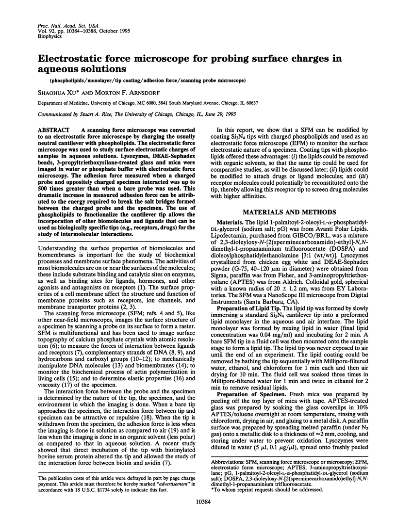
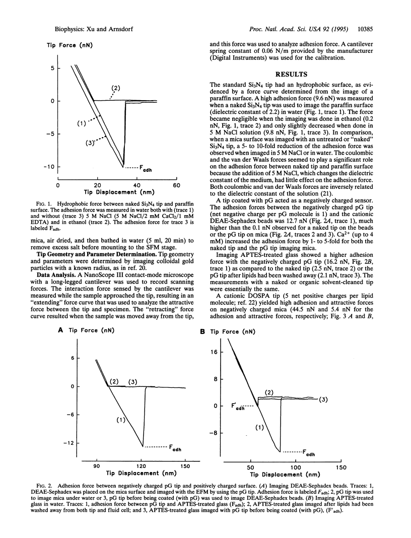
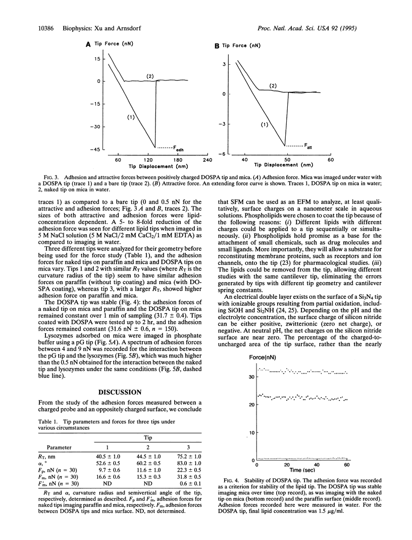
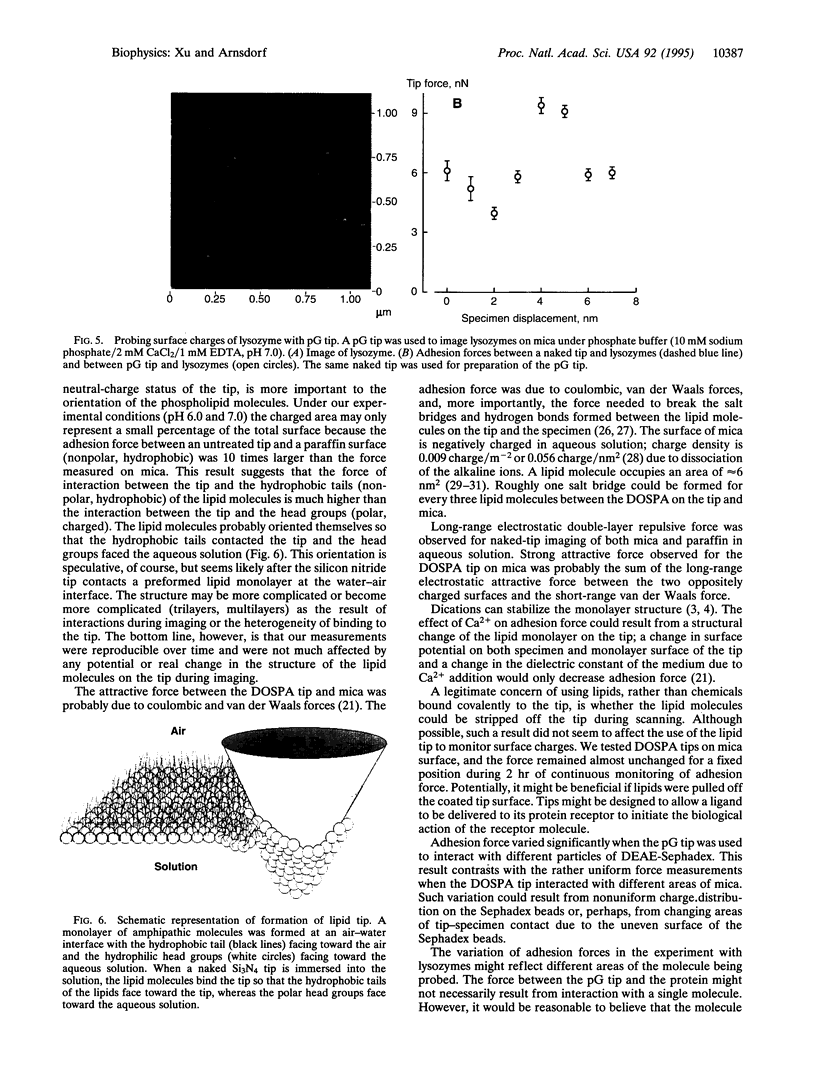
A scanning force microscope was converted to an electrostatic force microscope by charging the usually neutral cantilever with phospholipids. The electrostatic force microscope was used to study surface electrostatic charges of samples in aqueous solutions. Lysozymes, DEAE-Sephadex beads, 3-propyltriethoxysilane-treated glass and mica were imaged in water or phosphate buffer with electrostatic force microscopy. The adhesion force measured when a charged probe and oppositely charged specimen interacted was up to 500 times greater than when a bare probe was used. This dramatic increase in measured adhesion force can be attributed to the energy required to break the salt bridges formed between the charged probe and the specimen. The use of phospholipids to functionalize the cantilever tip allows the incorporation of other biomolecules and ligands that can be used as biologically specific tips (e.g., receptors, drugs) for the study of intermolecular interactions.
Full text
PDF
Images in this article

Selected References
These references are in PubMed. This may not be the complete list of references from this article.
- Binnig G, Quate CF, Gerber C. Atomic force microscope. Phys Rev Lett. 1986 Mar 3;56(9):930–933. doi: 10.1103/PhysRevLett.56.930. [DOI] [PubMed] [Google Scholar]
- Burnham NA, Dominguez DD, Mowery RL, Colton RJ. Probing the surface forces of monolayer films with an atomic-force microscope. Phys Rev Lett. 1990 Apr 16;64(16):1931–1934. doi: 10.1103/PhysRevLett.64.1931. [DOI] [PubMed] [Google Scholar]
- Butt H. J. Measuring electrostatic, van der Waals, and hydration forces in electrolyte solutions with an atomic force microscope. Biophys J. 1991 Dec;60(6):1438–1444. doi: 10.1016/S0006-3495(91)82180-4. [DOI] [PMC free article] [PubMed] [Google Scholar]
- Florin E. L., Moy V. T., Gaub H. E. Adhesion forces between individual ligand-receptor pairs. Science. 1994 Apr 15;264(5157):415–417. doi: 10.1126/science.8153628. [DOI] [PubMed] [Google Scholar]
- Frisbie C. D., Rozsnyai L. F., Noy A., Wrighton M. S., Lieber C. M. Functional group imaging by chemical force microscopy. Science. 1994 Sep 30;265(5181):2071–2074. doi: 10.1126/science.265.5181.2071. [DOI] [PubMed] [Google Scholar]
- Hansma H. G., Vesenka J., Siegerist C., Kelderman G., Morrett H., Sinsheimer R. L., Elings V., Bustamante C., Hansma P. K. Reproducible imaging and dissection of plasmid DNA under liquid with the atomic force microscope. Science. 1992 May 22;256(5060):1180–1184. doi: 10.1126/science.256.5060.1180. [DOI] [PubMed] [Google Scholar]
- Haydon D. A., Hladky S. B. Ion transport across thin lipid membranes: a critical discussion of mechanisms in selected systems. Q Rev Biophys. 1972 May;5(2):187–282. doi: 10.1017/s0033583500000883. [DOI] [PubMed] [Google Scholar]
- Henderson E., Haydon P. G., Sakaguchi D. S. Actin filament dynamics in living glial cells imaged by atomic force microscopy. Science. 1992 Sep 25;257(5078):1944–1946. doi: 10.1126/science.1411511. [DOI] [PubMed] [Google Scholar]
- Hoh J. H., Lal R., John S. A., Revel J. P., Arnsdorf M. F. Atomic force microscopy and dissection of gap junctions. Science. 1991 Sep 20;253(5026):1405–1408. doi: 10.1126/science.1910206. [DOI] [PubMed] [Google Scholar]
- Lal R., John S. A. Biological applications of atomic force microscopy. Am J Physiol. 1994 Jan;266(1 Pt 1):C1–21. doi: 10.1152/ajpcell.1994.266.1.C1. [DOI] [PubMed] [Google Scholar]
- Lee G. U., Chrisey L. A., Colton R. J. Direct measurement of the forces between complementary strands of DNA. Science. 1994 Nov 4;266(5186):771–773. doi: 10.1126/science.7973628. [DOI] [PubMed] [Google Scholar]
- Levine Y. K., Wilkins M. H. Structure of oriented lipid bilayers. Nat New Biol. 1971 Mar 17;230(11):69–72. doi: 10.1038/newbio230069a0. [DOI] [PubMed] [Google Scholar]
- Ohnesorge F., Binnig G. True atomic resolution by atomic force microscopy through repulsive and attractive forces. Science. 1993 Jun 4;260(5113):1451–1456. doi: 10.1126/science.260.5113.1451. [DOI] [PubMed] [Google Scholar]
- Radmacher M., Tillmann R. W., Gaub H. E. Imaging viscoelasticity by force modulation with the atomic force microscope. Biophys J. 1993 Mar;64(3):735–742. doi: 10.1016/S0006-3495(93)81433-4. [DOI] [PMC free article] [PubMed] [Google Scholar]
- Roth N. S., Lefkowitz R. J., Caron M. G. Structure and function of the adrenergic receptor family. Adv Exp Med Biol. 1991;308:223–238. doi: 10.1007/978-1-4684-6015-5_20. [DOI] [PubMed] [Google Scholar]
- Tao N. J., Lindsay S. M., Lees S. Measuring the microelastic properties of biological material. Biophys J. 1992 Oct;63(4):1165–1169. doi: 10.1016/S0006-3495(92)81692-2. [DOI] [PMC free article] [PubMed] [Google Scholar]
- Tsao Y. H., Evans D. F., Wennerström H. Long-range attractive force between hydrophobic surfaces observed by atomic force microscopy. Science. 1993 Oct 22;262(5133):547–550. doi: 10.1126/science.8211182. [DOI] [PubMed] [Google Scholar]
- Xu S., Arnsdorf M. F. Calibration of the scanning (atomic) force microscope with gold particles. J Microsc. 1994 Mar;173(Pt 3):199–210. doi: 10.1111/j.1365-2818.1994.tb03442.x. [DOI] [PubMed] [Google Scholar]
- Xu S., Cramer W. A., Peterson A. A., Hermodson M., Montecucco C. Dynamic properties of membrane proteins: reversible insertion into membrane vesicles of a colicin E1 channel-forming peptide. Proc Natl Acad Sci U S A. 1988 Oct;85(20):7531–7535. doi: 10.1073/pnas.85.20.7531. [DOI] [PMC free article] [PubMed] [Google Scholar]
- Yang J., Tamm L. K., Tillack T. W., Shao Z. New approach for atomic force microscopy of membrane proteins. The imaging of cholera toxin. J Mol Biol. 1993 Jan 20;229(2):286–290. doi: 10.1006/jmbi.1993.1033. [DOI] [PubMed] [Google Scholar]