Plasmacytomas are localized tumors of monoclonal plasma cells without evidence of significant bone marrow infiltration or systemic myeloma, comprising ∼2–10% of all plasma cell neoplasms.1 Medullary plasmacytomas classically occur in the bone of the vertebrae or skull, whereas extramedullary plasmacytomas occur in the soft tissues, most frequently in the head and neck region involving the sinuses or rhinopharynx.1 Both medullary and extramedullary plasmacytomas occur in males more often than in females with a median age at diagnosis of 54 years.1 Furthermore, plasmacytomas in general are fairly radiosensitive, such that the current treatment of choice is localized radiation therapy except in cases of neurologic compromise or bone instability that require surgical resection.2 There is no evidence thus far that adjuvant systemic chemotherapy offers additional clinical benefit.2 Despite these similarities, medullary and extramedullary plasmacytomas are considered distinct clinical entities owing to differences in their predisposition to evolve into multiple myeloma (MM). Medullary plasmacytomas are traditionally thought to have a higher risk of progression, also with a lower 5-year survival rate of 33% once they have progressed to MM (as opposed to 5-year survival rate of 100% in extramedullary plasmacytomas that progress to MM).2 Yet, very little is known regarding genetic or molecular factors that distinguish these two groups of plasmacytomas and influence their clinical outcome.
MicroRNAs (miRNAs) are small non-coding RNAs of ∼22 nt in length, which regulate target genes through posttranscriptional degradation or translational repression.3 Downstream programs impact cell cycle progression, apoptosis, proliferation and angiogenesis among others, and through these mechanisms, contribute to hematopoiesis and human cancers.4 Multiple recent studies have linked deregulated miRNA expression to the pathogenesis and progression of MM.5, 6, 7 In one study, miRNA expression profiling of CD138+ bone marrow plasma cells from MM patients, normal donors and those with monoclonal gammopathy of undetermined significance (MGUS) identified selective upregulation of miRNAs including miR-19a and -19b in MM patient samples. miR-19a and -19b were shown to suppress the expression of SOCS-1, a negative regulator of IL-6/STAT3 pathway.6 Another study proposed a ‘MM-specific microRNA signature' defined by upregulation of miR-222, -221, -382, -181a and -181b, and downregulation of miR-15a and -16, in which the latter functionally inhibit AKT3, ribosomal protein S6 as well as angiogenic VEGF (vascular endothelial growth factor).7 These miRNA–mRNA–protein networks add another layer of complexity to the pathogenesis of MM. To date, no published study has examined the global miRNA expression differences between plasmacytoma and MM, or between the intra- versus extramedullary plasmacytomas, at least in part due to the limited availability of plasmacytoma patient samples.
Here, we analyzed 15 patients with plasmacytomas to gain insight into miRNA expression pattern differences between medullary and extramedullary plasmacytomas. On the basis of the diagnostic criteria for localized plasmacytoma, 11 patients were classified as having medullary plasmacytoma and 4 patients as extramedullary. Patient characteristics are summarized in Supplementary Table 1. The median age at diagnosis was 61 and 57 years in the medullary and extramedullary groups, respectively, largely consistent with the reported age range in the literature. The patient population consisted of nine males and six females (with male-to-female ratio of 8:3 in the medullary and 1:3 in the extramedullary groups). Although limited in some cases by loss to follow-up, at least 4 of the 11 medullary plasmacytoma patients definitively progressed to develop MM, whereas only 1 of the 4 extramedullary plasmacytoma patients progressed.
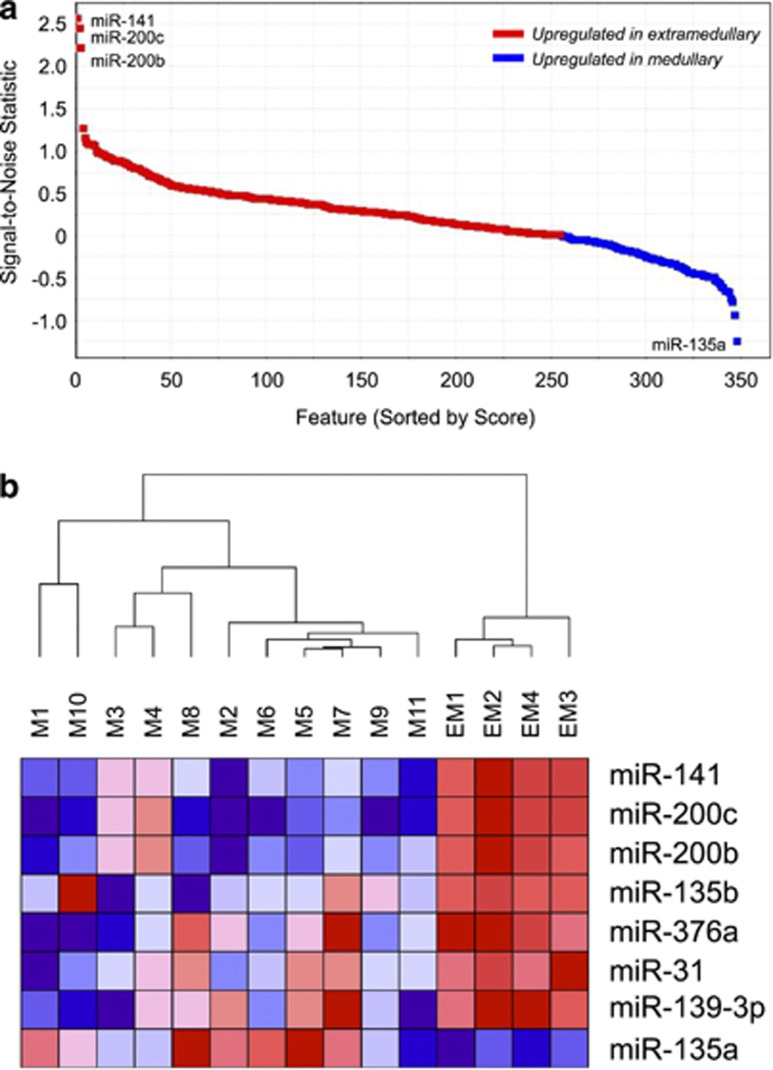
For our miRNA microarray analysis, miRNAs derived from the above patient population were profiled using the TaqMan Human MicroRNA Low Density Arrays Version 2.0 (Applied Biosystems, Grand Island, NY, USA), consisting of a total of 667 unique assays against human miRNAs. Comparative marker selection analysis8 (signal-to-noise ratio score cutoff of ±0.5) of the miRNA microarray data set after missing value imputation and normalization revealed seven miRNAs that were significantly upregulated in the extramedullary versus medullary plasmacytomas: miR-141, miR-200c, miR-200b, miR-135b, miR-139-3p, miR-376a and miR-31 (Figure 1a; Supplementary Table 2). In addition, one miRNA was identified as significantly downregulated in the extramedullary versus medullary plasmacytoma: miR-135a. All miRNAs had a fold change of at least 1.5 and a q-value cutoff of 0.05 (Supplementary Figure 1).
Figure 1.
Candidate miRNAs differentially expressed in medullary and extramedullary plasmacytomas. (a) Comparative marker selection score plot for miRNAs differentially expressed between medullary and extramedullary plasmacytomas. Red, upregulated in extramedullary plasmacytomas; blue, upregulated in medullary plasmacytomas. (b) Hierarchical clustering dendrogram of miRNA expression for the 15 analyzed plasmacytoma patient samples. Red, upregulated; blue, downregulated.
Notably, hierarchical clustering of our patient samples using these eight candidate miRNAs and Pearson correlation showed very clear linkage segregation between the two phenotypes of plasmacytomas (Figure 1b). This demonstrates that our eight miRNAs could represent a candidate miRNA signature to define the plasmacytoma classes. Indeed, unsupervised hierarchical clustering using all miRNAs analyzed in our data set also showed segregation between medullary and extramedullary plasmacytomas (data not shown).
We next predicted target genes for the above candidate miRNAs using the in silico algorithm TargetScan. The list of predicted targets was subsequently entered as input to the PANTHER gene ontogeny database to query for gene pathways enriched in this target gene list. As a control, the same number of miRNAs were selected at random using a computational random number generator (miR-125b, miR-138, miR-185, miR-224, miR-330-3p, miR-487b, miR-520c-3p and miR-766), and their targets and pathways were analyzed in parallel. Several pathways were more significantly enriched in the candidate miRNA target gene list (Table 1; Supplementary Table 3), including FGF, TGF-beta and angiogenesis pathways. Angiogenesis (which in turn is regulated by FGF and TGF-beta pathways, among others) is known to have a pivotal role in MM pathogenesis, and more specifically, in mediating the progression of plasmacytoma to MM.9, 10 Our finding proposes the notion that differential regulation of angiogenesis pathways may further be biologically relevant to the variant propensity of medullary versus extramedullary plasmacytomas to progress to MM, similar to their role in regulating the progression of MGUS to MM.9 Our analysis also discerns PI3 kinase and apoptosis pathways to be more highly enriched in the candidate miRNA target gene list, suggesting differential regulation of growth and proliferative capacity in extramedullary versus medullary plasmacytoma.
Table 1. TargetScan-predicted target genes for candidate miRNAs differentially expressed between extramedullary versus medullary plasmacytomas.
| Pathway | No. in ref. | No. in list | Expected | +/−a | P-value (Bonferroni-corrected) |
|---|---|---|---|---|---|
| Unclassified | 19 446 | 1985 | 2132.42 | − | 1.51E−17 |
| Gonadotropin-releasing hormone receptor pathway | 228 | 76 | 25 | + | 1.96E−14 |
| EGF receptor signaling pathway | 123 | 43 | 13.49 | + | 1.89E−08 |
| FGF signaling pathway | 115 | 40 | 12.61 | + | 9.54E−08 |
| PDGF signaling pathway | 132 | 42 | 14.47 | + | 4.65E−07 |
| Angiogenesis | 152 | 44 | 16.67 | + | 3.01E−06 |
| Ras pathway | 73 | 24 | 8.01 | + | 6.33E−04 |
| TGF-beta signaling pathway | 95 | 28 | 10.42 | + | 7.92E−04 |
| Insulin/IGF pathway–protein kinase B signaling cascade | 37 | 16 | 4.06 | + | 1.00E−03 |
| Apoptosis signaling pathway | 113 | 31 | 12.39 | + | 1.04E−03 |
| Axon guidance mediated by netrin | 34 | 15 | 3.73 | + | 1.54E−03 |
| Insulin/IGF pathway–mitogen-activated protein kinase kinase/MAP kinase cascade | 32 | 14 | 3.51 | + | 3.31E−03 |
| Integrin signaling pathway | 175 | 40 | 19.19 | + | 3.57E−03 |
| Metabotropic glutamate receptor group III pathway | 56 | 19 | 6.14 | + | 4.11E−03 |
| PI3 kinase pathway | 47 | 17 | 5.15 | + | 4.97E−03 |
Abbreviation: ref., reference.
Only pathways with Bonferroni-corrected P<0.005 are listed. Pathways listed in bold type are unique to the candidate miRNA list (not overlapping in the control miRNA list, Supplementary Table 3).
‘+' indicates that pathways are overrepresented in the analyzed miRNA target gene list compared with the ‘ref.' Homo sapiens gene list. ‘−' indicates that pathways are underrepresented.
In parallel, the top candidate differentially regulated miRNAs are individually known to be involved in the regulation of tumor cell growth, proliferation and metastatic capacity. For example, miR-200b and miR-200c, two of the top three ranked miRNAs in our analysis, serve as tumor suppressors by downregulating VEGF and angiogenesis pathways, thereby reducing tumor metastatic potential.11, 12 Furthermore, miR-139 has been implicated in targeting CXCR4 in cancer.13 SDF-1alpha/CXCR4 axis is an established player in bone marrow homing via regulation of chemotaxis, motility, and expansion and migration of myeloma cells.14 Differential miRNA-mediated regulation of these pathways may have a role in the plasmacytoma biology and prognosis.
In summary, we performed miRNA microarray profiling to identify a set of miRNAs differentially expressed between medullary versus extramedullary plasmacytomas. To the best of our knowledge, this is the first published study to date that offers insight into the role of miRNAs in plasmacytoma biology. It was beyond the scope of this investigation to precisely define how each candidate miRNA contributes to the distinct clinical characteristics of medullary and extramedullary plasmacytomas. However, our study proposes that regulation of a particular set of miRNAs may affect the plasmacytomas' differential propensity to progress to MM by influencing their proliferative and angiogenic capacity.
Additional studies are required to confirm our findings with a larger patient sample size, ideally with balanced sample number in each class and with longer clinical follow-up. A larger study will allow for a more rigorous statistical analysis to assess differential miRNA expression. It will also permit a more in-depth analysis comparing plasmacytomas that ultimately do or do not progress to myeloma, and to further compare differential miRNA expression between each plasmacytoma class and MM. Our current data set was not powered to achieve the aforementioned analyses. Interestingly, 2 of the 11 patients with medullary plasmacytoma in our data set progressed to myeloma within 1 year. It is not entirely clear based on clinical parameters alone what could explain the relatively rapid progression. Although one of the two patients was diagnosed at the advanced age of 81 years (Supplementary Table 1), which is a known poor prognostic factor for progression, the other was diagnosed at 61 years of age. In cases like these, examining for miRNA expression differences that may contribute to the prognosis would provide helpful biological insights. With regards to elucidating miRNA differences between plasmacytomas versus MM, one study previously showed that miR-223 expression was lacking in extramedullary plasmacytoma compared with MM, suggesting that miR-223(−) phenotype could represent one distinguishing factor between the two.15 However, this study examined the expression of only a very limited subset of miRNAs (namely, miR-30a, -93, -181b and -223) using quantitative real-time reverse transcriptase PCR, and excluded patients with clinical diagnosis of medullary plasmacytoma. It will be important to continue to investigate how the global miRNA expression profiles differ between extramedullary and medullary plasmacytoma and MM. This will expand our knowledge of the biology of these plasma cell neoplasms, refine how we assess the risk for progression and thereby identify patients who may benefit from adjuvant novel or targeted therapies.
The authors declare no conflict of interest.
Footnotes
Supplementary Information accompanies this paper on Blood Cancer Journal website (http://www.nature.com/bcj)
Supplementary Material
References
- Galieni P, Cavo M, Avvisati G, Pulsoni A, Falbo R, Bonelli MA, et al. Solitary plasmacytoma of bone and extramedullary plasmacytoma: two different entities. Ann Oncol. 1995;6:687–691. doi: 10.1093/oxfordjournals.annonc.a059285. [DOI] [PubMed] [Google Scholar]
- Kilciksiz S, Karakoyun-Celik O, Agaoglu FY, Haydaroglu A. A review for solitary plasmacytoma of bone and extramedullary plasmacytoma. ScientificWorldJournal. 2012;2012:895765. doi: 10.1100/2012/895765. [DOI] [PMC free article] [PubMed] [Google Scholar]
- Bartel D. MicroRNAs: genomics, biogenesis, mechanism, and function. Cell. 2004;116:281–297. doi: 10.1016/s0092-8674(04)00045-5. [DOI] [PubMed] [Google Scholar]
- Iorio MV, Croce CM. MicroRNAs in cancer: small molecules with a huge impact. J Clin Oncol. 2009;27:5848–5856. doi: 10.1200/JCO.2009.24.0317. [DOI] [PMC free article] [PubMed] [Google Scholar]
- Dimopoulos K, Gimsing P, Grønbæk K. Aberrant microRNA expression in multiple myeloma. Eur J Haematol. 2013;91:95–105. doi: 10.1111/ejh.12124. [DOI] [PubMed] [Google Scholar]
- Pichiorri F, Suh SS, Ladetto M, Kuehl M, Palumbo T, Drandi D, et al. MicroRNAs regulate critical genes associated with multiple myeloma pathogenesis. Proc Natl Acad Sci USA. 2008;105:12885–12890. doi: 10.1073/pnas.0806202105. [DOI] [PMC free article] [PubMed] [Google Scholar]
- Roccaro AM, Sacco A, Thompson B, Leleu X, Azab F, Runnels J, et al. MicroRNAs 15a and 16 regulate tumor proliferation in multiple myeloma. Blood. 2009;113:6669–6680. doi: 10.1182/blood-2009-01-198408. [DOI] [PMC free article] [PubMed] [Google Scholar]
- Gould J, Getz G, Monti S, Reich M, Mesirov MR. Comparative gene marker selection suite. Bioinformatics. 2006;22:1924–1925. doi: 10.1093/bioinformatics/btl196. [DOI] [PubMed] [Google Scholar]
- Rajkumar SV, Mesa RA, Fonseca R, Schroeder G, Plevak MF, Dispenzierl A. Bone marrow angiogenesis in 400 patients with monoclonal gammopathy of undetermined significance, multiple myeloma, and primary amyloidosis. Clin Cancer Res. 2002;8:2210–2216. [PubMed] [Google Scholar]
- Kumar S, Fonseca R, Dispenzieri A, Lacy MQ, Lust JA, Wellik L, et al. Prognostic value of angiogenesis in solitary bone plasmacytoma. Blood. 2003;101:1715–1717. doi: 10.1182/blood-2002-08-2441. [DOI] [PubMed] [Google Scholar]
- Choi YC, Yoon S, Jeong Y, Yoon J, Baek K. Regulation of vascular endothelial growth factor signaling by miR-200b. Mol Cells. 2011;32:77–82. doi: 10.1007/s10059-011-1042-2. [DOI] [PMC free article] [PubMed] [Google Scholar]
- Shi L, Zhang S, Wu H, Zhang L, Dai X, Hu J, et al. MiR-200c increases the radiosensitivity of non-small-cell lung cancer cell line A549 by targeting VEGF-VEGFR2 pathway. PLoS One. 2013;8:e78344. doi: 10.1371/journal.pone.0078344. [DOI] [PMC free article] [PubMed] [Google Scholar]
- Luo HN, Wang ZH, Sheng Y, Zhang Q, Yan J, Hou J, et al. MiR-139 targets CXCR4 and inhibits the proliferation and metastasis of laryngeal squamous carcinoma cells. Med Oncol. 2014;31:789. doi: 10.1007/s12032-013-0789-z. [DOI] [PubMed] [Google Scholar]
- Bladé J, Fernández de Larrea C, Rosiñol L, Cibeira MT, Jiménez R, Powles R. Soft-tissue plasmacytomas in multiple myeloma: incidence, mechanisms of extramedullary spread, and treatment approach. J Clin Oncol. 2011;29:3805–3812. doi: 10.1200/JCO.2011.34.9290. [DOI] [PubMed] [Google Scholar]
- Yu S-C, Chen S-U, Lu W, Liu T-Y, Lin C-W. Expression of CD19 and lack of miR-223 distinguish extramedullary plasmacytoma from multiple myeloma. Histopathology. 2011;58:896–905. doi: 10.1111/j.1365-2559.2011.03793.x. [DOI] [PubMed] [Google Scholar]
Associated Data
This section collects any data citations, data availability statements, or supplementary materials included in this article.


