The cadherin gene FAT1, located on chromosome 4q34-35 (ref. 1) within a region frequently deleted in human cancers,2 encodes a large protein with 34 extracellular cadherin repeats.3 In solid tumors, aberrant expression of FAT1 was found to be associated with disease progression.4 Although the gene was originally cloned from a human T-cell acute lymphoblastic leukemia (T-ALL) cell line,4 FAT1 just recently gained interest owing to its altered gene expression levels and the detection of somatic mutations identified by next-generation sequencing (NGS) in acute leukemia.2, 5, 6, 7 FAT1 was shown to be aberrantly expressed in pediatric patients with acute leukemia, whereas hematopoietic progenitors from healthy donors lacked FAT1 expression.5, 8 In addition, a recent report correlated high FAT1 expression with a higher probability of relapse in a small cohort of pediatric patients with B-cell precursor acute lymphoblastic leukemia (BCP-ALL) based on an in silico analysis comprising two BCP-ALL data sets including 32 and 27 patients.5
With the emergence of NGS, it has become obvious that FAT1 is not only aberrantly expressed in various tumors, but also frequently mutated in solid tumors.7, 9 Morris et al.2 were able to link the mutational inactivation of FAT1 to the loss of its tumor suppressor capacity and the activation of the WNT pathway. In summary, these data make FAT1 an interesting candidate for disease monitoring, risk stratification and the development of targeted therapies. Herein, we investigated FAT1 expression in a large, homogenously treated cohort of adult acute leukemia patients, and explored the mutation status of FAT1 and its clinical significance.
We analyzed FAT1 expression by real-time PCR in different cell populations of healthy donors, various leukemia cell lines, a small cohort of acute myeloid leukemia (AML; n=13), in 112 adult T-ALL samples and in 129 adult BCP-ALL samples (Supplementary Methods). We examined the clinical and molecular characteristics with respect to FAT1 expression in this large cohort of adult ALL patients using specimens sent to the reference laboratory of the German Study Group for adult ALL (GMALL; n=231). Of these, 180 patients were enrolled into the trials GMALL 06/99 and 07/03 with available clinical follow-up. The treatment strategy of the GMALL trials has been described previously (Supplementary Methods). We were able to confirm the reported expression pattern for FAT1 in different cell lines (Supplementary Figure S1).5 The cell line BE13 showed the lowest, nearly absent, expression of FAT1 and was used as a cutoff to define samples with a high expression (FAT1pos) compared with a lower/absent expression (FAT1neg). We also investigated the expression of FAT1 in different cell populations from healthy donors. Unselected bone marrow (BM), CD34+ progenitors, peripheral blood and CD3+ T cells from healthy donors lacked FAT1 expression (Supplementary Figure S2), whereas FAT1 expression was highly expressed in BM-derived mesenchymal stromal cells (BMSC) from healthy donors (Supplementary Figure S2). In contrast, FAT1 was aberrantly expressed in adult leukemia: 23% of AML and 32% of BCP-ALL patients expressed FAT1 and were defined as FAT1pos. The highest percentage of FAT1pos patients was found within the T-ALL cohort (54%, Supplementary Figure S2).
FAT1 expression was correlated with a more mature leukemic immunophenotype. In BCP-ALL, patients with a preB-ALL or a common ALL immunophenotype were in 57% and 26% classified as FAT1pos compared with only 9% of pro-B-ALL patients (see Supplementary Table S2). In T-ALL, a genotype–phenotype association was even more striking: 74% of patients with thymic T-ALL were FAT1pos compared with 45% of patients with mature T-ALL and only 4% of early T-ALL patients (see Supplementary Table S1). In accordance with the predominance of FAT1 expression in more mature T-ALL, FAT1pos patients had a higher rate of clonal T-cell receptor rearrangement and a lower expression of the stem cell-associated genes IGFBP7, BAALC and MN1 (Supplementary Table S1). Likewise, FAT1pos T-ALL patients showed higher white blood cell counts (WBC; ≥30 000/μl at diagnosis) compared with FAT1neg T-ALL patients (78% vs 42%, P<0.01, Supplementary Table S1). No significant differences were observed between the FAT1pos versus FAT1neg groups in age or sex among T-ALL patients.
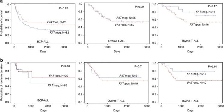
Regarding response to a standard induction therapy, we found no differences between FAT1pos and FAT1neg BCP-ALL patients (Supplementary Table S2). In T-ALL, FAT1neg patients failed to achieve a complete remission more frequently (3/25) after induction therapy compared with FAT1pos patients (0/50, P=0.04, Supplementary Table S1). In contrast to the in silico data of pediatric patients,5 we found no differences in BCP-ALL or T-ALL between FAT1pos and FAT1neg patients regarding overall survival and remission duration (Figure 1). However, in the prognostic favorable subgroup of thymic T-ALL, we observed an inferior overall survival for FAT1pos patients, although not statistically significant.
Figure 1.
Overall survival (a) and duration of remission (b) for patients with BCP-ALL, overall T-ALL and the standard-risk subgroup of thymic T-ALL enrolled into GMALL trials.
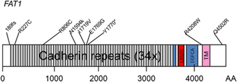
On the basis of the high frequency of FAT1 expression in T-ALL and recurrent FAT1 mutations in early T-cell precursor (ETP)-ALL,7 we examined 68 T-ALL samples for the presence of FAT1 mutations by target enrichment and NGS (Supplementary Methods). Interestingly, FAT1 mutations were detectable in a considerable number of adult T-ALL patients (8/68, 12%, Supplementary Table S3). One patient carried two point mutations within FAT1. All mutations were missense mutations, one leading to a frameshift and another encoding a stop codon (Supplementary Table S3). Mutations were predominantly located within the cadherin domains (Figure 2). T-ALL patients with FAT1 mutations (FAT1mut) did not differ from T-ALL patients carrying a wild-type FAT1 (FAT1wt) regarding sex, age, WBC and expression of specific cell surface antigens associated with an early differentiation stage. FAT1 mutations were present in early T-ALL (3/12, 25%) and in thymic T-ALL (5/41, 12%), but absent in T-ALL with a mature immunophenotype (0/15, NS, Supplementary Table S4). Expression of FAT1, was more common in FAT1wt T-ALL patients than in FAT1mut patients (55% vs 25%, P=0.15). No differences were observed in overall survival and remission duration between FAT1mut and FAT1wt patients (Supplementary Figure S3).
Figure 2.

Protein domain plot of FAT1 with mutations (n=9) found in 8 of 68 T-ALL patients. One patient carried two mutations. Changes are annotated in Supplementary Table S3.
Although there are increasing data on the genetic characterization of ALL, only few molecular markers have been integrated into risk stratification for individualized therapies. The postulated correlation of high FAT1 expression with inferior outcome in pediatric BCP-ALL5 could not be confirmed in our cohort of adult BCP-ALL. The most obvious reasons for these conflicting results might be different therapeutic approaches and large age differences between pediatric and adult patients as shown for other prognostic markers.10 Also limitations of in silico analyses of cohorts including low number of patients might at least in part explain these conflicting findings. Although FAT1 might not have a prognostic value, its expression and mutation profile make it an interesting candidate for minimal residual disease monitoring, the development of targeted therapies, and improved understanding of leukemogenesis in different ALL subgroups.
In addition to its potential role in leukemogenesis, it is tempting to speculate about the role of FAT1 in the interaction of leukemic cells with the microenvironment. It is known that, FAT1 is associated with cell migration, polarity and cell–cell adhesion and direct interaction with β-catenin.2, 4, 11 As we found a high FAT1 expression in BMSC, FAT1 might have a role in the stabilization of the interaction of leukemic cells with the bone marrow niche and/or thymic homing. This might also explain the significantly higher expression of FAT1 in the more differentiated subgroups of T-ALL and BCP-ALL. On the other hand, inactivating mutations of FAT1 in different human cancers have been linked to the inability to bind β-catenin and deregulated activation of the WNT pathway.2 These mechanisms might have a role in solid cancer leading to higher treatment sensitivity and evasion of tumor metastasis.2, 12 Interestingly, in gingiva-buccal oral squamous cell cancer, FAT1 mutations occur in addition to mutations in NOTCH1 and MLL2, a spectrum very similar to the one observed in ETP-ALL.7, 12 Deregulation of the WNT pathway has been linked to leukemogenesis in T-ALL.13, 14 The previously unreported FAT1 mutation rate of 15% in adult T-ALL stresses the importance of the WNT pathway in T-ALL.
In conclusion, we explored the pattern of FAT1 expression and its mutation status in a large, homogenously treated cohort of adult ALL patients. Our analysis revealed an aberrant expression predominantly in mature BCP-ALL and thymic T-ALL and a high rate of FAT1 mutations in T-ALL. Further studies should explore a link to WNT pathway activation and potential therapeutic implications.
Acknowledgments
We thank Liliana H Mochmann for critical reading of the manuscript. This work was supported by grants from the Deutsche Krebshilfe (Mildred Scheel Professur) and Gutermuth-Stiftung to CDB, Deutsche Krebshilfe grant 70-2657-Ho2 to DH and partly BMBF 01GI 9971 to DH and NG, and Deutsche Krebshilfe grant 109031 to PAG.
The authors declare no conflict of interest.
Footnotes
Supplementary Information accompanies this paper on Blood Cancer Journal website (http://www.nature.com/bcj)
Supplementary Material
References
- Dunne J, Hanby AM, Poulsom R, Jones TA, Sheer D, Chin WG, et al. Molecular cloning and tissue expression of FAT, the human homologue of the Drosophila fat gene that is located on chromosome 4q34-q35 and encodes a putative adhesion molecule. Genomics. 1995;30:207–223. doi: 10.1006/geno.1995.9884. [DOI] [PubMed] [Google Scholar]
- Morris LG, Kaufman AM, Gong Y, Ramaswami D, Walsh LA, Turcan S, et al. Recurrent somatic mutation of FAT1 in multiple human cancers leads to aberrant Wnt activation. Nat Genet. 2013;45:253–261. doi: 10.1038/ng.2538. [DOI] [PMC free article] [PubMed] [Google Scholar]
- Mahoney PA, Weber U, Onofrechuk P, Biessmann H, Bryant PJ, Goodman CS. The fat tumor suppressor gene in Drosophila encodes a novel member of the cadherin gene superfamily. Cell. 1991;67:853–868. doi: 10.1016/0092-8674(91)90359-7. [DOI] [PubMed] [Google Scholar]
- Sadeqzadeh E, de Bock CE, Thorne RF. Sleeping Giants: Emerging Roles for the Fat Cadherins in Health and Disease. Med Res Rev. 2013;34:190–221. doi: 10.1002/med.21286. [DOI] [PubMed] [Google Scholar]
- de Bock CE, Ardjmand A, Molloy TJ, Bone SM, Johnstone D, Campbell DM, et al. The Fat1 cadherin is overexpressed and an independent prognostic factor for survival in paired diagnosis-relapse samples of precursor B-cell acute lymphoblastic leukemia. Leukemia. 2012;26:918–926. doi: 10.1038/leu.2011.319. [DOI] [PubMed] [Google Scholar]
- Katoh M. Function and cancer genomics of FAT family genes (review) Int J Oncol. 2012;41:1913–1918. doi: 10.3892/ijo.2012.1669. [DOI] [PMC free article] [PubMed] [Google Scholar]
- Neumann M, Heesch S, Schlee C, Schwartz S, Gokbuget N, Hoelzer D, et al. Whole-exome sequencing in adult ETP-ALL reveals a high rate of DNMT3A mutations. Blood. 2013;121:4749–4752. doi: 10.1182/blood-2012-11-465138. [DOI] [PubMed] [Google Scholar]
- Coustan-Smith E, Song G, Clark C, Key L, Liu P, Mehrpooya M, et al. New markers for minimal residual disease detection in acute lymphoblastic leukemia. Blood. 2011;117:6267–6276. doi: 10.1182/blood-2010-12-324004. [DOI] [PMC free article] [PubMed] [Google Scholar]
- Morris LG, Ramaswami D, Chan TA. The FAT epidemic: a gene family frequently mutated across multiple human cancer types. Cell Cycle. 2013;12:1011–1012. doi: 10.4161/cc.24305. [DOI] [PMC free article] [PubMed] [Google Scholar]
- Pui CH, Relling MV, Downing JR. Acute lymphoblastic leukemia. N Engl J Med. 2004;350:1535–1548. doi: 10.1056/NEJMra023001. [DOI] [PubMed] [Google Scholar]
- Hou R, Liu L, Anees S, Hiroyasu S, Sibinga NE. The Fat1 cadherin integrates vascular smooth muscle cell growth and migration signals. J Cell Biol. 2006;173:417–429. doi: 10.1083/jcb.200508121. [DOI] [PMC free article] [PubMed] [Google Scholar]
- India Project Team of the International Cancer Genome Consortium Mutational landscape of gingivo-buccal oral squamous cell carcinoma reveals new recurrently-mutated genes and molecular subgroups. Nat Commun. 2013;4:2873. doi: 10.1038/ncomms3873. [DOI] [PMC free article] [PubMed] [Google Scholar]
- Mochmann LH, Bock J, Ortiz-Tanchez J, Schlee C, Bohne A, Neumann K, et al. Genome-wide screen reveals WNT11, a non-canonical WNT gene, as a direct target of ETS transcription factor ERG. Oncogene. 2011;30:2044–2056. doi: 10.1038/onc.2010.582. [DOI] [PubMed] [Google Scholar]
- Behrens J, von Kries JP, Kuhl M, Bruhn L, Wedlich D, Grosschedl R, et al. Functional interaction of beta-catenin with the transcription factor LEF-1. Nature. 1996;382:638–642. doi: 10.1038/382638a0. [DOI] [PubMed] [Google Scholar]
Associated Data
This section collects any data citations, data availability statements, or supplementary materials included in this article.


