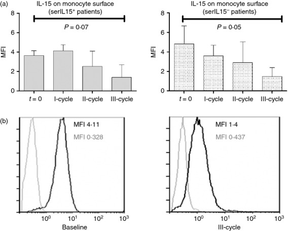
Figure 3.

(a) Interleukin-15 (IL-15) expression (mean fluorescence intensity; MFI) on CD14+ monocytes from rheumatoid arthritis patients at baseline and after each rituximab cycle. Patients were segregated according to serum IL-15 levels: serIL-15+ (baseline and I cycle n = 25, II-cycle n = 17 and III-cycle n = 13) and serIL-15− (baseline and I-cycle n = 8, II-cycle n = 6 and III-cycle n = 4) patients. P-values correspond to the comparison of IL-15 expression (MFI) on monocytes after third cycle of treatment with the baseline (t = 0) by Wilcoxon test for paired samples. Results were expressed as the mean and error bars represent SD. (b) IL-15 expression (black line) on monocytes from a representative patient at baseline and after the third cycle. Grey line represents the corresponding isotype control.
