Figure 1.

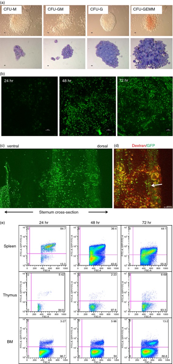
Embryonic stem (ES) cell-derived haematopoietic progenitor cells (HPCs) form colony-forming units (CFUs) and engraft into the bone marrow. (a) HPCs were derived from the 129SvJ ES cells and used to form CFUs. The cells formed multi-lineage haematopoietic cells (upper row) which were stained by the Giemsa stain (lower panel). (b) Engraftment of the HPCs into the bone marrow was studied by two-photon excitation microscopy 24, 48 and 72 hr post-transplantation. Sternums of transplanted mice were surgically cut open and subsequently imaged. After 24 hr, the population of HPCs was quite low. The density of the HPCs in the bone marrow increased with time. These experiments were repeated three times. (c) The transplanted HPCs were polarized towards the dorsal side of the sternum; however, it is still unclear why this is the case. (d) Two hours before mice were killed, they were intravenously injected with Dextran Texas Red to visualize blood vessels. Interestingly, HPCs were detected perivascularly and within the sinusoids (arrow head) of the bone marrow. (e) To investigate the distribution of the HPCs in lymphoid tissues post transplantation, mice were killed 24, 48 or 72 hr post transplantation and the spleen, thymus and bone marrow cells were recovered. The cells were analysed by flow cytometry for green fluorescent cells. Initially, most HPCs were in the spleen, but had drastically redistributed to other organs after 24 hr.
