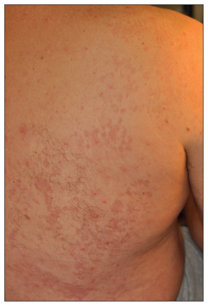
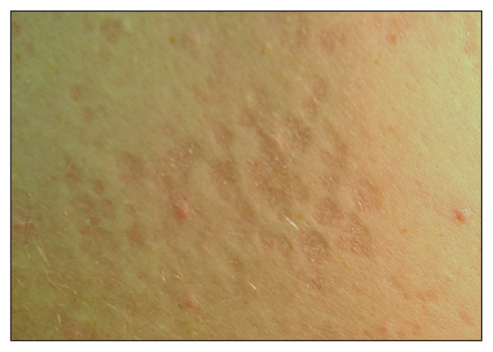
A 47-year-old man with a 25-year history of small plaque psoriasis treated with betamethasone dipropionate cream presented with well-demarcated areas of skin atrophy, confined to his back (Figure 1). The atrophic areas were mildly erythematous and showed slight scaling (Figure 2). Skin scrapings from these areas were examined microscopically using 15% potassium hydroxide with Parker blue–black ink. Thick fungal hyphae and grape-like bunches of spores were seen, consistent with Malassezia yeast.
Figure 1:
Pink, atrophic, scaly plaques on the back of a 47-year-old man.
Figure 2:
Close-up view of the patient’s skin under tangential lighting, showing mild scaling and atrophy.
Although pityriasis (tinea) versicolor is common, the atrophic form is unusual, with the first case reported in 1971.1 The atrophy is limited to the area affected by the pityriasis versicolor. Case reports have suggested an association with the use of topical corticosteroids. The fungus, which is confined to the stratum corneum and follicles, is thought to disrupt the skin barrier, allowing better absorption of the steroid, which induces atrophy. Corticosteroid-induced atrophy is usually associated with epidermal atrophy and telangiectasis. Our patient did not have this feature.
In a case series involving 12 patients with atrophying pityriasis versicolor, long-term topical corticosteroid use was reported for only one patient.2 In these cases, the atrophy was thought to have been induced directly by Malassezia yeast, which produce diffusible products that enhance dermal elastolysis, possibly by activating tissue histiocytes. Degeneration of elastic fibres has been noted on skin biopsies.3
Following treatment of Malassezia, the atrophy usually resolves. Our patient had a complete recovery following treatment with systemic ketoconazole.
The presence of fine scaling overlying the atrophy allows clinicians to distinguish this form of cutaneous atrophy from other causes of skin atrophy, especially anetoderma. Examination using potassium hydroxide confirms the diagnosis.
Footnotes
Competing interests: None declared.
This article has been peer reviewed.
References
- 1.De Graciansky P, Mery F. Atrophie sur pityriasis versicolor après corticotherapie locale prolongee [article in French]. Bull Soc Fr Dermatol Syphiligr 1971;78:295 [Google Scholar]
- 2.Crowson AN, Magro CM. Atrophying tinea versicolor: a clinical and histological study of 12 patients. Int J Dermatol 2003;42: 928–32 [DOI] [PubMed] [Google Scholar]
- 3.Romano C, Maritati E, Ghilardi A, et al. A case of pityriasis versicolor atrophicans. Mycoses 2005;48:439–41 [DOI] [PubMed] [Google Scholar]


