Abstract
The accepted importance of circulatory impairment to sickle cell anemia remains to be verified by in vivo experimentation. Intravital microscopy studies of blood flow in patients are limited to circulations that can be viewed noninvasively and are restricted from deliberate perturbations of the circulation. Further knowledge of sickle blood flow abnormalities has awaited an animal model of human sickle cell disease. We compared blood flow in the mucosal–intestinal microvessels of normal mice with that in transgenic knockout sickle cell mice that have erythrocytes containing only human hemoglobin S and that exhibit a degree of hemolytic anemia and pathological complications similar to the human disease. In sickle cell mice, in addition to seeing blood flow abnormalities such as sludging in all microvessels, we detected decreased blood flow velocity in venules of all diameters. Flow responses to hyperoxia in both normal and sickle cell mice were dramatic, but opposite: Hyperoxia promptly slowed or halted flow in normal mice but markedly enhanced flow in sickle cell mice. Intravital microscopic studies of this murine model provide important insights into sickle cell blood flow abnormalities and suggest that this model can be used to evaluate the causes of abnormal flow and new approaches to therapy of sickle cell disease.
Introduction
The importance of vaso-occlusion to the pathophysiology of sickle cell disease (SCD) is axiomatic, despite limited in vivo evidence to support this conviction. A rigorous understanding of the processes that modulate circulatory impairment requires a method for their examination in vivo during informative perturbations.
In the first written report of SCD in 1910, Herrick (1) speculated that the clinical consequences of the disorder were related to the “peculiar elongated and sickle-shaped red blood corpuscles” that he and his intern, Dr. Irons, had observed. Thirty years later, the noumenal “vicious cycle of erythrostasis” of Ham and Castle (2) provided indomitable support for the importance of hypoxia, red blood cell (RBC) sickling, and abnormal blood flow. Despite decades of research on the effects of hypoxia on RBC sickling and on sickle hemoglobin (Hb S) gelation (3), there was no direct evidence that oxygen deprivation caused rheologic impairment of sickle RBC (SS RBC) until the landmark report by Harris et al. (4) in 1956. Our current wealth of information on the in vitro effect of hypoxia on RBC sickling and Hb S gelation (3, 5) stands in marked contrast to limited in vivo observations consisting of a few assessments of microscopic flow — in 1947, Knisely et al. (6) included SCD among 61 disorders associated with “sludged blood” flow; in 1961, Fink et al. (7) reported abnormal conjunctival flow; and more recently, Lepowsky et al. (8) observed abnormal nailbed flow — and to the laser-Doppler demonstration of abnormal periodic flow in forearm skin capillaries by Rodgers et al. (9). Recently, we have used computer-assisted intravital videomicroscopy to characterize the abnormal microvascular morphometry and flow in the perilimbal conjunctivae of subjects with SCD (10).
Despite the inarguable importance of characterizing blood flow in human patients with SCD, such in vivo studies are limited to circulations that can be examined noninvasively. Moreover, the use of deliberate perturbations to discern determinants of abnormal blood flow is not feasible in these patients. Animal models were predicted to overcome both limitations. Studies of human SS RBC infused into other species in vivo or into animal tissues ex vivo provided meaningful information on the contributions of hypoxia and of different SS RBC subpopulations to sickle blood flow (11, 12). In particular, these studies defined that vaso-occlusion is a two-step process initiated by adherence of reticulocyte-enriched SS RBC and completed by trapping of dense SS RBC (13). Despite such contributions, trans-species studies are limited by the short survival of the human SS RBC infused and by the potential interspecies differences in adhesion molecules and procoagulants. The goal in developing models of human SCD has been to create a transgenic animal containing only human Hb S within host RBC membranes (12, 14–16). Recently, two groups (17, 18) independently developed superior transgenic knockout strains of SCD mice that contain only human Hb S within the RBC and exhibit the hematologic, and many of the clinical, abnormalities of human SCD. Intravital videomicroscopic studies on these animals, unlike studies of patients, are not restricted to noninvasive approaches and are adaptable to informative perturbations.
Traditional wisdom predicted that vaso-occlusion should occur in the microcirculation (3, 5), and this has been confirmed by ex vivo studies (13). The physiological vasoconstrictive responses of arterioles to oxygen administration (19) have been studied in a transgenic animal model of SCD (20). However, this model is significantly divergent from SCD in humans, having persistent mouse hemoglobin in the RBC, lacking anemia, and requiring artificial levels of hypoxia to elicit characteristic SS RBC pathobiologic responses (21). Because our SCD mice have only human Hb S in their RBC and conform closely to the SCD of humans, the findings we report here on blood flow properties of the SCD mouse represent the first intravital characterization of flow abnormalities and responses to oxygen administration of a relevant vascular bed in an in vivo setting comparable to human SCD. Further analysis of microcirculatory flow in these SCD mice is predicted to complement in vitro studies on SS RBC rheology, furnish important insights into the blood flow abnormalities of SCD, and guide the development of new therapies for the disorder.
Methods
Transgenic knockout SCD mice.
Two groups of eight mice each, ranging in weight from 18 to 35 g, were used in the primary comparisons of this study. One group, the controls, were C57BL /6 mice obtained from Charles River Laboratories (Wilmington, Massachusetts, USA). The SCD test group were produced through successive rounds of breeding a transgenic strain that expresses human α1-, Gγ-, Aγ-, and βS-globin with knockout strains heterozygous for the deletions of the murine α- and β-globin genes (17). Adult SCD mice have only human Hb S in their RBC. Under conditions of ambient oxygen tension, these animals have a degree of hemolytic anemia similar to that of human patients with SCD and exhibit pathologic features highly concordant with the human condition.
An additional group of four animals, identical in genetic background to the SCD mice, was also studied. These mice, heterozygous for human Hb S (Hb AS mice), were produced by the same successive rounds of breeding as the SCD mice. Their RBC contain 40%–60% human Hb S, the remainder being a hybrid hemoglobin consisting of human α-globin and murine β-globin. These mice lacked the severe hematologic abnormalities of the SCD mice and had RBC that did not sickle appreciably under hypoxic conditions (unpublished data). Animal studies were undertaken with consent of the Committees on Animal Research of both the Lawrence Berkeley National Laboratory and the University of California–Davis Medical Center.
Intravital microscopy and analysis.
Studies of blood flow using computer-assisted intravital videomicroscopy were conducted using a prototype intravital microscope, fabricated from infinity-corrected Olympus (metallurgic series) optical elements (Scientific Co., Sunnyvale, California, USA), a DC mercury burner light source, and appropriate filters for heat filtering and light-intensity calibration. The microscope was equipped with epi-brightfield and epi-fluorescence illumination capabilities. The microcirculation was viewed via a high-resolution, low-light-level CCD videocamera (model 6415-3000; Cohu Inc., San Diego, California, USA) (22). Mice were anesthetized with an intraperitoneal injection of sodium pentobarbital at a dose of 0.06–0.075 μg/g body weight (Nembutal, 0.6 g/dl; American Pharmaceutical, Arcadia, California, USA). The mucosal–intestinal microvessels were viewed after making a midline incision in the ventral abdominal wall and carefully externalizing the viscera using a moistened sterile cotton tip applicator. Special precautions were taken not to stretch the mesentery or touch it with metallic instruments. The mesentery and viscera were moistened repeatedly with 37°C saline during the procedure. View of blood flow was enhanced by using reverse epi-fluorescence illumination (22) after intramuscular injection of 0.25 ml sodium fluorescein (25% Fundescein; IOLABS, Claremont, California, USA). Studying the mucosal–intestinal microvessels affords the best view of relevant microcirculatory flow. The intestine has a high metabolic rate similar to rates of the spleen, bone marrow, and brain, which are common target organs in SCD. Our studies of blood flow in the mucosal–intestinal microvessels are thus predicted to afford information that may be more relevant to SCD pathophysiology than that from previous studies of the relatively low-metabolism circulatory beds of the nailbed and skin (8, 9) or of the mesentery alone. This is particularly true of the nailbed circulation, in which the closed-loop circulation does not resemble the end-organ circulations of relevant SCD target organs, and the arterioles and venules have exceptionally large diameters (8). Compared with laser-Doppler studies of human skin (9), the advantages of intravital videomicroscopy of animal models include the capacities to deliberately perturb circulatory determinants deliberately, to track visually the flow and adherence of specific cells, and to assess directly the tissue and organ outcomes of circulatory impairment. In addition, studying vessels of the intestine generates data on flow parameters that are not as vulnerable to physical disturbances (e.g., stretching) as are vessels of the mesentery. Invasive study of the mucosal–intestinal microvessels requires sacrifice of the animal, which was accomplished by anesthetic overdose at the end of the study. Data were stored on videotape and analyzed using the VASCAN and VASVEL image analysis software developed at University of California–Davis to frame-capture, digitize, and objectively measure morphometric and flow characteristics (23). Videotapes were coded for subsequent blinded analysis.
After characterizing the flow parameters of blinded video sequences, the videotapes were uncoded. Flow in all the vessels was analyzed. Because of the particular importance of venules to vaso-occlusion, venular flow parameters were selected to provide uniformity to flow comparisons. Venules were categorized into three groups according to internal diameter, as follows: small venules had diameters <20 μm; medium venules had diameters of 20–40 μm; and larger venules had diameters of 40–80 μm. Blood flow velocity was assessed by determining the velocity of RBC in the vessel luminal center (Vrbc) using imaging procedures and an adaptation of the method of Wayland and Johnson (24).
Baseline vessel morphology and blood flow data were obtained from mice breathing ambient air with a fraction of inhaled oxygen of 21% (normoxia). Effects of hyperoxia on these variables were determined by studying mice whose heads were enclosed in a chamber into which oxygen was flowing at a rate of 15 liters/min, which is predicted to provide a fraction of inhaled oxygen of 100% (hyperoxia). Five vessels, each arbitrarily selected from blinded videotapes of four control and four SCD mice, were analyzed for blood flow parameters during normoxia and 100 s after the initiation of hyperoxia. Measurement of Vrbc and internal diameter of the vessel using VASVEL and VASCAN enabled calculation of the mean red cell velocity across the vessel diameter (Vmean) as Vrbc/1.6 using the method of Baker and Wayland (25, 26), the wall shear rate as 8Vmean/D according to the method of Lipowsky et al. (27), and the volumetric flow rate (Q) as the product of Vrbc multiplied by cross-sectional area of the vessel (πD2/4) as described by Lipowsky et al. (8). Statistical analyses were made with the two-tailed Student's t test.
Results
Standard intravital transmitted brightfield videomicroscopy provided a view of blood flow in mesenteric vessels. However, because of the abundance of mesenteric fat in these animals, a clear view of most blood flow, especially capillary flow, was obscured during overall viewing of the mesenteric circulation. Furthermore, transillumination brightfield viewing limits study to vessels in the middle portions of the mesentery where the circulatory and metabolic rates are low. In marked contrast to these limitations of brightfield transillumination are the advantages of the reverse epi-fluorescence illumination that we developed (22). Using this protocol, within three to four minutes after fluorescein injection, we obtained an excellent view of mucosal–intestinal arterioles, capillaries, and venules, a vascular bed having high circulatory and metabolic rates. To provide uniformity to our comparisons, we chose to make all flow measurements and comparisons on the venules, the vascular site where vaso-occlusion has been found to occur in trans-species studies of SCD (28, 29).
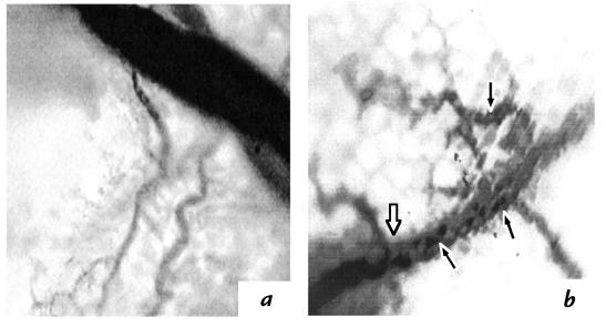
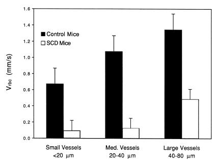
Videotapes of the blood flow revealed remarkably consistent differences in the mucosal–intestinal microvessels of control and SCD mice under normoxic conditions and in flow responses to hyperoxia. In the eight control mice, flow uniformly was rapid and even in the capillaries, arterioles, and venules of all sizes. The eight SCD mice, on the other hand, had slowing, sludging, irregularity, and stasis of flow in most vessels of all types and sizes. In medium and large vessels in which flow was nearly stopped, a back-and-forth (antegrade–retrograde) flow phenomenon was observed. These observations are reflected in frame-captured images in which the even blood flow of control mice could be distinguished from the sludged and stopped blood flow of SCD mice (Fig. 1). VASCAN and VASVEL imaging software were used to analyze blood flow velocities in vessels as a function of vessel diameter. In control mice, >95% of the small vessels had smooth and measurable flow velocity. By comparison, ∼50% of small vessels in SCD mice had trickle to no flow (or no measurable flow velocity). Overall in SCD mice, in most of the small vessels in which flow persisted, it was substantially reduced. Quantitative blood flow velocities within representative small, medium, and large venules of control and SCD mice were compared from selected images on blinded videotapes (Fig. 2). Vrbc in venules from SCD mice was strikingly lower than Vrbc in control mice, a relationship that was highly significant in small (P < 0.0002), medium (P = 0.0002), and large (P < 0.0003) venules.
Figure 1.
Frame-captured images from videotaped intravital microscopy of the mesenteric microcirculations of control and SCD mice. (a) Image of the microcirculation in a control mouse. Regular flow is indicated by uniformity of flow presentation in large (top right) and small (bottom) vessels, which is noticeably different from that in b. (b) Image of the microcirculation in an SCD mouse. Occluded blood flow is indicated by the abrupt transition between the dark, sludged (SS RBC–rich) blood below the open arrow and the lighter blood above it. Clumps of SS RBC apparently adherent to the wall of the same vessel also are visible (filled arrows). SCD, sickle cell disease; SS RBC, sickle red blood cell.
Figure 2.
Blood flow velocities in control and SCD mice. The bar graph demonstrates paired mean Vrbc in small, medium, and large venules of control and SCD mice. The error bars indicate SD; the ordinate is blood flow velocity, Vrbc (mm/s); for each bar, n = 10.
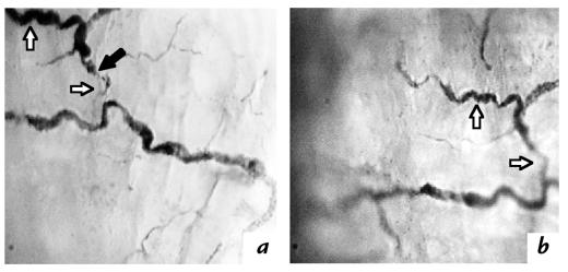
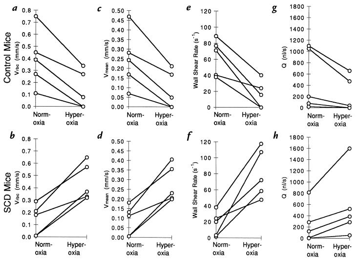
Visual inspection of blood flow revealed that initiation of hyperoxia resulted in prompt (within 10–100 seconds), albeit opposite, flow responses in control and SCD mice. In control mice, there was a rapid reduction of flow in most vessels and cessation in a minority of the smaller vessels. In contrast, blood never slowed during hyperoxia in the SCD mice. Rather, it nearly always was accelerated. These findings are demonstrated in frame-captured images from videotapes in which the abnormal, compromised flow of normoxic SCD blood was seen to improve within 100 seconds of hyperoxia (Fig. 3). To further define the influence of hyperoxia on Vrbc, after decoding the tapes to determine the experimental group, we compared this parameter within five venules each from control and SCD mice in representative vessels from blinded videotapes. The venules selected had mean (and range) diameters of 31.4 (14.5–55.8) μm for the control group and 35.8 (14.9–59.8) μm for the SCD group. Vrbc was measured under normoxic conditions and 100 seconds after the initiation of hyperoxia (Fig. 4). In control mice, hyperoxia resulted in consistent decreases in Vrbc (P < 0.008). Conversely, in SCD mice, hyperoxia resulted in consistent increases in Vrbc (P < 0.004). We used these measured Vrbc data to calculate Vmean, wall shear rate, and Q for each of the data points (Fig. 4). In comparing these 10 selected vessels, the flow parameters of hyperoxic SCD mice improved to levels matching those of normoxic control mice (Fig. 4).
Figure 3.
Frame-captured images from videotaped intravital microscopy of the mesenteric microcirculations of SCD mice during normoxic and hyperoxic conditions. (a) Sludged flow of normoxic SCD blood indicated by discontinuous columns of RBC. A site of vaso-occlusion (filled arrow) has apparently distended the vessel proximally (top open arrow) and vacated the vessel of blood distally (bottom open arrow). (b) Improved flow of hyperoxic SCD blood indicated by less distention of the proximal vessel (top open arrow) and return of blood flow distally (bottom open arrow). RBC, red blood cell.
Figure 4.
Changes in venular blood flow parameters of SCD and control mice induced by hyperoxia. Data pairs representing flow parameter values are shown as open circles connected by lines, the slope of which reflects the change in the parameter induced by hyperoxia. The top panels contain data from control mice, and the bottom panels contain data from SCD mice. (a and b) Vrbc data; (c and d) Vmean data; (e and f) wall shear rate; (g and h) volume metric flow rate (Q). The units for each parameter are shown in the ordinate labels. For both the control and SCD mice, n = 5.
We observed a modest (e.g., ∼5%) contraction of diameter in some medium and small arterioles of control mice (data not shown), which may relate to the downstream slowing of blood flow observed. Verification of that association will require improved imaging software and focusing the videotaping specifically on arterioles. We postulate that the slowing of flow we observed in control mice was due to hyperoxia-induced constriction of arterioles (19) proximal in the circulation to the majority of the vessels we videotaped and studied. No changes in arteriolar diameters were observed in SCD mice. The increase in velocity in this group was likely due to the salutary rheologic effect of oxygen on Hb S polymerization, SS RBC sickling, and rheologic properties of blood (30).The net improvement in blood flow parameters is most likely the result of rheologic improvements outweighing undetected arteriolar constriction or of the combined influences of the same rheologic improvements and the arteriolar unresponsiveness reported for a different SCD mouse model (20). The hyperoxia-induced changes in both control and SCD mice were reversed with resumption of normoxia.
To rule out the possibility that the changes in blood flow parameters we observed were not the result of inherited differences in the vascular properties or susceptibility to physical manipulation (e.g., stretching) or contact of any instrument with the mesentery, we also studied blood flow in transgenic knockout Hb AS mice that have the identical genetic background as the SCD mice as a result of similar rounds of crossbreeding. Visual inspection of the mesenteric–intestinal microcirculations of these mice (n = 4) revealed flow characteristics indistinguishable from those of normal C57BL/6 controls and markedly different from those of the SCD mice. None of the Hb AS mice were observed to have the slowed, sludged, or intermittent characteristic of SCD mice. In response to hyperoxia, these mice experienced slowing of blood flow velocity similar to that observed in normal control mice. In no instance did the blood flow velocity increase in the manner observed in SCD mice.
Discussion
The findings we report validate the notion that the clinical manifestations of SCD are the result of circulatory impairment (2, 31). To our knowledge, they constitute the first detailed in vivo characterization of blood flow abnormalities in a circulatory environment highly relevant to human SCD. Previous human studies have been limited to circulations accessible noninvasively and restricted from circulatory perturbations that facilitate analysis of the determinants of blood flow. Animal models are not encumbered by these factors, but they have other limitations (12). Although improved transgenic models increasingly have approached the hematologic and clinical abnormalities of SCD (12, 16), until recently artificial hypoxia was required to evoke a condition resembling SCD (21). A previous report (20) of microcirculatory abnormalities in transgenic sickle mice used a model with these limitations. Thus, the development of our transgenic knockout SCD mouse (17) and that of Ryan et al. (18), both of which have only human Hb S within RBC and hematologic and clinical abnormalities concordant with those of patients with SCD, represent an excellent opportunity for the study of the blood flow abnormalities of SCD in vivo. Our findings further validate these animals as models for studies of SCD.
A major finding of the present study is that of abnormal blood flow in mucosal–intestinal microvessels of all sizes. The extreme sludging, halted flow, and the resultant antegrade–retrograde flow seen, and the measured Vrbc in small, medium, and large venules of the microcirculation, differed remarkably from the blood flow observed in normal control mice. These abnormal flow characteristics are likely related to the RBC abnormalities that account for their hypoxia-induced sickling in vitro and for the hemolytic anemia (mean hematocrits of 28.7%, and reticulocyte counts of 26.8%, compared with mean control values of 43.6% and 3.4%, respectively) of these SCD mice (17). The reported splenic, cardiac, and renal hypertrophy; renal fibrosis, atrophy, infarcts, and cysts; pulmonary vascular congestion; splenic sinusoidal congestion; and hepatic and renal iron deposits of these mice may be attributed to these RBC abnormalities (17) and to the blood flow abnormalities reported here.
A second major finding derives from our studies of the effect of hyperoxia on blood flow parameters. We found that in control mice, all parameters were decreased by hyperoxia, and that in SCD mice, the same parameters were enhanced to a level resembling normoxic control mice. We speculate that the former is related to arteriolar constriction (19) proximal to our field of study and that the latter was due to improved rheologic properties of SS RBC (30). Oxygen inhalation is commonly used to treat complications of SCD, despite a paucity of controlled clinical data to support the practice (32, 33), evidence that it may suppress erythropoiesis (34), and the possible association of pain crisis with its cessation (34). The remarkable improvement in flow parameters of SCD mice induced by hyperoxia suggests that oxygen inhalation may indeed have therapeutic utility in SCD, which is consistent with the changes in blood cell populations and rheology in vivo during oxygen administration (32, 34). Confirmation of the benefits of oxygen inhalation on sickle blood flow parameters awaits verification by controlled clinical trial. Considering the detrimental potential of long-term oxygen inhalation (34–36), alternative approaches to oxygen delivery such as the use of artificial oxygen-carrying blood substitutes may be considered (37, 38).
The knowledge base of SCD is vast. Yet many of the accepted principles remain to be confirmed in vivo. The potential of this SCD mouse model for defining the determinants that modulate SCD blood flow provides enormous opportunity for elucidating important mechanisms. This approach affords exciting new possibilities for the development of improved SCD therapies based on objective evaluations of strategies for improving blood flow.
Acknowledgments
The authors express their appreciation to Neil Matsui, Marcos Intaglietta, and Peter Chen for insightful discussions, and to N. Matsui for assistance with preparing the figures. This work was supported in part by a Professional Development Award from the Department of Medicine, San Francisco General Hospital and the University of California (to S.H. Embury); a grant from the National Heart, Lung, and Blood Institute (NHLBI) (HL31579; to N. Matsui); a Professional Development Award from the University of California, discretionary gifts to the Biomedical Engineering Division; and a grant from the NHLBI (NIH-R29-HL-51181; principal investigator Ted Wun) to A.T.W. Cheung.
References
- 1.Herrick JB. Peculiar elongated and sickle-shaped red blood corpuscles in a case of severe anemia. Arch Int Med. 1910;6:517–521. [PMC free article] [PubMed] [Google Scholar]
- 2.Ham TH, Castle WB. Relationship of increased hypotonic fragility of erythrostasis to the mechanisms of hemolysis in certain anemias. Trans Assoc Am Phys. 1940;55:127–132. [Google Scholar]
- 3.Dean J, Schechter AN. Sickle cell anemia: molecular and cellular basis of therapeutic approaches. N Engl J Med. 1978;299:752–870. doi: 10.1056/NEJM197810052991405. [DOI] [PubMed] [Google Scholar]
- 4.Harris JW, Brewster HH, Ham TH, Castle WB. Studies on the destruction of red blood cells. X. The biophysics and biology of sickle-cell disease. Arch Int Med. 1956;97:145–168. doi: 10.1001/archinte.1956.00250200021002. [DOI] [PubMed] [Google Scholar]
- 5.Eaton WA, Hofrichter J. Sickle cell hemoglobin polymerization. Adv Prot Chem. 1990;40:63–279. doi: 10.1016/s0065-3233(08)60287-9. [DOI] [PubMed] [Google Scholar]
- 6.Knisely MH, Bloch EH, Eliot TS, Warner L. Sludged blood. Science. 1947;106:431–440. doi: 10.1126/science.106.2758.431. [DOI] [PubMed] [Google Scholar]
- 7.Fink AI, Funahashi T, Robinson M, Watson RJ. Conjunctival blood flow in sickle-cell disease. Arch Ophthalmol. 1961;66:824–829. doi: 10.1001/archopht.1961.00960010826008. [DOI] [PubMed] [Google Scholar]
- 8.Lipowsky HH, Sheikh NU, Katz DM. Intravital microscopy of capillary microdynamics in sickle cell disease. J Clin Invest. 1987;80:117–127. doi: 10.1172/JCI113036. [DOI] [PMC free article] [PubMed] [Google Scholar]
- 9.Rodgers GP, et al. Periodic microcirculatory flow in patients with sickle-cell disease. N Engl J Med. 1984;311:1534–1538. doi: 10.1056/NEJM198412133112403. [DOI] [PubMed] [Google Scholar]
- 10.Cheung ATW, et al. Development of a severity index to objectively characterize microvascular abnormalities in sickle cell disease. Blood. 1997;90(Suppl. 1):127A. (Abstr.) [Google Scholar]
- 11.Castro O, Orlin J, Rosen MW, Finch SC. Survival of human sickle-cell erythrocytes in heterologous species: response to variations in oxygen tension. Proc Natl Acad Sci USA. 1973;70:2356–2359. doi: 10.1073/pnas.70.8.2356. [DOI] [PMC free article] [PubMed] [Google Scholar]
- 12.Fabry, M. 1994. Transgenic animal models. In Sickle cell disease: basic principles and clinical practice. S.H. Embury, R.P. Hebbel, N. Mohandas, and M.H. Steinberg, editors. Raven Press. New York, NY. 105–120.
- 13.Kaul DK, Fabry ME, Nagel RL. Microvascular sites and characteristics of sickle cell adhesion to vascular endothelium in shear flow conditions: pathophysiological implications. Proc Natl Acad Sci USA. 1989;86:3356–3360. doi: 10.1073/pnas.86.9.3356. [DOI] [PMC free article] [PubMed] [Google Scholar]
- 14.Trudel M, et al. Towards a transgenic mouse model of sickle cell disease: hemoglobin SAD. EMBO J. 1991;10:3157–3165. doi: 10.1002/j.1460-2075.1991.tb04877.x. [DOI] [PMC free article] [PubMed] [Google Scholar]
- 15.Trudel M, et al. Towards a mouse model for sickle cell disease: HB SAD. Nouv Rev Fr Hematol. 1990;32:407–408. [PubMed] [Google Scholar]
- 16.Nagel RL. A knockout of a transgenic mouse — animal models of sickle cell anemia. N Engl J Med. 1998;339:194–195. doi: 10.1056/NEJM199807163390310. [DOI] [PubMed] [Google Scholar]
- 17.Paszty C, et al. Transgenic knockout mice with exclusively human sickle hemoglobin and sickle cell disease. Science. 1997;278:876–878. doi: 10.1126/science.278.5339.876. [DOI] [PubMed] [Google Scholar]
- 18.Ryan TM, Ciavatta DJ, Townes TM. Knockout-transgenic mouse model of sickle cell disease. Science. 1997;278:873–876. doi: 10.1126/science.278.5339.873. [DOI] [PubMed] [Google Scholar]
- 19.Messina EJ, et al. Increases in oxygen tension evoke arteriolar constriction by inhibiting endothelial prostaglandin synthesis. Microvasc Res. 1994;48:151–160. doi: 10.1006/mvre.1994.1046. [DOI] [PubMed] [Google Scholar]
- 20.Kaul DK, et al. In vivo demonstration of red cell–endothelial interaction, sickling and altered microvascular response to oxygen in the sickle transgenic mouse. J Clin Invest. 1995;96:2845–2853. doi: 10.1172/JCI118355. [DOI] [PMC free article] [PubMed] [Google Scholar]
- 21.Fabry ME, et al. A second generation transgenic mouse model expressing both hemoglobin S (HbS) and HbS-Antilles results in increased phenotypic severity. Blood. 1995;86:2419–2428. [PubMed] [Google Scholar]
- 22.Cheung ATW, et al. Microcirculation and metastasis in a new mouse mammary tumor model system. Int J Oncol. 1997;11:69–77. [PubMed] [Google Scholar]
- 23.Cheung ATW, et al. Conjunctival microcirculation in sickle cell disease (SCD): a computer-assisted intravital study. Microcirculation. 1997;4:164. (Abstr.) [Google Scholar]
- 24.Wayland K, Johnson P. Erythrocyte velocity measurements in microvessels by a two-slit method. J Appl Physiol. 1967;22:333–337. doi: 10.1152/jappl.1967.22.2.333. [DOI] [PubMed] [Google Scholar]
- 25.Baker M, Wayland H. On-line volume flow rate and velocity profile measurement for blood in microvessels. Microvasc Res. 1974;7:131–143. doi: 10.1016/0026-2862(74)90043-0. [DOI] [PubMed] [Google Scholar]
- 26.Seki J, Lipowsky HH. In vivo and in vitro measurements of red cell velocity under epifluorescence microscopy. Microvasc Res. 1989;38:110–124. doi: 10.1016/0026-2862(89)90020-4. [DOI] [PubMed] [Google Scholar]
- 27.Lipowsky HH, Usami S, Chien S. In vivo measurements of “apparent viscosity” and microvessel hematocrit in the mesentery of the cat. Microvasc Res. 1980;19:297–319. doi: 10.1016/0026-2862(80)90050-3. [DOI] [PubMed] [Google Scholar]
- 28.Kaul DK, Fabry ME, Nagel RL. Erythrocytic and vascular factors influencing the microcirculatory behavior of blood in sickle cell anemia. Ann NY Acad Sci. 1989;565:316–326. doi: 10.1111/j.1749-6632.1989.tb24179.x. [DOI] [PubMed] [Google Scholar]
- 29.Kaul DK, et al. Erythrocytes in sickle cell anemia are heterogeneous in their rheological and hemodynamic characteristics. J Clin Invest. 1983;72:22–29. doi: 10.1172/JCI110960. [DOI] [PMC free article] [PubMed] [Google Scholar]
- 30.Chien S. Rheology of sickle cells and erythrocyte content. Blood Cells. 1977;3:283–303. [Google Scholar]
- 31.Embury, S.H., Hebbel, R.P., Mohandas, N., and Steinberg, M.H. 1994. Pathogenesis of vasoocclusion. In Sickle cell disease: basic principles and clinical practice. S.H. Embury, R.P. Hebbel, N. Mohandas, and M.H. Steinberg, editors. Raven Press. New York, NY. 311–326.
- 32.Zipursky A, et al. Oxygen therapy in sickle cell disease. Am J Pediatr Hematol Oncol. 1992;14:222–228. doi: 10.1097/00043426-199208000-00007. [DOI] [PubMed] [Google Scholar]
- 33.Khoury H, Grimsley E. Oxygen inhalation in nonhypoxic sickle cell patients during vaso-occlusive crisis [letter] Blood. 1995;86:3998. [PubMed] [Google Scholar]
- 34.Embury SH, et al. Oxygen inhalation by subjects with sickle cell anemia. Effects on endogenous erythropoietin kinetics erythropoiesis. N Engl J Med. 1984;311:291–295. doi: 10.1056/NEJM198408023110504. [DOI] [PubMed] [Google Scholar]
- 35.Fridovich I. Oxygen toxicity: a radical explanation. J Exp Biol. 1998;201:1203–1209. doi: 10.1242/jeb.201.8.1203. [DOI] [PubMed] [Google Scholar]
- 36.Putman E, van Golde LM, Haagsman HP. Toxic oxidant species and their impact on the pulmonary surfactant system. Lung. 1997;175:75–103. doi: 10.1007/pl00007561. [DOI] [PubMed] [Google Scholar]
- 37.Gonzalez P, et al. A phase I/II study of polymerized bovine hemoglobin in adult patients with sickle cell disease not in crisis at the time of study. J Invest Med. 1997;45:258–264. [PubMed] [Google Scholar]
- 38.Hess JR. Alternative oxygen carriers. Curr Opin Hematol. 1996;3:492–497. doi: 10.1097/00062752-199603060-00016. [DOI] [PubMed] [Google Scholar]


