Abstract
Zoophilic fungal infections are a prevalent disease in tropical countries and clinicians must consider them in the differential diagnosis of pruritic skin lesions. We report a clinical case of Trichophyton erinacei skin infection after recreational exposure to an Asian Elephant. As far as we were able to search the literature, it is the first case described after contact with elephants
Keywords: Trichophyton erinacei, Zoonotic fungal infections, Elephants
Travellers are at risk of acquiring zoonotic diseases, especially in destinations with outdoor activities that allow contact with exotic animals.
A 26-year old Caucasian woman presented to our clinic with a 10 days history of highly pruritic erythematous plaques on her knees and inner thighs (Fig. 1). She was medicated by her general practitioner with a topical association of gentamicin with betamethasone dipropionate, reporting only a slight improvement in pruritus. The partner had no lesions. They had return from a trip to Thailand in the previous month, where she rode an elephant in a national park. She reported that the plaques had appeared on the skin surface that had contact with the animal. She denied also contact with other animals, namely hedgehogs in national parks, and had no pets. Oral empirical treatment was started with 100 mg itraconazole bid for 10 days, with a rapid response being observed.
Figure 1.

Erythematous plaques on the knees and thighs, with peripheral scales.
Skin scales were digested with 20% KOH and incubated for 30 minutes in room temperature and hyaline septate hyphae through the host tissue were observed in both bright-field and phase-contrast microscopy. Colonies grown on Sabouraud dextrose agar were cottony, white, and expanding. Colony reverse became brilliant lemon yellow.
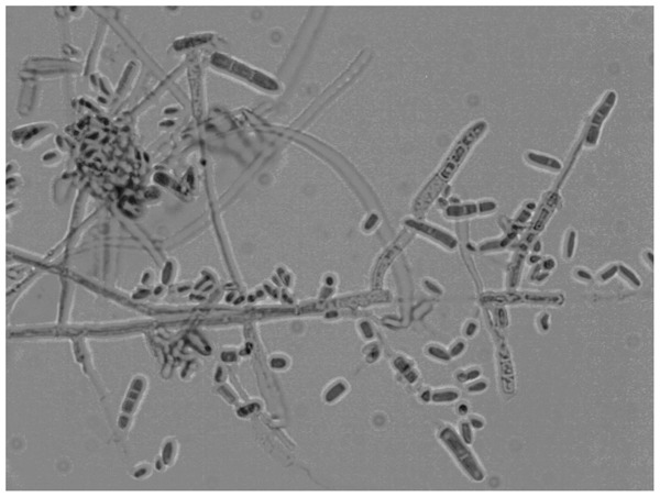
Direct microscopic examination of the scales showed hyaline septate hyphae and a cottony, white colony grew on the Sabouraud dextrose agar. Microscopically microconidia were abundant (Fig. 2), first widely interspaced, but later close together, liberated by deterioration of supporting hyphae.
Figure 2.

Microscopically microconidia were abundant, large slender, and clavate, borne on the slides of hyphae. Smooth and thin-walled macroconidia were also present, cylindrical to clavate, variable between 2 and 6 cells.
Arthroconidia and macroconidia were also present and, 4 weeks later, chlamydospores were also observed. Morphological features were compatible with Trichophyton erinacei. The DNA sequence of nuclear ribosomal ITS region of the isolated fungus was also in accordance with Trichophyton erinacei 5.8 rRNA gene, ITS1 and ITS2 DNA, and strain CBS 677.86 (GenBank accession No. Z97997.1).
Trichophyton erinacei is a zoophilic dermatophytosis that is mainly reported after contact with hedgehogs.1 Most recent published cases come from Asia, especially when owned as exotic pets,2–5 and Oceania but reports from South America6 and Europe7 also exist. Canine and feline infections have been reported8 but, as far as we were able to search the literature, no previous report of human infection after recreational contact with elephants was described. Our patient reported that she rode the elephant with a seat but reported also that she had contact with the animal skin surface on the same areas where lesions developed.
Besides infection from hedgehogs, transmission to humans can occur by indirect infection from soil, hedgehog nests, among humans, or through animal vectors,1,2 as it possibly occurred in our patient.
Human infection with this zoophilic fungi causes highly pruritic lesions that are usually confined to the skin surface that had contact with the animal,3 as the thigh plaques in our patient. Treatment options in published cases included mostly oral agents as itraconazole,2,3,7 terbinafine,6,7 and griseofulvin,9 all obtaining lesion clearing. Zoophilic dermatophytosis must be considered in the differential diagnosis of skin lesions in travellers, since recreational activities with exotic animals are increasingly popular.
References
- 1.Quaife RA. Human infection due to the hedgehog fungus, Trichophyton mentagrophytes var. erinacei. J Clin Pathol. 1966;19:177–78. doi: 10.1136/jcp.19.2.177. [DOI] [PMC free article] [PubMed] [Google Scholar]
- 2.Lee DW, Yang JH, Choi SJ, Won CH, Chang SE, Lee MW, et al. An unusual clinical presentation of tinea faciei caused by Trichophyton mentagrophytes var. erinacei. Pediatr Dermatol. 2011;28:210–12. doi: 10.1111/j.1525-1470.2011.01391.x. [DOI] [PubMed] [Google Scholar]
- 3.Rhee DY, Kim MS, Chang SE, Lee MW, Choi JH, Moon KC, et al. A case of tinea manuum caused by Trichophyton mentagrophytes var. erinacei: the first isolation in Korea. Mycoses. 2009;52:287–90. doi: 10.1111/j.1439-0507.2008.01556.x. [DOI] [PubMed] [Google Scholar]
- 4.Mochizuki T, Takeda K, Nakagawa M, Kawasaki M, Tanabe H, Ishizaki H. The first isolation in Japan of Trichophyton mentagrophytes var. erinacei causing tinea manuum. Int J Dermatol. 2005;44:765–8. doi: 10.1111/j.1365-4632.2004.02180.x. [DOI] [PubMed] [Google Scholar]
- 5.Hsieh CW, Sun PL, Wu YH. Trichophyton erinacei infection from a hedgehog: a case report from Taiwan. Mycopathologia. 2010;170:417–21. doi: 10.1007/s11046-010-9333-2. [DOI] [PubMed] [Google Scholar]
- 6.Concha M, Nicklas C, Balcells E, Guzmán AM, Poggi H, León E, et al. The first case of tinea faciei caused by Trichophyton mentagrophytes var. erinacei isolated in Chile. Int J Dermatol. 2012;51:283–85. doi: 10.1111/j.1365-4632.2011.04995.x. [DOI] [PubMed] [Google Scholar]
- 7.Schauder S, Kirsch-Nietzki M, Wegener S, Switzer E, Qadripur SA. From hedgehogs to men. Zoophilic dermatophytosis caused by Trichophyton erinacei in eight patients. Hautarzt. 2007;58:62–7. doi: 10.1007/s00105-006-1100-4. [DOI] [PubMed] [Google Scholar]
- 8.Fairley RA. The histological lesions of Trichophyton mentagrophytes var erinacei infection in dogs. Vet Dermatol. 2001;12:119–22. doi: 10.1046/j.1365-3164.2001.00235.x. [DOI] [PubMed] [Google Scholar]
- 9.Jury CS, Lucke TW, Bilsland D. Trichophyton erinacei: an unusual cause of kerion. Br J Dermatol. 1999;141:606–7. doi: 10.1046/j.1365-2133.1999.03092.x. [DOI] [PubMed] [Google Scholar]


