Abstract
The aim of this study is to validate optical coherence tomography (OCT) in assessing human articular cartilage by means of histological analyses. Twenty resected human femoral head specimens were evaluated with OCT and histological analysis. OCT and histological evaluation was performed according to the Bear and the Mankin criteria. OCT grades and Mankin scores (total score and sub-score structure) were correlated and intra-/inter-observer agreement for repeated OCT evaluations was tested by interclass-correlation coefficient (ICC) analysis. OCT grades and Mankin scores were correlated [Spearman correlation=0.742 (total) and 0.656 (structure), P<0.001], revealing significant differences between the histological scores in various OCT grades of cartilage degeneration (P<0.001). Intra-observer (ICC 0.930) and inter-observer (ICC 0.933) reliability was high (P<0.001). OCT appears to be reliable in the assessment of human articular cartilage. Further studies on intra-operative cartilage evaluation by OCT are necessary to substantiate its applicability in clinical routine.
Key words: optical coherence tomography, reliability, hip, cartilage
Introduction
Osteoarthritis (OA) is characterized by degenerative changes in cartilage, bone, and soft tissues. As the number of individuals with OA continues to rise, and with the aim to reduce or delay the onset of this debilitating disease via timely intervention, early detection of cartilage damage is required. Whereas conventional imaging techniques such as radiography or standard magnetic resonance imaging (MRI) have limitations in their ability to depict early cartilage changes such as subtle alterations of the collagen architecture, optical coherence tomography (OCT), with its ability to assess the microstructure of cartilage tissue in near realtime microscopic resolution images, may help to overcome this limitation. With this technique it is possible to detect even subtle subsurface cartilage changes without removal of cartilage tissue in high image resolution that is comparable to low-power histology. Although several in vitro and in vivo studies indicate the potential of this unique technique,1-5 the potential of OCT to detect various histologically approved grades of cartilage degeneration is insufficiently documented. The current data lack histologically controlled studies that elaborate on the potential of OCT to detect various grades of cartilage degeneration. The purpose of this study was to evaluate the reliability of OCT in the assessment of human articular cartilage. Therefore, we conducted a histological controlled in vitro study including femoral head samples with various grades of cartilage degeneration.
Materials and Methods
Study sample
Twenty resected femoral head specimens were collected from 20 patients (seven males, 13 females, mean age: 60.9±9.6 years) who underwent total hip arthroplasty for the treatment of symptomatic OA. Four pins (Ethicon, Norderstedt, Germany) were inserted in an area of morphologically intact appearing cartilage to define a region of interest (ROI) for subsequent correlation of OCT and histology. The study was approved by the local ethics committee and all subjects gave informed consent to the study.
Optical coherence tomography
OCT is a non-contact imaging technology which measures multiple wavelengths of a reflected light spectrum thus producing high resolution cross-sectional and three-dimensional (3D) images with 40.000 scans per second. OCT was originally employed to visualize pathologies within the retina and has recently been used in interventional cardiology and orthopedic surgery.
In this present study, OCT imaging was performed using a clinical OCT scanning system (Spectralis, Heidelberg Engineering, Heidelberg, Germany), which uses a spectral domain technology (also referred to as Fourier domain OCT).6
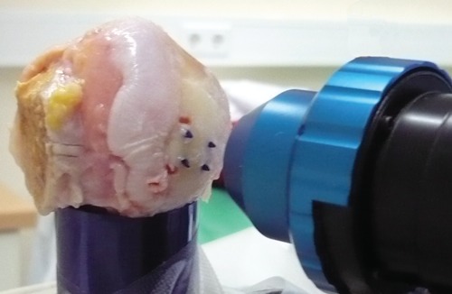
The femoral head specimens were placed stably in front of the OCT-device (Figure 1). A series of A-scans generating two- and 3D data sets were obtained. Notably, this system creates a series of cross-sectional images with high image resolution (axial resolution of 5 to 7 µm) that is comparable to low-power histology, while the depth of the OCT penetration is approximately 2 mm.6,4,7
Figure 1.

A femoral head is mounted on the chin rest of the Spectralis OCT (optical coherence tomography) scanning system (Heidelberg Engineering). Four Ethipins® mark the area of interest on the morphologically intact cartilage.
Histology
The cartilage-bone blocks were dehydrated using standardized techniques for non-decalcified sectioning in methacrylate, as described previously.8 Coronal sections were then cut along the long axis of the pins, which generated 10 to 18 serial sections per specimen (264 histological sections in total), with a final section thickness of approximately 50 μm.
The histological sections were stained with toluidine blue (0.1% toluidine blue in 0.1% sodium tetraborate, Merck, Darmstadt, Germany) and destained for 30 minutes in tap water to assess cartilage structure and composition. This procedure was followed by dehydration and embedding in DePex (Merck KG, Darmstadt, Germany). Digital image evaluation was performed using a binocular light microscope (Olympus BX50, Olympus, Hamburg, Germany) with a color CCD camera (Color View III, Olympus), and imaging software (analySIS FIVE, Soft Imaging System, Münster, Germany).
Imaging analysis
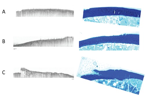
The OCT images were graded according to a modified classification system reported by Bear et al.,9 in which a grade A indicates an intact surface and an obvious birefringence while grade B changes correspond to an intact surface with no birefringence, and a grade C represents an irregular articular surface and/or subsurface voids (Figure 2). For reliability assessment, all OCT measurements were repeated by the senior author (CZ) and by a second observer (KVH).
Figure 2.

Optical coherence tomography (left) and equivalent histological sections with conventional toluidine blue staining (right) showing grades A-C according to the classification system of Bear et al.9
The histological sections were staged by one expert (MH) according to the Mankin classification scale that grades surface morphology, cellularity, toluidine staining, and tidemark integrity, in which a total score of 14 can be assigned (Table 1).10 In the present study, the Mankin score structure and the total Mankin score were used for further analyses.
Table 1.
Cartilage evaluation according to the Mankin system.
| Criteria | Score | Histological finding |
|---|---|---|
| Structure | 0 | Smooth intact surface |
| 1 | Slight surface irregularities | |
| 2 | Pannus/surface fibrillation | |
| 3 | Clefts into transitional zone | |
| 4 | Clefts into radial zone | |
| 5 | Clefts into calcified zone | |
| 6 | Total disorganization | |
| Cells | 0 | Uniform cell distribution |
| 1 | Diffuse cell proliferation | |
| 2 | Cell clustering | |
| 3 | Cell loss | |
| Toluidin staining | 0 | Uniform staining |
| 1 | Minor discoloration | |
| 2 | Moderate discoloration | |
| 3 | Severe discoloration | |
| 4 | Total discoloration | |
| Tidemark integrity | 0 | Intact |
| 1 | Vascularity |
Statistical analyses
SPSS® software (Version 19.0; SPSS, Inc., Chicago, IL, USA) was utilized for statistical analyses that included a Spearman’s rho analysis to evaluate the correlation between OCT and histology measures, and a Wilcoxon signed-rank test to compare the various OCT grades according to their corresponding Mankin scores. Intra- and inter-observer reproducibility was assessed for OCT grading by performing intra-class correlation (ICC) analysis (pair-wise correlation, absolute agreement). In this study, all tests were conducted at the 5% significance level (P=0.05).
Results
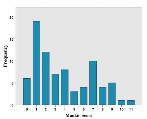
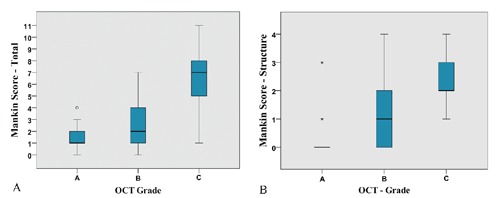
A total of 80 corresponding OCT and histology regions were identified. Figure 3 demonstrates the frequency distribution of the various grades of cartilage degeneration in these regions. The Spearman’s rho correlation analyses revealed a correlation between the OCT grade and the total Mankin score (Spearman correlation=0.742, P<0.001) and between the OCT grade and the Mankin score for structure (Spearman correlation=0.656, P<0.001). Furthermore, significant differences were noted between the Mankin scores in various OCT grades of cartilage degeneration (Figure 4). ICC analysis revealed high intra-observer (ICC=0.930 P<0.001) and high inter-observer reproducibility (ICC=0.933, P<0.001) for OCT grading.
Figure 3.

Frequency of histological grades (according to Mankin) in histological analyses.
Figure 4.

A) Significant correlation between total Mankin score and optical coherence tomography (OCT) grade; B) significant correlation between Mankin score structure and OCT grade.
Discussion and Conclusions
OA is a progressive degenerative disease in which the diagnosis of early cartilage damage is essential to prevent further joint destruction and associated debilitation. In addition to standard imaging modalities such as plain radiography and MRI, direct visual cartilage inspection by minimally invasive arthroscopy or open surgical evaluation is considered the gold standard for the grading of cartilage damage albeit even the invasive methods are limited in macroscopic surface evaluation and subjective tactile probing. Histological assessment may reveal early structural and compositional changes throughout the cartilage tissue. However, histological evaluation is not standard practice in clinical routine as it requires the removal of tissue for cartilage evaluation. In vivo, minimally-invasive OCT could fill this diagnostic gap, provided that structural changes are reliably depicted in both superficial and deeper cartilage zones.
This in vitro study was conducted to evaluate the potential of OCT for human cartilage assessment, utilizing the gold standard histology for comparison. In this work, we noted a correlation between the OCT and histology measures. Furthermore, significant differences between the various OCT grades of cartilage damage were observed. The reliability of the OCT measurements in this study was confirmed by a high intra- and inter-reader reproducibility (ICC>0.930, P<0.001).
The outcome of this study is quite similar to those of previous reports that revealed encouraging results regarding the identification of subtle degenerative cartilage changes by using OCT.1-3,5,7,11-15 Li et al.15 found a correlation of structural features detected with OCT and corresponding histology in six patients with OA. The study by Chu et al.11 demonstrated the correlation between OCT, arthroscopy, and the highly sensitive MRI assessment modality T2 relaxation mapping in the assessment of articular cartilage. Similar to our study, OCT correlated with histological cartilage assessment in 45 cartilage cores harvested from nine human osteoarthritic tibial plateaus (kappa value: 0.80).4 In an in vitro study including histology and polarized microscopy, Bear et al.1 observed a correlation between chondrocyte viability and OCT cartilage signal intensity ratio after applying impaction in eight bovine tibial plateaus. These observations indicate the potential of OCT to identify even subtle non-visible acute cartilage changes related to impact injury. The study by Herrmann et al.7 showed the correlation between the ultrastructure of articular cartilage and the polarization state of the incident light of OCT, and that, in this regard, birefringence could be evaluated using polarization-sensitive OCT (PS-OCT). These findings are similar to those reported by Drexler et al.,12 who revealed that the collagen disorganization found in osteoarthritic articular cartilage was detectable by PS-OCT as a loss of normal birefringence.
Our study has limitations. Although pins were utilized to provide additional landmarks to identify identical OCT and histological section planes, minor differences between the corresponding OCT and histological regions led to a mismatch in in-plane orientation and differences in image resolution could not be avoided. Furthermore, image selection for analyses of different regions was performed manually and was, therefore, operator-dependent. In this in vitro study, OCT was conducted on a clinical scanner that is routinely used for scanning the anterior segment of the eye and the retina. Therefore, the observations of this study may not be generalized and the ability of in vivo cartilage OCT assessment (for example, by means of arthroscopic instruments with an integrated OCT device) certainly needs to be further assessed. Finally, in our study, polarized microscopy was not available for comparison and minor changes of the collagen ultrastructure might have been missed.
In summary, the correlation between OCT and the diagnostic gold standard histology and the high reproducibility of the OCT assessment in this study point towards the reliability of the OCT technique for assessing early cartilage degeneration. Because of the high sensitivity for subsurface cartilage degeneration, OCT may be a useful addition in the development of innovative therapeutic strategies that aim to prevent ongoing cartilage destruction. Further studies that involve intraoperative OCT analyses are needed to prove the clinical value of this technique.
Funding Statement
Funding: This study was funded by a research grant from the German Osteoarthritis Aid (Deutsche Arthrose-Hilfe e.V.). The authors have full control of all primary data. Other data from these femoral head specimens were used in previous studies (Zilkens C, et al. Validity of gradient-echo three-dimensional delayed gadolinium-enhanced magnetic resonance imaging of hip joint cartilage: a histologically controlled study. Eur J Radiol 2013;82:e81-6. Bittersohl B, et al. T2* mapping of hip joint cartilage in various histological grades of degeneration. Osteoarthritis Cartilage 2012;20:653-60).
References
- 1.Bear DM, Szczodry M, Kramer S, et al. Optical coherence tomography detection of subclinical traumatic cartilage injury. J Orthop Trauma 2010;24:577-82 [DOI] [PMC free article] [PubMed] [Google Scholar]
- 2.Cernohorsky P, de Bruin DM, van Herk M, et al. In-situ imaging of articular cartilage of the first carpometacarpal joint using co-registered optical coherence tomography and computed tomography. J Biomed Opt 2012;17:060501. [DOI] [PubMed] [Google Scholar]
- 3.Chu CR, Izzo NJ, Irrgang JJ, et al. Clinical diagnosis of po lly treatable early articular cartilage degeneration using optical coherence tomography. J Biomed Opt 2007;12:051703. [DOI] [PubMed] [Google Scholar]
- 4.Chu CR, Lin D, Geisler JL, et al. Arthroscopic microscopy of articular cartilage using optical coherence tomography. Am J Sports Med 2004;32:699-709 [DOI] [PubMed] [Google Scholar]
- 5.Zheng K, Martin SD, Rashidifard CH, et al. In vivo micron-scale arthroscopic imaging of human knee osteoarthritis with optical coherence tomography: comparison with magnetic resonance imaging and arthroscopy. Am J Orthop (Belle Mead, NJ) 2010;39:122-5 [PMC free article] [PubMed] [Google Scholar]
- 6.Barteselli G, Bartsch DU, El-Emam S, et al. Combined depth imaging technique on spectral-domain optical coherence tomography. Am J Ophthalmol 2013;155:727-732 [DOI] [PMC free article] [PubMed] [Google Scholar]
- 7.Herrmann JM, Pitris C, Bouma BE, et al. High resolution imaging of normal and osteoarthritic cartilage with optical coherence tomography. J Rheumatol 1999;26:627-35 [PubMed] [Google Scholar]
- 8.Zilkens C, Miese FR, Crumbiegel C, et al. Magnetic resonance imaging and histology of ovine hip joint cartilage in two age populations: a sheep model with assumed healthy cartilage. Skeletal Radiol 2013;42:699-705 [DOI] [PubMed] [Google Scholar]
- 9.Bear DM, Williams A, Chu CT, et al. Optical coherence tomography grading correlates with MRI T2 mapping and extracellular matrix content. J Orthop Res 2010;28:546-52 [DOI] [PMC free article] [PubMed] [Google Scholar]
- 10.Mankin HJ, Dorfman H, Lippiello L, Zarins A. Biochemical and metabolic abnormalities in articular cartilage from osteoarthritic human hips. II. Correlation of morphology with biochemical and metabolic data. J Bone Joint Surg Am 1971;53:523-37 [PubMed] [Google Scholar]
- 11.Chu CR, Williams A, Tolliver D, et al. Clinical optical coherence tomography of early articular cartilage degeneration in patients with degenerative meniscal tears. Arthritis Rheum 2010;62:1412-20 [DOI] [PMC free article] [PubMed] [Google Scholar]
- 12.Drexler W, Stamper D, Jesser C, et al. Correlation of collagen organization with polarization sensitive imaging of in vitro cartilage: implications for osteoarthritis. J Rheumatol 2001;28:1311-8 [PubMed] [Google Scholar]
- 13.Rashidifard C, Vercollone C, Martin S, et al. The application of optical coherence tomography in musculoskeletal disease. Arthritis 2013:563268. [DOI] [PMC free article] [PubMed] [Google Scholar]
- 14.Szczodry M, Coyle CH, Kramer SJ, et al. Progressive chondrocyte death after impact injury indicates a need for chondroprotective therapy. Am J Sports Med 2009;37:2318-22 [DOI] [PMC free article] [PubMed] [Google Scholar]
- 15.Li X, Martin S, Pitris C, et al. High-resolution optical coherence tomographic imaging of osteoarthritic cartilage during open knee surgery. Arthritis Res Ther 2005;7:R318-23 [DOI] [PMC free article] [PubMed] [Google Scholar]


