
Figure 2.
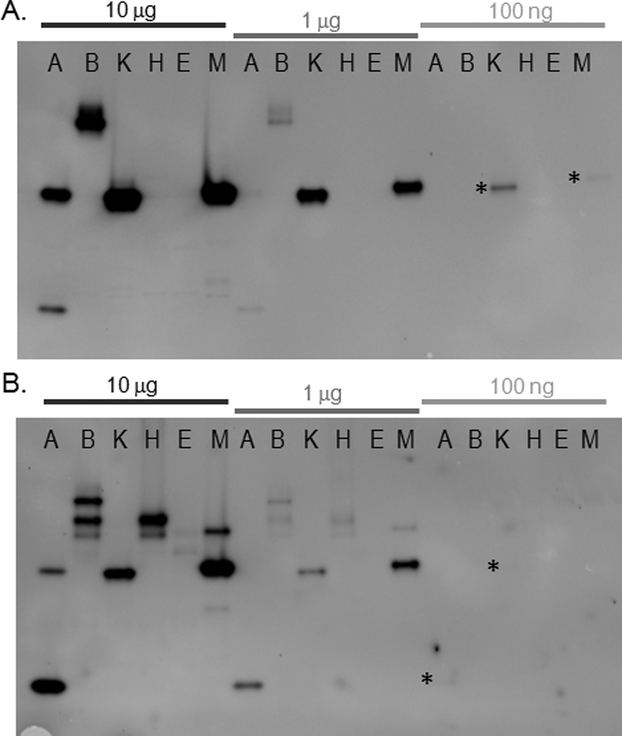
Sensitivity of sulfatase activity detection in mycobacterial lysates. Different amounts of mycobacterial lysates were separated by native gel, followed by incubation for 15 min with (A) MFS (50 µM) or (B) RS (25 µM). For the most faint bands, which were visible upon adjustment of the image contrast, an asterisk (*) is shown to the left of the band location. Lane designations are: A: M. avium; B: M. bovis (BCG); K: M. kansasii; H: M. tuberculosis (H37Rv); E: M. tuberculosis (Erdman); M: M. marinum.