Abstract
Reproductive and endocrine disruption is commonly reported in aquatic species exposed to complex contaminant mixtures. We previously reported that Atlantic killifish (Fundulus heteroclitus) from the chronically contaminated Newark Bay, NJ, exhibit multiple endocrine disrupting effects, including inhibition of vitellogenesis (yolk protein synthesis) in females and false negative vitellogenin biomarker responses in males. Here, we characterized the effects on estrogen signaling and the transcriptional regulation of estrogen–responsive genes in this model population. First, a dose–response study tested the hypothesis that reproductive biomarkers (vtg1, vtg2, chg H, chg Hm, chg L) in Newark Bay killifish are relatively less sensitive to 17β–estradiol at the transcriptional level, relative to a reference (Tuckerton, NJ) population. The second study assessed expression for various metabolism (cyp1a, cyp3a30, mdr) and estrogen receptor (ER α, ER βa, ER βb) genes under basal and estrogen treatment conditions in both populations. Hepatic metabolism of 17β–estradiol was also evaluated in vitro as an integrated endpoint for adverse effects on metabolism. In the third study, gene methylation was evaluated for promoters of vtg1 (8 CpGs) and vtg2 (10 CpGs) in both populations, and vtg1 promoter sequences were examined for single nucleotide polymorphism (SNPs). Overall, these studies show that multi–chemical exposures at Newark Bay have desensitized all reproductive biomarkers tested to estrogen. For example, at 10 ng/g 17β–estradiol, inhibition of gene induction ranged from 62% to 97% for all genes tested in the Newark Bay population, relative to induction levels in the reference population. The basis for this recalcitrant phenotype could not be explained by a change in 17β– estradiol metabolism, nuclear estrogen receptor expression, promoter methylation (gene silencing) or SNPs, all of which were unaltered and normal in the Newark Bay population. The decreased transcriptional sensitivity of estrogen–responsive genes is suggestive of a broad effect on estrogen receptor pathway signaling, and provides insight into the mechanisms of the endocrine disrupting effects in the Newark Bay population.
Keywords: biomarkers, choriogenin, endocrine disruption, estrogen, killifish, vitellogenin
1.0. Introduction
Adverse reproductive effects are frequently reported in aquatic species living within contaminated environments (Tyler et al., 1998). Chemicals that disrupt hormonal pathways and modulate gene expression can result in deleterious effects throughout the hypothalamus– pituitary–gonad–liver axis (Rempel and Schlenk, 2008). We have previously characterized a population of Atlantic killifish (Fundulus heteroclitus) from the historically polluted Newark Bay, NJ (USA), which exhibit reproductive dysfunction and abnormal biomarker responses indicative of complex endocrine disruption (Bugel et al., 2010, 2011). Integrated biomarkers are commonly used for ecological risk assessments (Amiard–triquet et al., 2013). Most biomarkers are physiologically relevant, apical endpoints that respond predictably to single chemicals or simple mixtures (e.g. cytochrome P4501A, metallothionein, vitellogenin). However, environmental exposures typically involve complex mixtures with diverse mechanisms that may result in atypical biomarker responses (Celander, 2011). Atlantic killifish are a model teleost widely used for comparative ecotoxicological studies pertaining to exposures and effects, adaptations and tolerance, endocrine disruption, and population genetics (Burnett et al., 2007). In the present study, we used the Newark Bay killifish population as a model to study mechanisms of endocrine disruption associated with chronic exposure to complex mixtures.
Newark Bay and the interconnected greater New York–New Jersey Harbor Estuary have a long history of contamination by PAHs, PCBs, heavy metals, polychlorinated dibenzo–p– dioxins and furans, and other emerging chemicals of concern (Panero et al., 2005; Muñoz et al., 2006; Valle et al., 2007). Reproductively active female killifish from Newark Bay exhibit reduced expression levels of vitellogenin, correlated to reduced fecundity and inhibited vitellogenin–dependent follicular development (Bugel et al., 2010, 2011). In most oviparous vertebrates (i.e. birds, amphibians, fish, etc.), vitellogenins are hepatically derived phosphoglycolipoprotein precursors to egg yolk proteins that serve as growth substrate during embryogenesis (e.g. amino acids, lipids, sugars) (Arukwe and Goksøyr. 2010). Vitellogenins are highly expressed during oogenesis, and expression directly correlates with fecundity (Miller et al., 2007; Thorpe et al., 2007). In Newark Bay killifish, vitellogenin protein levels are much less responsive to induction by 17β–estradiol (E2), relative to a reference population (Bugel et al., 2011). Thus, a functional desensitization of the vitellogenin pathway likely contributes to the adverse reproductive effects observed in the female population and undermines the use of vitellogenin as a biomarker in males.
Expression of vitellogenin and other estrogen–responsive genes (e.g. choriogenin egg envelope genes) are regulated by 17β–estradiol activation of estrogen receptors (ER), which dimerize and bind to cis-regulatory estrogen–responsive elements to induce transcription (Menuet et al., 2005). In killifish, three estrogen receptors have been identified (ER α, ER βa, ER βb), although the exact transcriptional role of each for different estrogen–responsive genes is not yet known (Greytak and Callard, 2007). In zebrafish, vitellogenin transcription is regulated primarily by ER α, and secondarily by ER βb, with no clear role of ER βa (Griffin et al., 2013). Epigenetic mechanisms are also important to vitellogenin gene regulation. When transcriptionally active, during spawning or when challenged with 17β–estradiol, CpG sites in promoters of vitellogenin genes are demethylated to facilitate high levels of induction (Saluz et al., 1988; Strömqvist et al., 2010). When transcriptionally inactive (i.e. in males or non–spawning females), CpG sites in promoters are methylated to maintain suppressed basal expression (gene silencing). In males, estrogen receptors are functionally expressed and sensitive to low levels of xeno–estrogens. Estrogen–responsive genes (e.g. vitellogenin, choriogenin) in males are therefore commonly used as universal biomarkers for exposures to endocrine disrupting compounds because of relatively low basal expression and high induction levels that can be achieved across a broad dosage range (Sumpter and Jobling, 1995; Lee et al., 2002; Pait and Nelson, 2003).
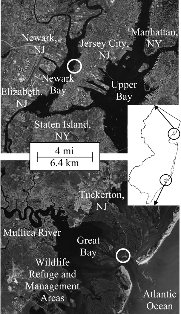
Three studies are presented here with the purpose of characterizing the endocrine disrupting effects influencing the transcriptional regulation of estrogen–responsive genes in the chemically impacted Newark Bay population, relative to a reference population from Tuckerton (Fig. 1). The first was a challenge study conducted using adult male killifish from Newark Bay and Tuckerton to compare the transcriptional sensitivity of various hepatic reproductive biomarker genes (chg H, chg Hm, chg L, vtg1, vtg2) to 17β–estradiol. This dose–response challenge study was used as an integrated endpoint that may be influenced by many potential effects on ER signaling that could result in adverse effects on ER–mediated gene expression. The second study evaluated 17β–estradiol metabolism (hepatic elimination activity) and expression levels for genes involved in nuclear estrogen receptor signaling (ER α, ER βa, ER βb) and E2 metabolism (cyp1a, cyp3a30, mdr). In the third study, we investigated differential methylation of CpG sites (gene silencing) in vtg promoters and single nucleotide polymorphisms (SNPs) in the promoter of vtg1 as possible explanations for the refractory sensitivity of various genes in Newark Bay killifish. Our overall hypothesis was that Newark Bay killifish are transcriptionally less sensitive to E2, which may correlate with changes in metabolism, receptor expression, or gene promoter methylation and sequence.
Fig. 1.
F. heteroclitus collection locations are indicated by circles at a reference site in Tuckerton, and the chemically impacted Newark Bay, NJ (USA) within the interconnected NY-NJ Harbor Estuary.
2.0. Materials and methods
2.1. Site selection, animal necropsy and husbandry protocols
All animal husbandry and handling methods were approved by the Rutgers University Animal Rights Committee in accordance with AALAC accreditation and NIH guidelines. Adult killifish (3–10 g, 5–9 cm) were collected and transported to the laboratory to be either sacrificed, or acclimated to laboratory conditions for 1 week prior to studies (20 ppt seawater, 20 ± 1 °C, 14:10 light:dark photoperiod, ground squid diet). Killifish were collected from two populations in New Jersey, USA (Fig. 1). The reference population was collected from a relatively pristine site in Tuckerton, NJ (Rutgers University Marine Field Station), and the chemically impacted population was collected from heavily contaminated Newark Bay, NJ (Richard Rutkowski Park, Bayonne, NJ). Animals were euthanized with MS–222 (tricaine methanesulphonate), weighed and measured. Livers were removed, weighed, snap frozen in liquid nitrogen and stored at −80 °C.
2.2. Dose-response for 17β–estradiol challenge
The sensitivity of various reproductive hepatic biomarker genes (chg H, chg Hm, chg L, vtg1, vtg2) to mRNA induction by a 17β–estradiol challenge was evaluated in adult male killifish from Tuckerton and Newark Bay (N = 7 per group). Graded doses of 17β–estradiol (≥ 98%, Sigma–Aldrich, St. Louis, MO) were injected intra–peritoneally with 10, 100 and 1000 ng E2/g body weight (parts–per–billion) in corn oil (10 µL/g body weight). These doses were equivalent to 37, 367, and 3671 picomoles E2/g body weight, respectively. Expression was measured in uninjected controls and 17β–estradiol treatment groups 4 days post–injection, the point at which circulating vitellogenin levels are maximally induced by E2 (Pait and Nelson, 2003). A preliminary E2 challenge study confirmed that 4 days post–injection leads to maximal induction of vtg1 in both populations (data not shown). Expression levels of hepatic genes involved in metabolism and estrogen signaling (cyp1a, cyp3a30, ER α, ER βa, ER βb, mdr) were also measured in uninjected controls and 100 ng E2/g treatment (N = 7 per group). Promoter methylation analysis was performed for vtg1 and vtg2 in the uninjected controls and 100 ng/g 17β–estradiol treatment group (N = 6 per group).
2.3. Analysis of mRNA expression by qRT PCR
Hepatic mRNA expression was evaluated using qRT–PCR methods adapted from Bugel et al. (2011, 2013). Briefly, total RNA was isolated from livers using TRIzol® (Invitrogen, Carlsbad, CA), DNAse treated (DNA–free, Ambion, Austin, TX), and reverse transcribed using High Capacity cDNA Reverse Transcription Kit (Applied Biosystems, Foster City, CA). qRT–PCR was performed using Applied Biosystems Power SYBR® Green PCR Master Mix with a StepOnePlus™ Real–Time PCR System (Applied Biosystems, Foster City, CA). Each gene was analyzed in duplicate and copy number was quantified using standard curves generated with PCR amplicons of each gene. For each gene and sample, expression was normalized using the ratio of the sample’s β–actin expression to the population’s median β–actin expression. Primers used are listed in Table 1, and were either developed previously (β–actin, cyp1a, ER α, ER βa, ER βb, vtg1) or newly validated (chg H, chg Hm, chg L, cyp3a30, mdr, vtg2) using criteria described by Bugel et al. (2010, 2011).
Table 1.
List of F. heteroclitus primers used for qRT-PCR, direct sequencing bisulphite PCR and SNP analysis.
| qRT–PCR Target | Gene | GenBank Accession | Size (bp) | Forward Primer (5′ →3′) | Reverse Primer (5′ →3′) |
| β–actin | β–actin | AY735154.1 | 136 | GCT CTG TGC AGA ACA ACC ACA CAT | TAA CGC CTC CTT CAT CGT TCC AGT |
| choriogenin H (zona pellucida 2) | chg H | AB533328.1 | 133 | CCC TGC CAC ACA TTG ACC TTG AAA | TCC TCC ATG ACA ACA GTC CCA CAA |
| choriogenin H minor | chg Hm | AB533329.1 | 136 | ATA CAC TGT GAT GCT GCT GTG TGC | CCT TGC TGC TAA CAA TGG TGG CTT |
| choriogenin L (zona pellucida 3) | chg L | AB533330.1 | 110 | TGT TTA CGT GGA CAG ATG TGT GGC | AGC CTG TGA CTC TGG CGT CAA TTA |
| cytochrome P450 1A | cyp1α | AF026800.1 | 258 | TGT TGC CAA TGT GAT CTG TG | CGG ATG TTG TCC TTG TCA AA |
| cytochrome P450 3A30 | cyp3α30 | AF105068 | 89 | ACC TGG ACT GCG TCA TCA ATG AGT | ATT TAT CTC CAC GGC TGC TTT GGC |
| estrogen receptor α | ER α | AY571785.1 | 195 | TTT CTT TCT GCA CCG GCA CAA TGG | GCT CCA TGC CTT TGT TGC TCA TGT |
| estrogen receptor βb | ER β α | AY570922.1 | 112 | ATC TTT CAC ATG CTA ATC GCC GCC | TCA GGC ACA TGT TGG AGT TGA GGA |
| estrogen receptor βb | ER βb | AY570923.1 | 162 | TTG ACG CTC TGG TTT GGG CTA TCT | ACA CAA GCA CCA CGT TCT TCC TCT |
| multidrug resistance transporter | mdr | AF099732.1 | 135 | ATG CAG ACC TTC CAG AAA GCG GAT | TGC AAA TGA CGA GCC TCT GGT AGA |
| vitellogenin 1 | vtg1 | U07055.2 | 234 | CAG CAC CAG GAA TAT CTC AG | GTG TAG AGT GTG TCT TCG AC |
| vitellogenin 2 | vtg2 | U70826.1 | 100 | CAA GCA GTA CAA CAC CAC | GAT GTA AGT AGG GAG TCT GG |
| Bisulphite PCR Target | Gene | Gene Region | Size (bp) | Forward Primer (5′ →3′) | Reverse Primer (5′ →3′) |
| vitellogenin 1 promoter set 1 | vtg1 | −1829 to −1634 | 195 | TTT TTA ATA TTG AGG GAA TAG TTT TGG | AAC CAA AAA ACC TAA CTT ACA ATT TC |
| vitellogenin 1 promoter set 2 | vtg1 | −1110 to −663 | 447 | TGT GAT GTA GGG ATA TTG GAA TTA GA | TCC ATC TTT TAA AAA AAA TTC CCT TA |
| vitellogenin 1 promoter set 3 | vtg1 | −143 to +583 | 726 | TTG ATG TAT GTT GTT TAA ATT AGT TGT TG | A TTA CCA AAC CTT CCT CAA AAA AAC C |
| vitellogenin 2 promoter set 1 | vtg2 | −1414 to −926 | 488 | GTA GAT TTT TAA TTG GAT TGG GGT T | TTC ACC ACC TTT ACT AAA AAA CCT TTA |
| vitellogenin 2 promoter set 2 | vtg2 | −256 to +280 | 536 | TGA TAA TGA AGG TAG TTG GGA TT | AAA ACT TAC CAT AAC TCA CCT AAT TAC C |
| SNP Sequencing | Gene | Gene Region | Size (bp) | Forward Primer (5′ →3′) | Reverse Primer (5′ →3′) |
| vitellogenin 1 promoter | vtg1 | −920 to +117 | 1037 | ATG CAC AGG ATG TCA TAT CTA | GGC ACA GAA GAA CTC CTA AA |
2.4. Quantitative DNA methylation analysis with bisulphite PCR and direct DNA sequencing
To analyze promoter methylation status for vtg1 and vtg2, the recently sequenced killifish genome (assembly v2b 2013.Mar, not yet published) and BLAST tools to search annotated genes were acquired from Mount Desert Island Biological Laboratory [http://www.mdibl.org/]. After identifying each gene, Clustal X2 alignment software was used to define exon and intron boundaries by aligning complete CDS of vtg1 (U07055.2) and vtg2 (U70826.1) to the genomic sequences. Putative estrogen–responsive elements (EREs) were identified in the 2 kb region upstream to the ATG start site using JASPAR [http://jasper.genereg.net], a transcription factor binding site prediction tool for vertebrates. Genomic sequences for vtg1 and vtg2 were then analyzed for CpG islands [http://cpgislands.usc.edu/] using flexible parameters (%GC = 50%, ObsCpG/ExpCpG = 0.6, Length = 200 bp, Gap = 100 bp). Regions of interest were chosen for methylation analysis 2 kb upstream and 1 kb downstream to the ATG start site.
DNA methylation was evaluated for CpG sites in the coding strands of vtg1 and vtg2 promoters using direct bisulphite PCR sequencing methods adapted from Lewin et al. (2004). Genomic DNA (gDNA) was isolated from uninjected control and E2–treated animal livers (N = 6 per group per population) using DNeasy Blood & Tissue Kit (Qiagen, Venlo, Netherlands). Two micrograms of gDNA were converted to bisulphite DNA (bisDNA) using EpiTect Fast Bisulfite Conversion Kit (Qiagen, Venlo, Netherlands). MethPrimer [http://www.urogene.org/methprimer/] was used to design primers specific to regions of interest in vtg1 and vtg2 (Table 1) (Li and Dahiya, 2002). Bisulphite PCR amplification was performed with 100 ng bisDNA using Epimark® Hot Start Taq DNA Polymerase (New England Biolabs, Ipswich, MA). SNP analysis for the vtg1 promoter was performed (N = 12 per population) by PCR amplification of the –920 to +117 region using primers listed in Table 1 with KOD Hot Start DNA polymerase (EMD Millipore, Billerica, MA). All PCR products were verified for size and specificity using gel electrophoresis, then purified using sodium acetate/ethanol precipitation prior to sequencing.
The Center for Genome Research & Biocomputing (CGRB) at Oregon State University (Corvallis, OR) performed all Sanger DNA sequencing. Briefly, all PCR products were sequenced using Big Dye Terminator 3.1 (Applied Biosystems, Foster City, CA) reaction mix and an Applied Biosystems 3730 DNA Analyzer with KB Basecaller Software. Epigenetic Sequencing Methylation Analysis software (ESME) was used to process *.ab1 sequencing trace files and quantify methylation at each CpG site as previously described (Lewin et al., 2004). Using these methods, an example sequencing trace shows that bisulfite conversion is complete for all non–CpG cytosines (Suppl. Fig. S1).
2.5. Hepatic E2 enzymatic elimination activity in vitro
An in vitro assay was used to measure 17β–estradiol elimination by hepatic S9 protein fractions as a surrogate measure of overall enzymatic activity in Tuckerton and Newark Bay male and female killifish (N = 8 per group). Livers were collected 3 days prior to a full moon, the peak level of circulating 17β–estradiol in females during spawning (Cerdá et al., 1996). Livers were homogenized on ice in Assay Buffer (100 mM Tris–HCl, 100 mM NaCl, 1 mM EDTA, 250 mM sucrose, pH 7.6), and centrifuged at 9,000 × g for 20 mins at 4 °C. Protein concentration of the S9 supernatant was quantified using modified Lowry Protein Assay (Pierce Biotechnology, Rockford, IL) and stored at −80 °C. Assay Buffer was used to prepare all reagents for the assay in 0.2 mL total, which contained 100 µg S9 protein, 5 µM 17β–estradiol, and 100 mM NADPH (NADPH Regeneration System, Promega, Fitchburg, WI). Prior to NADPH addition, 1 µL was diluted and saved on ice for analysis. Reactions were initiated by NADPH addition and incubated at 30 °C for 90 mins. Assays were then chilled to 4 °C, and 1 µL of the assay was diluted for analysis. All saved samples were immediately diluted 1:1500 and concentrations measured using Coat–A–Count Estradiol Radioimmunoassay (Siemens Medical Solutions Diagnostics, Los Angeles, CA). Using initial and final concentrations, hepatic E2 elimination activity was calculated as pmol 17β–estradiol loss per microgram protein per minute.
2.6. Statistical analyses
Statistical tests were performed using SigmaPlot™ (v. 11.0). For discrete data (SNPs), a Fisher’s Exact Test was performed. For single comparisons, a Student’s t–test was used (Mann– Whitney Rank Sum when equal variance failed). For multiple comparisons, a Two–Way ANOVA (Tukey’s post–hoc) was used to compare treatments between sites, and within each site. Gene expression data was log10 transformed for normality. All data was reported as mean ± standard deviation. Fold–changes in gene expression were calculated using mean values (treatment divided by control, or Tuckerton divided by Newark Bay). A p–value ≤ 0.05 was regarded as significantly different for all studies.
3.0. Results
3.1. Study 1: Evaluation of population differences in the sensitivity of biomarker genes to induction by 17β–estradiol
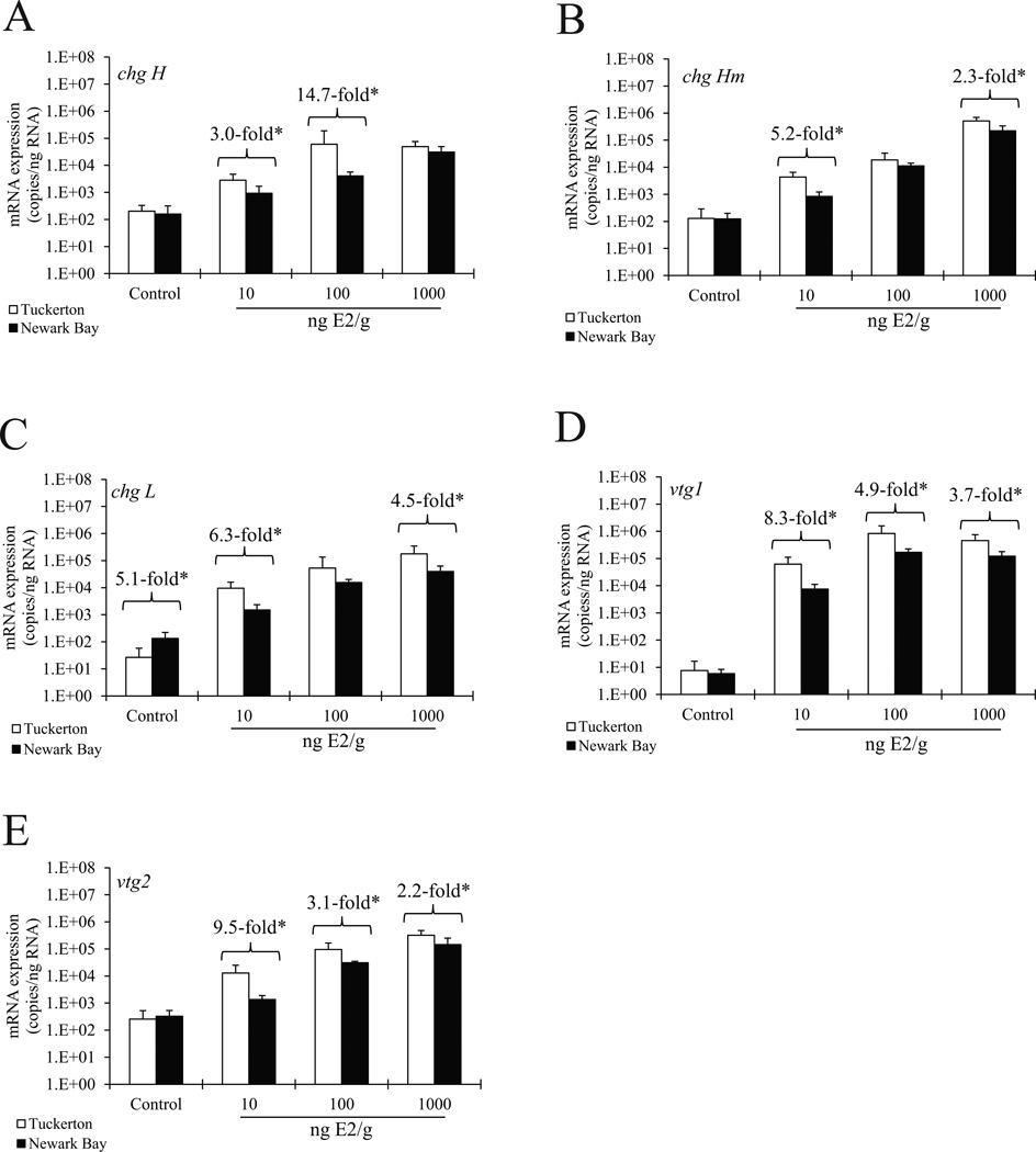
A challenge study was conducted to investigate population differences in the sensitivity of various reproductive biomarker genes (chg H, chg Hm, chg L, vtg1, vtg2) to graded doses of E2. Male killifish from Tuckerton and Newark Bay were of the same size class and were not significantly different for body and liver weights, and hepatosomatic index ratios (Suppl. Table 1, N = 28 per population, p ≤ 0.05, Student’s t –test). Overall, killifish from both populations responded in a dose–dependent manner, and expression levels were significantly higher than uninjected controls for all doses (Fig. 2, N = 7 per group, p ≤ 0.05, Two-Way ANOVA). There were no significant differences in basal mRNA expression levels measured in the population control groups for chg H, chg Hm, vtg1, and vtg2. However, control basal expression of chg L was 5.1–fold higher in the Newark Bay population relative to Tuckerton (Fig. 2C). Although all genes responded to E2 in the Newark Bay population, the magnitude of the gene induction and expression in response to various doses of E2 was lower than that of Tuckerton (Fig. 2). For example, Newark Bay vtg1 expression levels were significantly lower than those in the Tuckerton population by 8.3–, 4.9– and 3.7–fold at 10, 100, and 1000 ng E2/g (parts-per-billion), respectively (Fig. 2D). Similarly, vtg2 expression levels in Newark Bay killifish were 9.5–, 3.1–, and 2.2–fold significantly lower than levels in the Tuckerton population at 10, 100 and 1000 ng E2/g, respectively (Fig. 2E). For chg H, Newark Bay killifish had significantly lower expression levels only at 10 and 100 ng E2/g, while expression for chg Hm and chg L in Newark Bay were only significantly lower for 10 and 1000, but not 100 ng E2/g (Fig. 2A, 2B and 2C).
Fig. 2.
A 17β–estradiol dose-response study demonstrated differential hepatic mRNA expression between the reference Tuckerton population and the chemically impacted Newark Bay F. heteroclitus for (A) chg H, (B) chg Hm, (C) chg L, (D) vtg1 and (E) vtg2. Within both Tuckerton and Newark Bay populations, expression of all genes with all 17β–estradiol treatments was higher than respective uninjected controls. Overall, expression levels were significantly lower for all genes in E2 treatment groups for Newark Bay killifish compared to Tuckerton, except for 1000 ng E2/g (chg H) and 100 ng E2/g (chg Hm and chg L). Doses were nanograms of 17β– estradiol injected per gram body weight (parts-per-billion). *Fold-differences in mean expression between populations are shown for each treatment that was significantly differentially expressed between populations. Fold-inductions for each treatment over the population’s respective control are shown in Table 2. Data are reported as mean ± standard deviation (N = 7 per group). p ≤ 0.05 was regarded as significantly different (Two-Way ANOVA, Tukey’s post-hoc).
In addition to exhibiting significantly differential gene expression when treated with E2, there were major differences in fold induction levels for biomarker genes relative to each population’s respective uninjected controls (Table 2). For example, at 10 ng E2/g Tuckerton exhibited an 8185–fold induction for vtg1 over Tuckerton control fish, whereas Newark Bay exhibited a 1281–fold induction over Newark Bay control fish. Therefore, Newark Bay induction levels were approximately 6.4–fold lower than those in Tuckerton, corresponding to an 84.4% inhibition of induction. A similarly large inhibition in the Newark Bay population’s fold induction was observed in all genes that were differentially expressed when treated with E2 (Table 2).
Table 2.
Fold induction levels of genes in response to 17β-estradiol within each F. heteroclitus population.
| 17β–estradiol | |||
|---|---|---|---|
| 10 ng/g | 100 ng/g | 1000 ng/g | |
|
chg H |
|||
| Tuckerton fold | 14 | 295 | 242 |
| Newark Bay fold | 6 | 26 | 197 |
| Fold difference: | 2.4* | 11.4* | 1.2 |
| % inhibition: | 62.2% | 91.5% | – |
|
chg Hm |
|||
| Tuckerton fold | 34 | 144 | 3990 |
| Newark Bay fold | 7 | 91 | 1791 |
| Fold difference: | 5.0* | 1.6 | 2.2* |
| % inhibition: | 82.3% | – | 55.1% |
|
chg L |
|||
| Tuckerton fold | 360 | 2016 | 6726 |
| Newark Bay fold | 11 | 116 | 297 |
| Fold difference: | 32.1* | 17.3 | 22.6* |
| % inhibition: | 97.2% | – | 95.6% |
|
vtg1 |
|||
| Tuckerton fold | 8185 | 109423 | 60313 |
| Newark Bay fold | 1281 | 29057 | 20899 |
| Fold difference: | 6.4* | 3.8* | 2.9* |
| % inhibition: | 84.4% | 73.8% | 64.5% |
|
vtg2 |
|||
| Tuckerton fold | 50 | 375 | 1246 |
| Newark Bay fold | 4 | 96 | 444 |
| Fold difference: | 12.1* | 3.9* | 2.8* |
| % inhibition: | 93.6% | 74.7% | 64.5% |
Note: Fold inductions were calculated using mean expression in treatment relative to the respective population’s uninjected control for the data in Fig. 2 (N = 7 per group). Fold difference in induction levels between Tuckerton and Newark Bay are shown. Percent inhibition (decrease) of Newark Bay induction levels are shown relative to Tuckerton induction levels for significantly differentially expressed genes. Inhibition was calculated where 1-fold represents 100% inhibition (no induction relative to control) and the Tuckerton induction levels represent 0% inhibition (full induction). Control expression levels for all genes were not statistically different between populations, except for chg L which was 5.1 fold higher in the Newark Bay population. Percent inhibition was not calculated for treatment groups where induced expression levels were not significantly different. *Indicates significant difference for expression levels between populations in the 17β–estradiol treatment groups (p ≤ 0.05, Two-Way ANOVA, Tukey’s post-hoc).
3.2. Study 2: Hepatic ER signaling and E2 metabolism – analysis of gene expression and enzymatic E2 elimination
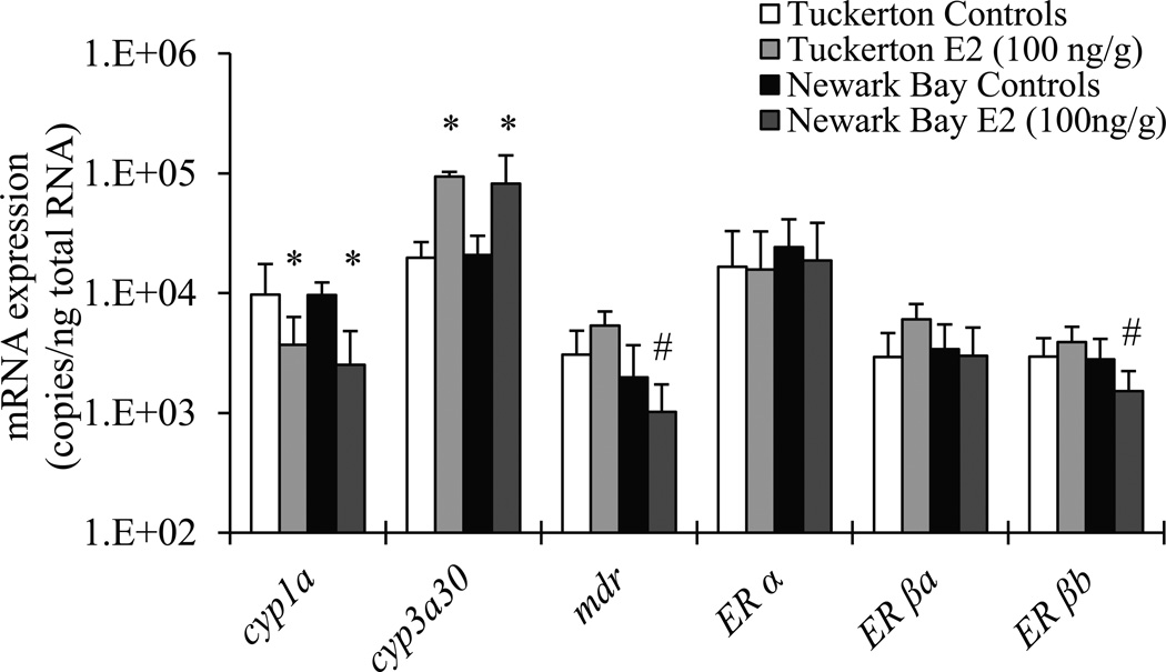
To investigate the etiology for the differential gene expression and decreased inducibility of estrogen–responsive genes in the Newark Bay population, we analyzed gene expression for genes involved in estrogen signaling and metabolism, as well as metabolic elimination activity of 17β–estradiol in vitro. First, we tested the hypothesis that expression levels of various genes critical to normal estrogen signaling and steroid metabolism in the liver would be differentially expressed between the Tuckerton and Newark Bay populations in uninjected control and estrogen treatment groups at 4 days post–injection. Using samples from the E2 challenge study (Study 1), expression was analyzed for cyp1a, cyp3a30, ER α, ER βa, ER βb and mdr in animals of the same size and weight class. Overall, basal expressions for all genes were statistically the same between Tuckerton and Newark Bay killifish (Fig. 3, N = 7 per group, p ≤ 0.05, Two-Way ANOVA). Treatment with 100 ng E2/g significantly altered the expression of two genes (cyp1a and cyp3a30), however, there were no significant differences for treatment expression levels between populations.
Fig. 3.
Basal mRNA expression of hepatic genes involved in drug metabolism (cyp1a, cyp3a30, mdr) and estrogen signaling (ER α, ER βa, ER βb) were not significantly different between populations of F. heteroclitus. Expression was evaluated in uninjected controls and animals injected with 100 ng nanograms of 17β–estradiol per gram body weight (parts-per-billion) at 4 days post-injection. Data are reported as mean ± standard deviation (N = 7 per group). *Indicates significant differences between control and E2 treated groups within the respective population. #Indicates significant differences between populations within the respective treatment group. p ≤ 0.05 was regarded as significantly different (Two-Way ANOVA, Tukey’s post-hoc).
We then tested the hypothesis that Newark Bay animals exhibit elevated levels of hepatic 17β–estradiol elimination activity using liver homogenates in an in vitro assay. Male and female killifish collected from Tuckerton and Newark Bay for the E2 metabolism study were of the same size class and did not differ significantly in regards to body weight, body length, and length to body weight ratios (N = 8 per group, p ≤ 0.05, Student’s t –test, Suppl. Table 2). Enzymatic elimination of E2 in vitro did not differ significantly between killifish from Tuckerton and Newark Bay for either gender, nor between genders within each site (Fig. 4, N = 8 per group, p ≤ 0.05, Two-Way ANOVA).
Fig. 4.
Hepatic 17β–estradiol enzymatic elimination activities using S9 fractions in vitro were not significantly different between F. heteroclitus populations, or between genders. Data are reported as mean ± standard deviation (N = 8 per group). p ≤ 0.05 was regarded as significantly different, although no significant differences were found (Two-Way ANOVA, Tukey’s post-hoc).
3.3 Study 3: Analysis of gene methylation and SNPs in vtg promoters
Gene methylation was evaluated in the coding strands for the promoter regions of vtg1 and vtg2 as a potential epigenetic mechanism for the decreased sensitivity of biomarker genes to E2 induction in the Newark Bay population. Prior to DNA methylation analysis, the genomic sequences for both genes were obtained and analyzed for various gene regulatory elements to determine regions of interest. To achieve this, the Mount Desert Island Biological Laboratory Fundulus Genomics Portal was used to search the sequenced killifish genome (assembly v2b 2013.Mar), and although vtg1 was not yet annotated, vtg2 was found to be annotated on Scaffold72. The Killifish Genome BLAST tool was used to locate vtg1 using the first 1 kb of the complete CDS (GenBank Accession U07055.2). Like vtg2, the first few kilobases of vtg1 were also found on Scaffold72. However, the gene was divided between two unjoined Scaffolds, and the latter half was located on Scaffold812. On Scaffold72, vtg1 was in close proximity to and directly downstream of vtg2 on the same coding strand, a likely result of a gene duplication event (Suppl. Figure S2). All exons, introns, ATG start sites, CpG sites, and putative EREs in the promoters were identified for these two genes (Suppl. Fig. S2). Neither gene had CpG islands in the promoter regions (2 kb upstream of the ATG start site), although several CpG sites were found in both promoters. Two CpG islands were however found within vtg2, and both in the latter half of the gene. These two CpG islands were unlikely to be important or directly involved in the regulation of transcription for these genes, and therefore they were not chosen as target regions for CpG methylation analysis.
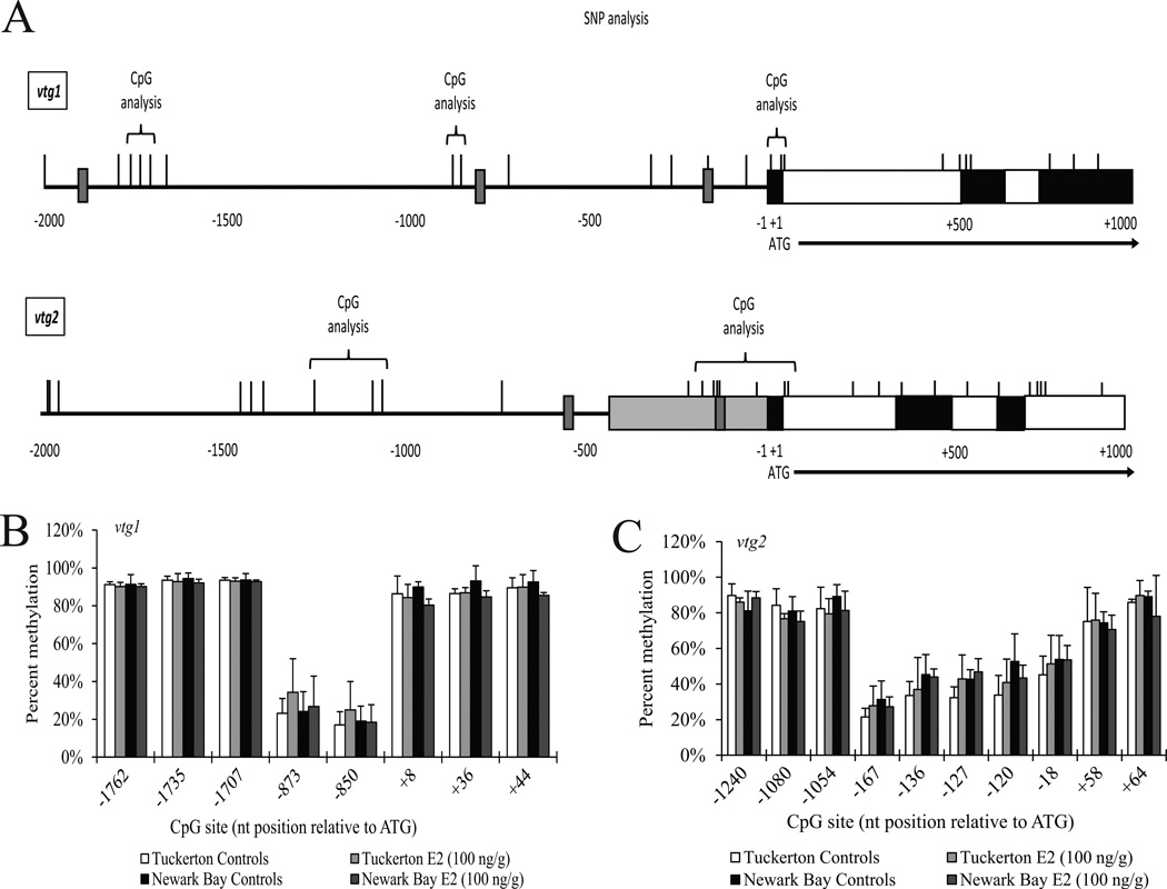
After annotating pertinent gene features for vtg1 and vtg2, DNA methylation was evaluated for CpG sites found in the promoters regions of uninjected control and E2 treated animals from both populations using samples from the E2 challenge study (Study 1). We tested the hypotheses that (1) CpG sites in the promoters of vtg1 and vtg1 would be hypermethylated in Newark Bay animals relative to Tuckerton, and (2) upon treatment with E2, CpG sites would demethylate in Tuckerton killifish, and significantly less so in Newark Bay animals. The regions considered most likely to be important and directly involved in the regulation of vtg transcription were 2 kb upstream and 1 kb downstream relative to the ATG start site (Fig. 5A). Overall, methylation levels varied greatly between CpG sites, although there were no significant differences between populations or between treatment groups for any specific CpG site tested (Figs. 5B and 5C, N = 6 per group, p ≤ 0.05, Two-Way ANOVA). For vtg1, CpG sites were hypermethylated at the three sites within exon 1 (> 80% methylation), and the three in the distal −1700 nt promoter region (> 90% methylation) (Fig. 5B). However, the two in the −850 nt promoter region were hypomethylated (< 35% methylation). Similarly for vtg2, CpG sites were hypermethylated at the two CpG sites within intron 1 (> 75% methylation), and the three in the distal –1000 nt promoter region (> 75% methylation) (Fig. 5C). Those found in the proximal – 100 nt promoter region exhibited varying levels of hypomethylation, ranging from 20–45% methylation.
Fig. 5.
Methylation analysis for vtg1 and vtg2 promoters in F. heteroclitus from two populations. (A) Gene maps show CpG sites identified in the 2 kb region upstream and 1 kb region downstream of the ATG transcriptional start site for vtg1 (top) and vtg2 (bottom). In the 1 kb downstream of the ATG site, exons are shown as black boxes and introns are shown as white boxes. In the 2 kb upstream of the ATG site, putative EREs are shown as small dark gray boxes, and a putative 5′–UTR for vtg2 is shown as a light gray shaded box. Putative EREs are also shown as small dark gray shaded boxes. CpG sites are shown as vertical lines, and those analyzed for methylation status are indicated with brackets. Methylation at these sites was quantified by bisulphite PCR and direct sequencing for (B) vtg1 and (C) vtg2. Overall, there were no significant differences in methylation status for any CpG site between populations (Tuckerton vs Newark Bay), or between treatments (Uninjected Control vs 100 ng E2/g body weight) at p ≤ 0.05 (Two-Way ANOVA, Tukey’s post-hoc). Data are reported as mean ± standard deviation (N = 6 per group).
To determine if there were sequence differences in vtg1 in Newark Bay killifish, we sequenced a 1 kb region upstream to the ATG start site that contained promoter regulatory elements such as putative EREs and CpG sites. Twelve single nucleotide polymorphisms (SNPs) were found in Tuckerton and Newark Bay killifish (Table 3). However, none were significantly more prevalent in Newark Bay animals relative to Tuckerton (Fisher’s Exact Test, p ≤ 0.05). Additionally, none of the twelve SNPs identified altered sequences of putative EREs or CpG sites (Suppl. Fig. S2).
Table 3.
Single Nucleotide Polymorphism analysis for vtg1 promoter in Tuckerton and Newark Bay killifish (F. heteroclitus).
| nt position | SNP | Tuckerton | Newark Bay | p–value |
|---|---|---|---|---|
| −670 | A:G | 11:1 | 8:4 | 0.32 |
| −666 | T:C | 10:2 | 8:4 | 0.64 |
| −632 | C:T | 11:1 | 7:5 | 0.16 |
| −567 | C:T | 12:0 | 9:3 | 0.22 |
| −566 | A:G | 10:2 | 6:6 | 0.19 |
| −380 | T:G | 10:2 | 12:0 | 0.48 |
| −201 | A:C | 12:0 | 9:3 | 0.22 |
| −81 | G:A | 9:3 | 6:6 | 0.40 |
| −61 | T:C | 7:5 | 6:6 | 1.00 |
| −24 | T:C | 8:4 | 4:8 | 0.22 |
| −22 | G:C | 12:0 | 10:2 | 0.48 |
| +18 | T:A | 12:0 | 10:2 | 0.48 |
Note: Numbers of SNPs are shown for each respective nucleotide position in the promoter region (nucleotides −920 to +117 relative to ATG start site). A total of 12 animals were sequenced from each population. p ≤ 0.05 was regarded as significantly different (Fisher’s Exact Test).
4.0. Discussion
In the present studies, we used a set of vitellogenin and choriogenin genes to study anti– estrogenic chemical effects on transcriptional regulation and estrogen sensitivity in Newark Bay killifish. In killifish, two hepatically derived vitellogenin genes (vtg1 and vtg2) are responsible for yolk protein biosynthesis, although the majority of yolk proteins are derived from vtg1 (LaFleur et al., 2005). Multiple choriogenin genes (chg H, chg Hm, chg L) are also hepatically expressed, and are involved in sperm binding and formation of the chorionic membrane, zona pellucida (Arukwe and Goksøyr. 2010). Our dose–response study demonstrated that all five of these genes are transcriptionally less sensitive to 17β–estradiol in the Newark Bay killifish population, with a robust inhibition of induction relative to Tuckerton control killifish (Fig. 2, Table 2). The desensitization of vtg1 and vtg2 to estrogen in our current studies has direct relevance to, and provides a transcriptional basis for, the previously reported reproductive effects and effects on vitellogenin expression in this population. Reproductively active female killifish from Newark Bay exhibit decreased vitellogenin expression levels, which correlates with an inhibition of vitellogenin–dependent growth of oocytes and reduced fecundity (Bugel et al., 2010, 2011). A similar dose–response study by Bugel et al. (2011) demonstrated that vitellogenin protein levels are also less inducible in this population and have similar magnitudes of inhibition to those reported in our current study. The relevance of the desensitization of choriogenins is not as clear, although we predict similar adverse effects on choriogenin expression and development of the chorionic membrane during folliculogenesis in Newark Bay killifish. The potential ramifications of such a disruption could include adverse effects on embryo viability due to decreased fertilization success and weakened membranes. Laboratory fertilization of killifish gametes demonstrated that embryos from Newark Bay are less viable, although a role for disrupted choriogenesis is not clear (Bugel et al., 2011). Overall, the anti–estrogenic effect altering the sensitivity of estrogen–responsive hepatic genes does not appear to be specific to any particular gene tested. It is therefore possible that other genes and other aspects of oogenesis are impaired similarly to the vitellogenin–dependent effects.
Vitellogenin biomarker responses in male killifish collected from Newark Bay are atypical and considered false negatives because expression levels in males are not elevated nor induced by sediment extracts (McArdle et al, 2004, Bugel et al., 2010, 2011). This was unexpected due to the presence of estrogenic contaminants throughout the NY–NJ Harbor, including PCBs, bisphenol A, phthalates, and synthetic estrogens (Litten, 2003). However, naïve male control killifish transplanted into Newark Bay exhibited a strong induction of vitellogenin after 1 month that was later undetectable at 2 months (Bugel et al., 2011). As a result of this attenuated expression over time, vitellogenin fails as a biomarker for exposure in this population. Newark Bay is therefore considered an immediately estrogenic environment with anti–estrogenic effects predominating from prolonged exposure. False negatives have been reported in other studies for vitellogenin, which can confound risk assessment and underestimate exposures (Bosker et al., 2010). In our current study, chg L (zona radiata, ZP3) was the only significant estrogen–responsive gene found to be basally elevated in the Newark Bay control group (5–fold), relative to the Tuckerton control group (Fig. 2, Table 2). Choriogenins, like vitellogenins, can be used as biomarkers of exposure to xeno–estrogens, although vitellogenin is more commonly used (Lee et al., 2002). All other genes (vtg1, vtg2, chg H, chg Hm) were not significantly elevated in the Newark Bay controls, which suggests that false negatives are more prevalent than previously reported. In this population, the desensitization and disruption of estrogen receptor transcriptional activity prohibits them from being used directly for evaluating exposures, although chg L successfully indicated exposure to estrogenic contaminants. In other species, chg L is more sensitive to estrogen than chg H, and choriogenins in general are more sensitive to estrogen than vitellogenins (Celius, 2000; Lee et al., 2002). These studies taken together suggest that chg L may be a sensitive biomarker that is less prone to false negative responses than vitellogenin. We therefore recommend that future studies measure choriogenin L concomitantly with vitellogenin in environments that may be co–contaminated by estrogenic and anti–estrogenic chemicals to minimize the potential for false negative results.
Many contaminants found in Newark Bay have been shown to inhibit the hepatic 17β– estradiol induction of vitellogenin in vitro, including aryl hydrocarbon receptor (AHR) ligands such as dioxins, furans, PAHs and PCBs (Bemanian et al., 2004; Gräns et al., 2010; Petersen and Tollsfsen, 2012). These ubiquitous chemicals are found in complex mixtures and exert similar cross–talk inhibition of vitellogenin expression in a number of teleosts, including zebrafish, rainbow trout, sea bass, and goldfish (Anderson et al., 1996b; Vaccaro et al., 2005; Yan et al., 2012; Bugel et al., 2013). AHR cross–talk inhibition of normal estrogen receptor functions may occur through several distinct mechanisms, including (1) direct competition for xenobiotic response elements (EREs and DREs) in gene promoters, (2) squelching of common cofactors, (3), AHR-dependent induction of inhibitory factors (e.g. miRNAs, inhibitory proteins), (4) proteasomic degradation of estrogen receptors, and (5) modulation of metabolic proteins (e.g. cytochrome P450s, multidrug resistance genes) (Safe and Wormke, 2003; Matthews and Gustafsson, 2006; Ohtake et al., 2008). Newark Bay killifish highly express Cyp1a protein indicative of elevated AHR activity, and we have previously reported an inverse relationship between Cyp1a and vtg1 expression (Bugel et al., 2010). The decreased transcriptional sensitivity of many estrogen-responsive genes to 17β–estradiol reported in the current studies is phenotypic of AHR-ER cross-talk inhibition. Abnormally high expression of hepatic cytochrome P450 enzymes and p-glycoproteins (multidrug resistance, mdr) can facilitate elimination of 17β–estradiol and decrease biological activity (Zhu and Conney, 1998; Mathieu et al., 2001). However, our current studies found no evidence suggesting that elevated levels of metabolic genes (cyp1a, cyp3a30, mdr) or decreased expression of nuclear estrogen receptors (ER α, ER βa, ER βb) are responsible for the desensitization to estrogen under basal conditions or estrogen treatment (Fig. 3). Previously, we reported elevated expression levels for cyp1a mRNA and protein in this Newark Bay killifish population (Bugel et al., 2010). However, in the current study, hepatic cyp1a levels were not elevated, which we believe may be due to the laboratory depuration period (1 week) prior to gene expression analysis. While we did not detect elevated mRNA levels for cyp1a, cyp3a30, or mdr, the protein and activity levels for these genes and others are not known for the current studies. 17β–estradiol metabolism is a complex process and although our gene set was not comprehensive and is limited, we also found no major differences in hepatic elimination rates in vitro (Fig. 4). Therefore, elevated hepatic metabolism is not expected to play a role in the decreased sensitivity of estrogen–responsive genes. Killifish from Newark Bay exhibit an altered AHR pathway that confers chemical tolerance and resilience to AHR–mediated toxicity (Prince and Cooper, 1995a,b; Nacci et al., 2010). Refractory phenotypes and altered AHR mechanisms have been characterized in other populations as well, including Elizabeth River (VA) and New Bedford Harbor (MA) (Bello et al., 2001; Whitehead et al., 2011; Clark et al., 2013). Future studies of chemically impacted fish populations such as these will be useful for exploring the relationship between altered AHR mediated processes and disruption of endocrine pathways.
Genetic mechanisms such as DNA methylation and SNPs are important to gene– environment interactions and can be useful for elucidating mechanism of toxicity in ecotoxicological studies (Vandegehuchte et al., 2013). For example, promoter methylation and SNPs have been explored as a mechanism for AHR–mediated resistance reported in various killifish populations (Timme–Laragy et al., 2005; Aluru et al., 2011; Reitzel et al., 2014). Global epigenome analyses have shown that CpG sites in promoters near transcriptional start sites and other regulatory elements (e.g. estrogen–responsive elements) play important roles in regulating gene expression through hypermethylation and gene silencing (Bird et al., 2002; Eckhardt et al., 2006). Using the recently sequenced killifish genome, we identified CpG sites and several putative EREs in promoters of vtg1 and vtg2 (Suppl. Fig. 2). However, we found no differential methylation between Newark Bay killifish and the control Tuckerton population for either gene under control or estradiol treated conditions (Fig. 5). It is therefore unlikely that these CpG sites play direct roles in gene silencing and the desensitization of vtg1 and vtg2 to estrogen in the Newark Bay population. Other intergenic or intragenic CpG sites may be involved, although their importance is increasingly unlikely with distance from the transcriptional start sites. Interestingly, neither gene was demethylated when treated with a concentration of estrogen that resulted in high induction levels at 4 days post–injection. This may suggest that (1) the sampling time point is too early to detect changes in methylation state, (2) demethylation occurs kinetically slower than maximal mRNA induction, or (3) the methylation responsive CpG site is found elsewhere. Studies in the chicken have demonstrated that CpG sites are demethylated either slower than, or in parallel with, the induction of vitellogenin gene expression (Meijlink et al., 1983; Saluz et al., 1988). Determining the kinetics of demethylation was beyond the scope of this study. In addition to methylation, we found no evidence for SNPs in the promoter of vtg1 in Newark Bay killifish (Table 3). Our studies do not support the hypothesis that epigenetic or genetic mechanisms (methylation or SNP) play a role for the desensitization of vtg1 and vtg2 to estrogen in the CpG sites evaluated. In the future, global genome and epigenome analyses with the newly sequenced killifish genome will facilitate these types of studies in a more comprehensive and unbiased fashion for mechanistic ecotoxicological studies.
5.0. Conclusions
Reproductive dysfunction and altered biomarker responses in killifish from Newark Bay, NJ, are the result of complex chemical exposures that exert anti–estrogenic effects (e.g. inhibition of vitellogenesis). Overall, we show that multiple estrogen–responsive genes (vtg1, vtg2, chg H, chg Hm, chg L) are transcriptionally desensitized to estrogen in a population of killifish from the chemically impacted Newark Bay compared to killifish of a relatively unpolluted reference site. The refractory transcriptional induction is indicative of a broad effect on global estrogen receptor pathway signaling. This suggests that the mechanism(s) responsible for endocrine disruption is pre–transcriptionally based and likely occurs prior to estrogen receptor transactivation of gene expression. However, our studies did not find a metabolic, genetic or epigenetic mechanism to explain this desensitization. Ultimately, chemicals that modulate gene regulation can result in deleterious effects such as those reported in Newark Bay killifish, which may pose a risk to reproductive function in females and undermines the direct use of exposure biomarkers such as vitellogenin in males (i.e. false negatives).
Supplementary Material
Highlights.
Reproductive biomarker genes in Newark Bay killifish are desensitized to estrogen
Gene desensitization indicates pre-transcriptional effects on estrogen signaling
Desensitization does not have a metabolic or epigenetic basis (gene methylation)
Modulation of vitellogenin and choriogenin genes correlates with reproductive impacts
choriogenin L appears less prone to false negatives and may be a sensitive biomarker
Acknowledgements
We wish to thank Craig Harvey for assistance with the E2 radioimmunoassay, Caprice Rosato at the Oregon State University Center for Genome Research & Biocomputing for assistance with bisulphite sequencing, and Michael Simonich for critical comments on the manuscript. Gene methylation studies were aided by early access to a preliminary draft of the F. heteroclitus genome sequence, which was supported by funding from the National Science Foundation [DEB-1120512].
Funding Information:
This work was partially carried out at Rutgers, The State University of New Jersey, with funding support to K.R.C. from the NJ Agricultural Experiment Station through Cooperative State Research, Education, and Extension Services [01201], The Environmental and Occupational Health Sciences Institute [ES05022], the NJ Water Resources Research Institute [2009NJ198B], NJ Department of Environmental Protection Agency, Division of Science, Research and Technology [SR09–019], and The National Oceanic and Atmospheric Administration [432742]. This work was also partially conducted at Oregon State University, with funding support to R.L.T. by U.S. National Institute of Environmental Health Sciences (NIEHS) Environmental Health Sciences Core Center grant [P30 ES000210], NIEHS Training grant T32 [ES007060], and an NIEHS Superfund Basic Research Program grant [P42 ES016465].
Abbreviations
- AHR
aryl hydrocarbon receptor
- chg
choriogenin
- cyp
cytochrome P450
- E2
17β–estradiol
- ER
estrogen receptor
- ERE
estrogen-responsive element
- mdr
multidrug resistance
- SNP
single nucleotide polymorphism
- vtg
vitellogenin
Footnotes
Publisher's Disclaimer: This is a PDF file of an unedited manuscript that has been accepted for publication. As a service to our customers we are providing this early version of the manuscript. The manuscript will undergo copyediting, typesetting, and review of the resulting proof before it is published in its final citable form. Please note that during the production process errors may be discovered which could affect the content, and all legal disclaimers that apply to the journal pertain.
All authors contributed to the study design and participated in writing of the manuscript. All authors have given final approval to the manuscript, and declare no competing financial interest.
References
- Aluru N, Karchner SI, Hahn ME. Role of methylation of AHR1 and AHR2 promoters in differential sensitivity to PCBs in Atlantic Killifish, Fundulus heteroclitus . Aquatic Toxicology. 2011;101(1):288–294. doi: 10.1016/j.aquatox.2010.10.010. [DOI] [PMC free article] [PubMed] [Google Scholar]
- Amiard-triquet C, Amiard JC, Rainbow PS, editors. Ecological Biomarkers: Indicators of Ecotoxicological Effects. Boca Raton, FL: CRC Press; 2013. p. 464. [Google Scholar]
- Anderson MJ, Olsen H, Matsumura F, Hinton DE. In vivo modulation of 17β- estradiol-induced vitellogenin synthesis and estrogen receptor in rainbow trout (Oncorhynchus mykiss) liver cells by β-naphthoflavone. Toxicology and Applied Pharmacology. 1996b;137:210–218. doi: 10.1006/taap.1996.0074. [DOI] [PubMed] [Google Scholar]
- Arukwe A, Goksøyr A. Eggshell and egg yolk proteins in fish: hepatic proteins for the next generation: oogenetic, population, and evolutionary implications of endocrine disruption. Comparative Hepatology. 2010;2:4. doi: 10.1186/1476-5926-2-4. [DOI] [PMC free article] [PubMed] [Google Scholar]
- Bello SM, Franks DG, Stegeman JJ, Hahn ME. Acquired resistance to Ah receptor agonists in a population of Atlantic killifish (Fundulus heteroclitus) inhabiting a marine Superfund site: in vivo and in vitro studies on the inducibility of xenobiotic metabolizing enzymes. Toxicological Sciences. 2001;60:77–91. doi: 10.1093/toxsci/60.1.77. [DOI] [PubMed] [Google Scholar]
- Bemanian V, Male R, Goksøyr A. The aryl hydrocarbon receptor-mediated disruption of vitellogenin synthesis in the fish liver: cross-talk between AhR and ERα-signaling pathways. Comparative Hepatology. 2004;3(2) doi: 10.1186/1476-5926-3-2. [DOI] [PMC free article] [PubMed] [Google Scholar]
- Bird A. DNA methylation patterns and epigenetic memory. Genes & Development. 2002;16:6–21. doi: 10.1101/gad.947102. [DOI] [PubMed] [Google Scholar]
- Bosker T, Munkittrick KR, MacLatchy DL. Challenges and opportunities with the use of biomarkers to predict reproductive impairment in fishes exposed to endocrine disrupting substances. Aquatic Toxicology. 2010;100:9–16. doi: 10.1016/j.aquatox.2010.07.003. [DOI] [PubMed] [Google Scholar]
- Bugel SM, White LA, Cooper KR. Impaired reproductive health of killifish (Fundulus heteroclitus) inhabiting Newark Bay NJ, a chronically contaminated estuary. Aquatic Toxicology. 2010;96(3):182–193. doi: 10.1016/j.aquatox.2009.10.016. [DOI] [PubMed] [Google Scholar]
- Bugel SM, White LA, Cooper KR. Decreased vitellogenin inducibility and 17β- estradiol levels correlated with reduced egg production in killifish (Fundulus heteroclitus) from Newark Bay, NJ. Aquatic Toxicology. 2011;105(1–2):1–12. doi: 10.1016/j.aquatox.2011.03.013. [DOI] [PMC free article] [PubMed] [Google Scholar]
- Bugel SM, White LA, Cooper KR. Inhibition of vitellogenin gene induction by 2,3,7,8-tetrachlorodibenzo-p-dioxin is mediated by aryl hydrocarbon receptor 2 (AHR2) in zebrafish (Danio rerio) Aquatic Toxicology. 2013;126:1–8. doi: 10.1016/j.aquatox.2012.09.019. [DOI] [PubMed] [Google Scholar]
- Burnett KG, Bain LJ, Baldwin WS, Callard GV, Cohen S, Di Giulio RT, Evans DH, Gómez-Chiarri Hahn ME, Hoover CA, Karchner SI, Katoh F, Maclatchy DL, Marshall WS, Meyer JN, Nacci DE, Oleksiak MF, Rees BB, Singer TD, Stegeman JJ, Towle DW, Van Veld PA, Vogelbein WK, Whitehead A, Winn RN, Crawford DL. Fundulus as the premier teleost model in environmental biology: opportunities for new insights using genomics. Comparative Biochemistry and Physiology - Part D: Genomics and Proteomics. 2007;2(4):257–286. doi: 10.1016/j.cbd.2007.09.001. [DOI] [PMC free article] [PubMed] [Google Scholar]
- Celander MC. Cocktail effects on biomarker responses in fish. Aquatic Toxicology. 2011;105(3–4):72–77. doi: 10.1016/j.aquatox.2011.06.002. [DOI] [PubMed] [Google Scholar]
- Celius T, Matthews JB, Giesy JP, Zacharewski TR. Quantification of rainbow trout (Oncorhynchus mykiss) zona radiata and vitellogenin mRNA levels using real-time PCR after in vivo treatment with estradiol-17β or α-zearalenol. Journal of Steroid Biochemistry and Molecular Biology. 2000;75:109–119. doi: 10.1016/s0960-0760(00)00165-5. [DOI] [PubMed] [Google Scholar]
- Cerdá J, Calman BG, LaFleur GJ, Jr, Limesand S. Pattern of vitellogenesis and follicle maturational competence during the ovarian follicular cycle of Fundulus heteroclitus . General and Comparative Endocrinology. 1996;103:24–35. doi: 10.1006/gcen.1996.0090. [DOI] [PubMed] [Google Scholar]
- Clark BW, Cooper EM, Stapleton HM, Di Giulio RT. Compound- and mixture-specific differences in resistance to polycyclic aromatic hydrocarbons and PCB-126 among Fundulus heteroclitus subpopulations throughout the Elizabeth River Estuary (Virginia, USA) Environmental Science & Technology. 2013;47(18):10556–10566. doi: 10.1021/es401604b. [DOI] [PMC free article] [PubMed] [Google Scholar]
- Eckhardt F, Lewin J, Cortese R, Rakyan VK, Attwood J, Burger M, Burton J, Cox TV, Davies R, Down TA, Haefliger C, Horton R, Howe K, Jackson DK, Kunde J, Koenig C, Liddle J, Niblett D, Otto T, Pettett R, Seemann S, Thompson C, West T, Rogers J, Olek A, Berlin K, Beck S. DNA methylation profiling of human chromosomes 6, 20 and 22. Nature Genetics. 2006;38(12):1378–1385. doi: 10.1038/ng1909. [DOI] [PMC free article] [PubMed] [Google Scholar]
- Gräns J, Wassmur B, Celander MC. One-way inhibiting cross-talk between arylhydrocarbon receptor (AhR) and estrogen receptor (ER) signaling in primary cultures of rainbow trout hepatocytes. Aquatic Toxicology. 2010;100:263–270. doi: 10.1016/j.aquatox.2010.07.024. [DOI] [PubMed] [Google Scholar]
- Greytak SR, Callard GV. Cloning of three estrogen receptors (ER) from killifish (Fundulus heteroclitus): differences in populations from polluted and reference environments. General and Comparative Endocrinology. 2007;150:174–188. doi: 10.1016/j.ygcen.2006.07.017. [DOI] [PubMed] [Google Scholar]
- Griffin LB, January KR, Ho KW, Cotter KA, Callard GV. Morpholino mediated knockdown of ERα, ERβa and ERβb mRNAs in zebrafish (Danio rerio) embryos reveals differential regulation of estrogen-inducible genes. Endocrinology. 2013;154:4158–4169. doi: 10.1210/en.2013-1446. [DOI] [PMC free article] [PubMed] [Google Scholar]
- LaFleur GJ, Jr, Raldúa D, Fabra M, Carnevali O, Denslow N, Wallace RA, Cerdá J. Derivation of major yolk proteins from parental vitellogenins and alternative processing during oocyte maturation in Fundulus heteroclitus . Biology of Reproduction. 2005;73:815–824. doi: 10.1095/biolreprod.105.041335. [DOI] [PubMed] [Google Scholar]
- Lee C, Na JG, Lee KC, Park K. Choriogenin mRNA induction in male medaka, Oryzias latipes as a biomarker of endocrine disruption. Aquatic Toxicology. 2002;61(3–4):233–241. doi: 10.1016/s0166-445x(02)00060-7. [DOI] [PubMed] [Google Scholar]
- Lewin J, Schmitt AO, Adorján P, Hildmann T, Piepenbrock C. Quantitative DNA methylation analysis based on four-dye trace data from direct sequencing of PCR amplificates. Bioinformatics. 2004;20(17):3005–3012. doi: 10.1093/bioinformatics/bth346. [DOI] [PubMed] [Google Scholar]
- Li LC, Dahiya R. MethPrimer: Designing primers for methylation PCRs. Bioinformatics. 2002;18(11):1427–1431. doi: 10.1093/bioinformatics/18.11.1427. [DOI] [PubMed] [Google Scholar]
- Litten S. New York/New Jersey Harbor Contaminant Assessment and Reduction Project (CARP) Albany, New York: New York State Department of Environmental Conservation; 2003. Available from: http://www.hudsonriver.org/ [Google Scholar]
- Mathieu MC, Lapierre I, Brault K, Raymond M. Aromatic hydrocarbon receptor (AhR).AhR nuclear translocator- and p53-mediated induction of the murine multidrug resistance mdr1 gene by 3-methylcholanthrene and benzo(a)pyrene in hepatoma cells. Journal of Biological Chemistry. 2001;276(7):4819–4827. doi: 10.1074/jbc.M008495200. [DOI] [PubMed] [Google Scholar]
- Matthews J, Gustafsson JA. Estrogen receptor and aryl hydrocarbon receptor signaling pathways. Nuclear Receptor Signaling. 2006;4:e016. doi: 10.1621/nrs.04016. [DOI] [PMC free article] [PubMed] [Google Scholar]
- McArdle ME, McElroy AE, Elskus AA. Enzymatic and estrogenic responses in fish exposed to organic pollutants in the New York-New Jersey (USA) Harbor Complex. Environmental Toxicology and Chemistry. 2004;4:953–959. doi: 10.1897/03-82. [DOI] [PubMed] [Google Scholar]
- Meijlink FC, Philipsen JN, Gruber M, Ab G. Methylation of the chicken vitellogenin gene: influence of estradiol administration. Nucleic Acids Research. 1983;11(5):1361–1373. doi: 10.1093/nar/11.5.1361. [DOI] [PMC free article] [PubMed] [Google Scholar]
- Menuet A, Adrio F, Kah O, Pakdel F. Regulation and function of estrogen receptors: comparative aspects. In: Melamed P, Sherwood N, editors. Hormones and Their Receptors in Fish Reproduction. Singapore: World Scientific Publishing Company; 2005. [Google Scholar]
- Miller DH, Jensen KM, Villeneuve DL, Kahl MD, Makynen EA, Durhan EJ, Ankley GT. Linkage of biochemical responses to population-level effects: a case study with vitellogenin in the fathead minnow (Pimephales promelas) Environmental Toxicology and Chemistry. 2007;26(3):521–527. doi: 10.1897/06-318r.1. [DOI] [PubMed] [Google Scholar]
- Muñoz GR, Panero MA, Powers CW. Pollution Prevention and Management Strategies for Dioxins in the New York/New Jersey Harbor. New York, NY: New York Academy of Sciences; 2006. Available from: http://www.nyas.org. [Google Scholar]
- Nacci DE, Champlin D, Jayaraman S. Adaptation of the estuarine fish Fundulus heteroclitus (Atlantic killifish) to polychlorinated biphenyls (PCBs) Estuaries & Coasts. 2010;33:853–864. [Google Scholar]
- Ohtake F, Baba A, Fujii-Kuriyama Y, Kato S. Intrinsic AhR function underlies cross-talk of dioxins with sex hormone signaling. Biochemical and Biophysical Research Communications. 2008;370:541–546. doi: 10.1016/j.bbrc.2008.03.054. [DOI] [PubMed] [Google Scholar]
- Pait AS, Nelson JO. Vitellogenesis in male Fundulus heteroclitus (killifish) induced by selected estrogenic compounds. Aquatic Toxicology. 2003;64(3):331–342. doi: 10.1016/s0166-445x(03)00060-2. [DOI] [PubMed] [Google Scholar]
- Panero M, Boehme S, Muñoz G. Pollution Prevention and Management Strategies for Polychlorinated Biphenyls in the New York/New Jersey Harbor. New York, NY: New York Academy of Sciences; 2005. Available from: http://www.nyas.org. [Google Scholar]
- Petersen K, Tollefsen KE. Combined effects of oestrogen receptor antagonists on in vitro vitellogenesis. Aquatic Toxicology. 2012;112–113:46–53. doi: 10.1016/j.aquatox.2012.01.023. [DOI] [PubMed] [Google Scholar]
- Prince R, Cooper KR. Comparison of the effects of 2,3,7,8-tetrachlorodibenzo-p-dioxin on chemically impacted and non-impacted subpopulations of Fundulus heteroclitus, I: TCDD toxicity. Environmental Toxicology and Chemistry. 1995a;14:579–587. 99. [Google Scholar]
- Prince R, Cooper KR. Comparison of the effects of 2,3,7,8-tetrachlorodibenzo-p-dioxin on chemically impacted and non-impacted subpopulations of Fundulus heteroclitus. II. Metabolic considerations. Environmental Toxicology and Chemistry. 1995b;14:589–596. [Google Scholar]
- Reitzel AM, Karchner SI, Franks DG, Evans BR, Nacci D, Champlin D, Vieira VM, Hahn ME. Genetic variation at aryl hydrocarbon receptor (AHR) loci in populations of Atlantic killifish (Fundulus heteroclitus) inhabiting polluted and reference habitats. BMC Evolutionary Biology. 2014;14:6. doi: 10.1186/1471-2148-14-6. [DOI] [PMC free article] [PubMed] [Google Scholar]
- Rempel MA, Schlenk D. Effects of environmental estrogens and antiandrogens on endocrine function, gene regulation, and health in fish. International Review of Cell and Molecular Biology. 2008;267:207–252. doi: 10.1016/S1937-6448(08)00605-9. [DOI] [PubMed] [Google Scholar]
- Safe S, Wormke M. Inhibitory aryl hydrocarbon receptor-estrogen receptor α cross-talk and mechanisms of action. Chemical Research in Toxicology. 2003;16(7):807–816. doi: 10.1021/tx034036r. [DOI] [PubMed] [Google Scholar]
- Saluz HP, Feavers IM, Jiricny J, Jost JP. Genomic sequencing and in vivo footprinting of an expression-specific DNase I-hypersensitive site of avian vitellogenin II promoter reveal a demethylation of a mCpG and a change in specific interactions of proteins with DNA. Proceedings of the National Academy of Sciences. 1988;85:6697–6700. doi: 10.1073/pnas.85.18.6697. [DOI] [PMC free article] [PubMed] [Google Scholar]
- Strömqvist M, Tooke N, Brunström B. DNA methylation levels in the 5′ flanking region of the vitellogenin I gene in liver and brain of adult zebrafish (Danio rerio)-sex and tissue differences and effects of 17α-ethinylestradiol exposure. Aquatic Toxicology. 2010;98:275–281. doi: 10.1016/j.aquatox.2010.02.023. [DOI] [PubMed] [Google Scholar]
- Sumpter JP, Jobling S. Vitellogenesis as a biomarker for estrogenic contamination of the aquatic environment. Environmental Health Perspectives. 1995;103(Suppl. 7):173–178. doi: 10.1289/ehp.95103s7173. [DOI] [PMC free article] [PubMed] [Google Scholar]
- Tyler CR, Jobling S, Sumpter JP. Endocrine disruption in wildlife: a critical review of the evidence. Critical Reviews in Toxicology. 1998;28(4):319–361. doi: 10.1080/10408449891344236. [DOI] [PubMed] [Google Scholar]
- Timme-Laragy AR, Meyer JN, Waterland RA, Di Giulio RT. Analysis of CpG methylation in the killifish CYP1A promoter. Comparative Biochemistry and Physiology Part C: Toxicology & Pharmacology. 2005;141(4):406–411. doi: 10.1016/j.cbpc.2005.09.009. [DOI] [PubMed] [Google Scholar]
- Thorpe KL, Benstead R, Hutchinson TH, Tyler CR. Associations between altered vitellogenin concentrations and adverse health effects in fathead minnow (Pimephales promelas) Aquatic Toxicology. 2007;85:176–183. doi: 10.1016/j.aquatox.2007.08.012. [DOI] [PubMed] [Google Scholar]
- Vaccaro E, Meucci V, Intorre L, Soldani G, Di Bello D, Longo V, Gervasi PG, Pretti C. Effects of 17β-estradiol, 4-nonylphenol and PCB 126 on the estrogenic activity and phase 1 and 2 biotransformation enzymes in male sea bass (Dicentrarchus labrax) Aquatic Toxicology. 2005;75(4):293–305. doi: 10.1016/j.aquatox.2005.08.009. [DOI] [PubMed] [Google Scholar]
- Vandegehuchte MB, Janssen CR. Epigenetics in an ecotoxicological context. Mutation Research/Genetic Toxicology and Environmental Mutagenesis. doi: 10.1016/j.mrgentox.2013.08.008. In press. [DOI] [PubMed] [Google Scholar]
- Valle S, Panera MA, Shor L, Powers CW. Pollution Prevention and Management Strategies for Polycyclic Aromatic Hydrocarbons in the New York/New Jersey Harbor. New York, NY: New York Academy of Sciences; 2007. Available from: http://www.nyas.org. [Google Scholar]
- Whitehead A, Pilcher W, Champlin D, Nacci D. Common mechanism underlies repeated evolution of extreme pollution tolerance. Proceedings of the Royal Society B: Biological Sciences. 2011;279(1728):427–433. doi: 10.1098/rspb.2011.0847. [DOI] [PMC free article] [PubMed] [Google Scholar]
- Yan Z, Lu G, He J. Reciprocal inhibiting interactive mechanism between the estrogen receptor and aryl hydrocarbon receptor signaling pathways in goldfish (Carassius auratus) exposed to 17β-estradiol and benzo[a]pyrene. Comparative Biochemistry and Physiology, Part C: Toxicology & Pharmacology. 2012;156(1):17–23. doi: 10.1016/j.cbpc.2012.03.001. [DOI] [PubMed] [Google Scholar]
- Zhu BT, Conney AH. Functional role of estrogen metabolism in target cells: review and perspectives. Carcinogenesis. 1998;19(1):1–27. doi: 10.1093/carcin/19.1.1. [DOI] [PubMed] [Google Scholar]
Associated Data
This section collects any data citations, data availability statements, or supplementary materials included in this article.


