



Abstract
Purpose
Bladder over-distension secondary to anatomic or functional obstruction can eventually lead to pathologic changes, including decreased elasticity and contractile dysfunction. We hypothesize that chronic bladder distension in a murine model will activate hypoxia-dependent signaling pathways despite intermittent relief of the distention.
Methods
Female C57Bl/6 mice were oophorectomized at 5–6 weeks and subjected to urethral catheterization and 90 minutes of bladder distension. Acute and chronic time points were evaluated. Bladder tissue was harvested for H&E staining and for immunohistochemical (IHC) staining with the hypoxia markers, glucose transporter-1 and Hypoxyprobe™-1. Bladder tissue was also harvested for RT-PCR and measurement of oxidative stress. Hypoxia PCR Arrays were performed to determine changes in gene expression. Oxidative stress was measured using F2-Isoprostanes (F2-IsoP). Functional changes in the bladder were evaluated using voided urine blots.
Results
After acute distension and 5 consecutive distensions, bladders exhibited marked inflammatory changes on H&E staining and evidence of tissue hypoxia by IHC. qRT-PCR demonstrated up-regulation of hypoxia and oxidative stress related genes including Hif1a, Arnt2, Ctgf, Gpx1, and Hmox1. Measurements of oxidative stress with F2-IsoP did not change. Voided urine blots before and after bladder distension demonstrated marked changes showing an overactive voiding pattern.
Conclusion
Chronic bladder distension is possible in the female mouse and does generate a hypoxic injury characterized functionally by increased voiding patterns. This bladder injury model might more closely replicate bladder dysfunction in patients with poor bladder emptying due to neurologic disease including patients who are noncompliant with intermittent catheterization.
Keywords: Hypoxia, Bladder dysfunction, Oxidative stress
INTRODUCTION
Understanding the mechanisms that underpin the functional and structural changes occurring in the bladder after obstruction is crucial to developing therapies to improve bladder function. The tissue response to hypoxia has been implicated as a potential mechanism of interest in the obstructed bladder.1, 2 Hypoxic tissue insult following lower urinary tract obstruction has been linked to bladder pathology including tissue fibrosis, and altered smooth muscle metabolism and contractility.3–5
The bladder routinely responds to intermittent physiologic periods of ischemia and reperfusion during voiding.6, 7 Prolonged and/or chronic deprivation of blood and oxygen with subsequent reperfusion is known to produce reactive oxygen species (ROS). Reperfusion events mediated by ROS may be more damaging than the ischemic event itself.8 This type of stimulus, termed oxidative stress, can have pathologic ramifications for bladder function.
We previously demonstrated tissue level hypoxia following partial bladder outlet obstruction (pBOO) in a mouse model of urethral ligation.1, 2 While urethral ligation is often used to study benign bladder obstruction, this model does not completely mimic what urologists observe clinically. We sought to evaluate a different method for achieving cyclic hypoxia followed by reperfusion in the bladder. We hypothesized that chronic over-distension, defined as intermittent periods of prolonged bladder distension, precipitates cycles of oxidative stress injury and creates a hypoxic environment.
MATERIALS AND METHODS
The Vanderbilt University Medical Center Institutional Animal Care and Use Committee approved all animal studies.
General Mouse Procedures
Female C57Bl/6 mice underwent bilateral oophorectomy at 5–6 weeks of age because estrogen can blunt the inflammatory process in the murine bladder.9 Two weeks later, mice were subjected to either 90 minutes of bladder over-distension or anesthesia (controls) and then sacrificed at acute and chronic time points. Data from multiple organ systems have shown that volatile anesthetics, such as isoflourane, can upregulate hypoxic pathways.10, 11 Experimental and control bladders were harvested for histologic and molecular analysis and measurement of tissue oxidative stress.
Bladder Distension
Under isoflourane anesthesia, the urethra was sterilely catheterized with a 24-gauge angiocatheter and connected to a gravity-dependent water column filled with sterile 0.9% saline at 37°C. If pericatheter leakage occurred, the water columns were continuously refilled to maintain appropriate water column height, thus ensuring a constant 60cmH20 pressure. This distending pressure was selected from our prior experience with murine urodynamics where control mice had an average leak-point pressure ranging from 30–40cmH20. After 90 minutes, bladders were drained. Acute time points included mice sacrificed immediately following (time 0), 3 (time 3), 6 (time 6), and 24 (time 24) hours after a single distension. Chronic time points included a single daily distension repeated consecutively for 3 or 5 days. Mice in these groups were sacrificed immediately after the last distension (Table 1).
Table 1. Distribution of Mice in Distension Experiments.
Mice for acute time points were sacrificed after a single distension and variable time of recovery denoted as Time 0 for sacrifice after no recovery period, Time 3 for sacrifice after 3 hours of recovery, etc. Control mice for acute time points included mice undergoing no anesthesia or 90 minutes of anesthesia without distension. Control mice for chronic time points underwent daily consecutive administration of anesthesia (3 days and 5 days, respectively) without distension. Mice undergoing distension for chronic time points underwent a single daily distension on consecutive days and were sacrificed immediately after the last distension.
| Acute Time Points Total N = 49 |
Chronic Time Points Total N = 39 |
|||||||
|---|---|---|---|---|---|---|---|---|
| Measurement | Control | Time 0 | Time 3 | Time 6 | Time 24 | Control | 3 Days | 5 Days |
| Total No./time Point | 15 | 8 | 9 | 8 | 9 | 20 | 7 | 12 |
| F2-Isoprsostane | 12 | 5 | 6 | 5 | 6 | 9 | 4 | 4 |
| Histology/IHC | 3 | 3 | 3 | 3 | 3 | 3 | -- | 3 |
| Voided Urine Blots | -- | -- | -- | -- | -- | 7* | -- | 8* |
| PCR Array | -- | -- | -- | -- | -- | 8 | 3 | 5 |
Mice used for multiple measurements.
Tissue Preparation, Staining and Immunohistochemistry
In animals used for histology, the dome and trigone were excised after formalin fixation creating a ring of bladder tissue which was dehydrated in 50% ethanol, processed, paraffin embedded, and sectioned at 5µm. Hematoxylin and eosin staining was performed. A single pathologist reviewed all slides and was blinded to study groups. The remaining bladders were stored in RNLater at −20°C (Qiagen, Venlo, Netherlands).
Immunohistochemistry for tissue-level hypoxia markers, glucose transporter-1 (Glut-1: EMD Millipore, Darmstadt, Germany) and Hypoxyprobe™-1 (HPI Inc, Burlington, MA), was performed. We have previously published the use of these markers in our lab.1, 2 Tissues were deparaffinized in xylene and rehydrated in a series of graded ethanol solutions. Antigen retrieval was performed by heating with sodium citrate (9 mM, pH 5.0) and endogenous peroxidase activity was blocked with Peroxidase Blocking Reagent (Dako, Carpinteria California). Sections were incubated overnight in a humidified chamber at 4°C with the primary antibody for Glut-1 and Hypoxyprobe™-1 (both 1:100 dilution). Tissues were visualized, followed by streptavidin horseradish peroxidase conjugated, secondary antibody incubation, and diaminobenzidine development.
Quantitative RT-PCR Array
RNA was isolated from bladder tissue using the RNeasy Plus Mini Kit (Qiagen, Venlo, Netherlands). RNA quantity was determined spectrophotometrically and quality verified on 2% agarose gel. cDNA was reverse transcribed using an RT2 First Strand Kit (SABiosciences, Qiagen). A SABiosciences commercial hypoxia qRT-PCR array (Plate #PAMM-032A, Qiagen) was used to amplify the molecular signature in 5-day distended mice in triplicate. In total, 84 genes were analyzed using SABiosciences qRT-PCR analysis software (sabiosciences.com) and represented by fold changes based on ΔΔCt analysis.
Oxidative Stress Measurement
Oxidative stress was measured using tissue levels of F2-Isoprostane (F2-IsoP). F2-IsoPs are stable end-products of lipid peroxidation created non-enzymatically by free radicals, thus providing an objective measure of oxidative stress. Snap frozen bladder tissue was analyzed by the Vanderbilt University Eicosanoid Core Laboratory. A simplified version of their protocol is: frozen bladder tissue was added to 20ml of ice-cold Folch solution (chloroform+methanol+BHT crystals) and tissue homogenized. For maximal extraction of lipids, the solution stood at room temperature for 1 hour. Aqueous 0.9% NaCl was added and centrifuged for 10 minutes to separate aqueous and organic layers. The organic layer was removed and evaporated under nitrogen stream until dry. Lipids were resuspended in 4ml methanol containing 0.005% BHT and 4ml aqueous KOH. This was incubated at 37°C for 20 minutes and then acidified to pH3 with 1N HCl and diluted to 80ml with pH3 water. Tissue samples were purified and quantified using gas chromatography and mass spectroscopy. F2-IsoP quantity was reported in nanograms of F2-IsoP per gram of tissue.12
Functional Bladder Assessment
To functionally assess the bladder, voided urine blots were obtained in the 5-day distension group before distension on days 1–5 and after distension on days 1–4. Mice were placed individually in metabolic cages with cellulose filter paper (Whatman International Ltd, Grade 3) placed over the metal grate for 2 hours with free access to water and food. Both distensions and urine blots were completed on the same day between 9am-3pm as diurnal variation in mouse voiding patterns has been reported.13 Urine blots were photographed under ultraviolet light and the area (mm2) and the number of individual voids was recorded. A standard curve plotting urine volume versus urine blot area was used to estimate voided volumes (r2=0.996) using Image J software (National Institute of Health).
Statistical Analysis
For qRT-PCR, mean fold change of genes was compared using a Student’s t-test of the replicate 2^(−ΔΔCT) values. A p-value ≤0.05 was considered significant. Tissue F2-Isop levels were compared using the Mann-Whitney U test for nonparametric data and p ≤0.05 was considered significant. Mean number of voids and mean urine volume was compared using ANOVA for repeated measures and by Student’s t-test.
RESULTS
Gross Pathologic Changes
Bladder weight increased significantly in mice undergoing 5 days of distension. Mean bladder weight in controls was 28.6mg (95% CI, 21.99–35.21) versus 58.6mg (95% CI, 49.24–65.26) in distended mice (p=0.0079). Bladder weight, as indexed by mouse body weight (mg bladder weight/kg body weight), was 1.34 (95% CI, 1.07–1.67) in controls versus 2.08 (95% CI, 2.33–3.28) in experimental animals (p=0.0079).
Histology
On H&E, mice at time 0 demonstrated diffuse hemorrhage in the lamina propria compared to controls (Fig 1a–b). At time 24, significant inflammatory infiltrate was observed throughout the bladder with edema in the lamina propria. The urothelium demonstrated enlarged nuclei (Fig 1c). Five-day distension animals demonstrated transmural and serosal inflammation with a marked increase in inflammatory cells. Areas of hemorrhage and necrosis were seen focally (Fig 1d–e). Tissue level hypoxia was demonstrable using both Hypoxyprobe™-1 and Glut-1. Among the acute group, hypoxia was most evident in time 24 animals. After 5 distensions, the urothelium and serosal layers exhibited positive membrane staining for Glut-1 in areas near hemorrhage (Fig 2a–b).
Figure 1. H&E staining.

H&E staining in bladders of control and experimental mice. 1A) Acute control mouse shows quiescent urothelium without evidence of inflammation. 1B) Subepithelial hemorrhage noted with margination of neutrophils (arrow) suggesting acute inflammation in time 0 mouse following single distension. 1C) Time 24 animal demonstrating diffuse, transmural inflammation with edema within the lamina propria, and evidence of urothelial hyperplasia. 1D) 5-day distension control bladder with normal appearance. 1E) 5-day chronic distension animal demonstrating transmural acute inflammation. All images reproduced from 200×.
Figure 2. IHC for hypoxia markers.

Glut-1 and Hypoxyprobe™-1 staining. 2A) Glut-1 antibody staining in a control mouse for the chronic distension group with non-specific tissue staining is seen primarily in the urothelium. 2B) 5-day distension animal with intense tissue staining for Glut-1 seen focally within the urothelium which demonstrates area of positive membrane staining (top portion of tissue) with additional areas of membrane staining in mesothelial cells along the serosa (lower edge of tissue). 2C) Nonspecific Hypoxyprobe™-1 staining within the urothelium of an acute control animal. 2D) Positive Hypoxyprobe™-1 staining in time 24 animal with intense staining seen particularly in the inflammatory cells in the bottom left. The urothelium has stained as well. Note reactive nuclei of the two urothelial cells (arrows). All images reproduced from 400×.
Quantitative RT-PCR Array
After 5 days of distension, qRT-PCR demonstrated upregulation of key hypoxia and oxidative stress related genes. Significant fold changes were noted for Hif1a (2.6 fold), Arylhydrocarbon receptor nuclear translocator2 (Arnt2) (4.07 fold), Connective tissue growth factor (Ctgf) (20.8 fold), Glutathione peroxidase-1 (Gpx1) (1.6 fold), and Hemoxygenase 1 (Hmox1) (4.8 fold) (Fig 3a–b). Significant upregulation of Collagen 1 alpha 1 (9.8 fold) and the inflammatory gene Interleukin-6 (145 fold) was noted.
Figure 3. Hypoxia Gene Array.
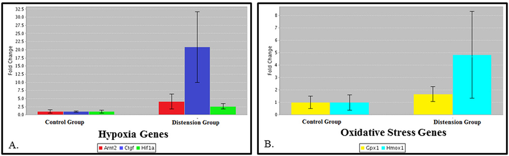
qRT-PCR results in 3 control mice and 3 5-day distension mice. 3A) A significant upregulation of genes associated with hypoxia observed (Hif1a, Ctgf, and Arnt2) (p < 0.05). 3B) Genes critical to clearance of ROS also significantly upregulated (Gpx1 and Hmox1) (p<0.05).
Oxidative Stress
In the acute phase, F2-IsoP levels increased with a slight peak at time 0 before returning to normal by time 24 (Fig 4a). After 3 and 5 days of consecutive over-distension, F2-IsoP levels were unchanged compared to controls (Fig 4b–c).
Figure 4. Measurement of Oxidative Stress.
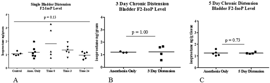
F2-IsoP measurements in acute and chronic distension animals. Horizontal lines represent group means. Fig 5A mild elevation in F2-Isop at time 0 with return to control levels by 24 hours. 1 way ANOVA revealed no statistical significance. Fig 4B and 4C shows identical F2-IsoP levels after 3 and 5 days of bladder distension compared to controls.
Bladder Function
Voided urine blots changed significantly over the course of 5 distensions characterized by increased voiding with gross hematuria (Fig 5). On days 1 and 4 prior to distension, the mean number of voids per animal was 2.8 ± 1.4 and increased to 21.2 ± 16.7, respectively (p=0.06). Mean volume per void decreased from 85.1µl ± 56.9 on day 1 pre-injury to 40.9µl ± 29.8 on day 4 pre-injury (p=0.37) (Fig 6).
Figure 5. Images of Voided Urine Blots.

Urine blots before and after distension in a single representative mouse. The bladder was removed immediately following the last distension on day 5 preventing post-distension urine blot.
Figure 6. Analysis of Voided Urine Blots – 5 day Chronic Distension.

Mean number of voids (lower portion of graph) and mean volume per void (upper portion of graph) over two hours before and after distension in 5 experimental mice. Means analyzed using one-way ANOVA for repeated measures.
DISCUSSION
We theorized that poor bladder emptying leads to over-distension, thus hypothesizing that repetitive and intermittent over-distension creates a hypoxic environment leading to oxidative stress events. We designed a mouse model of bladder distension to mimic a clinical scenario where a patient might experience periods of prolonged over-distension. A perfect example is a myelomeningocele patient that is noncompliant with intermittent catheterization.
Intermittent hypoxia may be a normal part of bladder function. Unlike the kidney, which maintains regions of low oxygen tension (pO2) under normal conditions (<10mmHg),14 the bladder muscle and mucosa maintain estimated levels of at least 25mmHg.15, 16 Several studies agree that brief periods of ischemia with subsequent reperfusion occur during and immediately after voiding in the healthy bladder. It is theorized that compression of intramural blood vessels creates this imbalance in bladder oxygen supply.17 Once the normal bladder empties, blood flow and thus oxygen delivery may actually rebound beyond pre-filling levels.6 These cyclic episodes of ischemia are likely brief (<30 seconds) and infrequent. While a compliant bladder is able to manage these small, intermittent periods of hypoxia, an obstructed bladder may lose the ability to compensate. The duration of post-contraction ischemia in the experimentally obstructed bladder may last up to 5 minutes.3
Using a model of chronic bladder distension, we induced tissue level hypoxia, infiltration of inflammatory cells, increases in hypoxia-related gene transcription, and demonstrable changes in bladder function. Upregulation of several specific hypoxia-related genes suggests a hypoxic response to chronic distension. Hif1a is the key monomer in the dimeric transcription factor Hypoxia Inducible Factor 1 (HIF1), which regulates cell response to hypoxia. Under hypoxic conditions, Hif1a accumulates and translocates to the nucleus to interact with Hif1b, forming functional HIF1.18 Arnt2 complexes with Hif1a in the nucleus and may aid transcriptional activity of HIF1.19 Ctgf is a profibrotic gene transcriptionally induced in renal epithelial cells by direct interaction between HIF1 and the Ctgf promoter.20 Hypoxia is also thought to induce collagen 1 transcription via HIF1. We found Collagen 1 alpha 1 mRNA to be elevated.
Initially, we were uncertain if repeated catheterization and distension would be possible without causing urethral or bladder perforation. These adverse events were uncommon. More commonly, ~20% of mice leaked around the catheter after 3 days of chronic distension, thus requiring frequent refilling of the distending water column to ensure a consistent pressure of 60cm H2O. In addition, maintenance of mouse body temperature was critical as early on several mice died from hypothermia (data not shown). With further refinement, this model could be applied to various aspects of bladder research, including: children with voiding dysfunction and postponement, early changes following bladder obstruction, mechanisms underlying hydrodistension for interstitial cystitis and chronic pelvic pain, and therapies to control bladder inflammation.
Measuring free radicals directly is daunting and various biomarkers have been used to quantify oxidative stress. Isoprostanes are a unique class of compounds now considered the “gold standard” for identifying oxidative stress caused by non-enzymatic free radical peroxidation of arachadonic acid.21 These molecules are similar in structure to prostaglandins and were discovered and characterized at our institution and various classes exist. One specific class, F2-IsoP, is detectable in all human tissues and fluid by mass spectroscopy. This isomer is a chemically stable metabolic end product and is readily quantifiable, thus an attractive marker of lipid peroxidation and in vivo oxidative stress.22 F2-IsoP may also mediate oxidative damage. Known bioactivity includes platelet activation and vascular smooth muscle contraction by ligand-activation of the thromboxane receptor.23
Our interest in oxidative stress stems from attempts to better quantify tissue response to pBOO. We found incremental and significant increases in tissue F2-IsoP levels over time in 4, 8, and 16-week animals following pBOO (unpublished data). In the current study, F2-IsoP levels appeared to be elevated at time 0 with a return to control levels by 24 hours. These data are how we chose our time points for distending bladders in a consecutive daily manner. In 3 and 5-day distension animals, no F2-IsoP increase was noted; however an oxidative stress event may still have occurred. We theorize that chronic distension increased the transcription of genes critical to cellular detoxifying mechanisms. Gpx1, an abundant cellular enzyme containing selenium, is critical to detoxifying hydrogen peroxide free radicals and was increased 2-fold.24 Hmox1, an inducible enzyme that plays a role in cellular free radical scavenging, was increased > 5-fold. In cardiac studies, heterozygous loss of Hmox1 resulted in greater susceptibility to ischemia/reperfusion injury.25
Over-distension has been shown to result in contractile and metabolic dysfunction of the bladder.26, 27 Previous investigators utilized single bladder distensions as a model of acute urinary retention, but no reports of chronic distension models are known. Following a single bladder distension, a burst in ROS has been documented in conjunction with an increase in pro-apoptotic gene transcription.26 Prolonged distension can result in injury to the neural pathways responsible for micturition, reduce bladder elasticity, alter the biochemical and neuronal responsiveness of the bladder, and lead to impaired micturition.28 We utilized voided urine blots, which have been shown to correlate with cystometric studies, as a measure of bladder function.29 Following 5 days of distension, mice developed progressively overactive voiding patterns with frequent, small volume voids. Pre- and post-distension voiding patterns worsened over time, yet the bladder displayed an ability to compensate overnight between distension insults. This appeared to wane until an apparent plateau was reached at day 4. We suspect this was a result of inflammation in the submucosal layers of the bladder, but the role of smooth muscle contractility alteration or changes in neuronal sensory response cannot be excluded. We cannot speculate how hypoxic injury may have affected neural pathways in the bladder; however, new research suggests that the urothelium functions as a sensory neural network all across the bladder lumen.30 Since the urothelium clearly demonstrated hypoxia, it is plausible that urothelial hypoxia may be driving the altered voiding pattern.
CONCLUSION
Experimental chronic over-distension of the mouse bladder creates a hypoxic environment that may mimic clinical disease states. Further studies are needed to demonstrate a true pathological injury but preliminary results from a relatively simple technique are promising. The application of this model will aid our investigation into the mechanisms by which hypoxia can alter bladder structure and function.
ABBREVIATIONS
- Arnt2
Arylhydrocarbon Receptor Nuclear Translocator 2
- F2-IsoP
F2-Isoprostane
- Glut-1
Glucose Transporter 1
- Gpx1
Glutathione peroxidase 1
- H&E
Hematoxylin and Eosin
- Hif1a
Hypoxia inducible factor 1 alpha
- HIF1
Hypoxia inducible factor 1
- Hmox1
Hemoxygenase 1
- IHC
Immunohistochemistry
- pBOO
Partial Bladder Outlet Obstruction
- qRT-PCR
Quantitative real time polymerase chain reaction
- ROS
Reactive oxygen species
REFERENCES
- 1.Drzewiecki BA, Anumanthan G, Penn HA, et al. Modulation of the hypoxic response following partial bladder outlet obstruction. J Urol. 2012;188:1549. doi: 10.1016/j.juro.2012.02.037. [DOI] [PubMed] [Google Scholar]
- 2.Woo LL, Tanaka ST, Anumanthan G, et al. Mesenchymal stem cell recruitment and improved bladder function after bladder outlet obstruction: preliminary data. J Urol. 2011;185:1132. doi: 10.1016/j.juro.2010.10.033. [DOI] [PubMed] [Google Scholar]
- 3.Greenland JE, Brading AF. The effect of bladder outflow obstruction on detrusor blood flow changes during the voiding cycle in conscious pigs. J Urol. 2001;165:245. doi: 10.1097/00005392-200101000-00072. [DOI] [PubMed] [Google Scholar]
- 4.Brading AF. Alterations in the physiological properties of urinary bladder smooth muscle caused by bladder emptying against an obstruction. Scand J Urol Nephrol Suppl. 1997;184:51. [PubMed] [Google Scholar]
- 5.Zderic SA, Butler S, Sliwoski J. Hypoxia Inducible Factor – the Tipping Point for Bladder Decompensation Following Partial Outlet Obstruction. Presented at the American Academy of Pediatrics - Section on Urology; October 15th, 2011; Boston, MA. [Google Scholar]
- 6.Azadzoi KM, Pontari M, Vlachiotis J, et al. Canine bladder blood flow and oxygenation: changes induced by filling, contraction and outlet obstruction. J Urol. 1996;155:1459. doi: 10.1016/s0022-5347(01)66307-9. [DOI] [PubMed] [Google Scholar]
- 7.Azadzoi KM, Tarcan T, Siroky MB, et al. Atherosclerosis-induced chronic ischemia causes bladder fibrosis and non-compliance in the rabbit. J Urol. 1999;161:1626. [PubMed] [Google Scholar]
- 8.Bratslavsky G, Kogan BA, Matsumoto S, et al. Reperfusion injury of the rat bladder is worse than ischemia. J Urol. 2003;170:2086. doi: 10.1097/01.ju.0000092144.48045.13. [DOI] [PubMed] [Google Scholar]
- 9.Martinez-Ferrer M, Iturregui JM, Uwamariya C, et al. Role of nicotinic and estrogen signaling during experimental acute and chronic bladder inflammation. Am J Pathol. 2008;172:59. doi: 10.2353/ajpath.2008.070529. [DOI] [PMC free article] [PubMed] [Google Scholar]
- 10.Hieber S, Huhn R, Hollmann MW, et al. Hypoxia-inducible factor 1 and related gene products in anaesthetic-induced preconditioning. Eur J Anaesthesiol. 2009;26:201. doi: 10.1097/eja.0b013e3283212cbb. [DOI] [PubMed] [Google Scholar]
- 11.Li QF, Zhu YS, Jiang H. Isoflurane preconditioning activates HIF-1alpha, iNOS and Erk1/2 and protects against oxygen-glucose deprivation neuronal injury. Brain Res. 2008;1245:26. doi: 10.1016/j.brainres.2008.09.069. [DOI] [PubMed] [Google Scholar]
- 12.Milne GL, Sanchez SC, Musiek ES, et al. Quantification of F2-isoprostanes as a biomarker of oxidative stress. Nat Protcol. 2007;2:221. doi: 10.1038/nprot.2006.375. [DOI] [PubMed] [Google Scholar]
- 13.Dorr W. Cystometry in mice--influence of bladder filling rate and circadian variations in bladder compliance. J Urol. 1992;148:183. doi: 10.1016/s0022-5347(17)36549-7. [DOI] [PubMed] [Google Scholar]
- 14.Brezis M, Rosen S. Hypoxia of the renal medulla--its implications for disease. N Engl J Med. 1995;332:647. doi: 10.1056/NEJM199503093321006. [DOI] [PubMed] [Google Scholar]
- 15.Badger WJ, Whitbeck C, Kogan B, et al. The immediate effect of castration on female rabbit bladder blood flow and tissue oxygenation. Urol Int. 2006;76:264. doi: 10.1159/000091631. [DOI] [PubMed] [Google Scholar]
- 16.Greenland JE, Hvistendahl JJ, Andersen H, et al. The effect of bladder outlet obstruction on tissue oxygen tension and blood flow in the pig bladder. BJU Int. 2000;85:1109. doi: 10.1046/j.1464-410x.2000.00611.x. [DOI] [PubMed] [Google Scholar]
- 17.Matsumoto S, Chichester P, Kogan BA, et al. Structural and vascular response of normal and obstructed rabbit whole bladders to distension. Urology. 2003;62:1129. doi: 10.1016/s0090-4295(03)00686-1. [DOI] [PubMed] [Google Scholar]
- 18.Higgins DF, Kimura K, Iwano M, et al. Hypoxia-inducible factor signaling in the development of tissue fibrosis. Cell Cycle. 2008;7:1128. doi: 10.4161/cc.7.9.5804. [DOI] [PMC free article] [PubMed] [Google Scholar]
- 19.Maltepe E, Keith B, Arsham AM, et al. The role of ARNT2 in tumor angiogenesis and the neural response to hypoxia. Biochem Biophys Res Commun. 2000;273:231. doi: 10.1006/bbrc.2000.2928. [DOI] [PubMed] [Google Scholar]
- 20.Higgins DF, Biju MP, Akai Y, et al. Hypoxic induction of Ctgf is directly mediated by Hif-1. Am J Physiol Renal Physiol. 2004;287:F1223. doi: 10.1152/ajprenal.00245.2004. [DOI] [PubMed] [Google Scholar]
- 21.Yin H, Porter NA. New insights regarding the autoxidation of polyunsaturated fatty acids. Antiox Redox Signaling. 2005;7:170. doi: 10.1089/ars.2005.7.170. [DOI] [PubMed] [Google Scholar]
- 22.Morrow JD, Zackert WE, Yang JP, et al. Quantification of the major urinary metabolite of 15-F2t-isoprostane (8-iso-PGF2alpha) by a stable isotope dilution mass spectrometric assay. Anal Biochem. 1999;269:326. doi: 10.1006/abio.1999.4008. [DOI] [PubMed] [Google Scholar]
- 23.Milne GL, Yin H, Hardy KD, et al. Isoprostane generation and function. Chem Rev. 2011;111:5973. doi: 10.1021/cr200160h. [DOI] [PMC free article] [PubMed] [Google Scholar]
- 24.Tsutsui H, Kinugawa S, Matsushima S. Oxidative stress and heart failure. Am J Physiol Heart Circ Physiol. 2011;301:H2181. doi: 10.1152/ajpheart.00554.2011. [DOI] [PubMed] [Google Scholar]
- 25.Yoshida T, Maulik N, Ho YS, et al. H(mox-1) constitutes an adaptive response to effect antioxidant cardioprotection: A study with transgenic mice heterozygous for targeted disruption of the Heme oxygenase-1 gene. Circulation. 2001;103:1695. doi: 10.1161/01.cir.103.12.1695. [DOI] [PubMed] [Google Scholar]
- 26.Yu HJ, Chien CT, Lai YJ, et al. Hypoxia preconditioning attenuates bladder overdistension-induced oxidative injury by up-regulation of Bcl-2 in the rat. J Physiol (Lond) 2004;554:815. doi: 10.1113/jphysiol.2003.056002. [DOI] [PMC free article] [PubMed] [Google Scholar]
- 27.Li WJ, Shin MK, Oh SJ. Time dependent bladder apoptosis induced by acute bladder outlet obstruction and subsequent emptying is associated with decreased MnSOD expression and Bcl-2/Bax ratio. J Korean Med Sci. 2010;25:1652. doi: 10.3346/jkms.2010.25.11.1652. [DOI] [PMC free article] [PubMed] [Google Scholar]
- 28.Tammela T, Lasanen L, Waris T. Effect of distension on adrenergic innervation of the rat urinary bladder. Urol Res. 1990;18:345. doi: 10.1007/BF00300785. [DOI] [PubMed] [Google Scholar]
- 29.Hodges SJ, Zhou G, Deng FM, et al. Voiding pattern analysis as a surrogate for cystometric evaluation in uroplakin II knockout mice. J Urol. 2008;179:2046. doi: 10.1016/j.juro.2007.12.039. [DOI] [PubMed] [Google Scholar]
- 30.Khandelwal P, Abraham SN, Apodaca G. Cell biology and physiology of the uroepithelium. Am J Physiol Renal Physiol. 2009;297:F1477. doi: 10.1152/ajprenal.00327.2009. [DOI] [PMC free article] [PubMed] [Google Scholar]